Introduction: Why 2D NMR for Peptide Characterization Is an Essential Tool in Therapeutic Development
When a therapeutic peptide progresses into the drug development pipeline, confirming the primary sequence and molecular mass is essential, but these data alone are insufficient for complete structural evaluation. Peptides adopt dynamic conformations in solution. They can form transient helical regions, establish intramolecular hydrogen bonds, and exist in multiple conformational states simultaneously. These structural behaviors directly influence receptor binding affinity, enzymatic stability, pharmacokinetics, and immunogenicity. Among currently available analytical technologies, 2D NMR for peptide characterization uniquely provides atomic-level structural information in a biologically relevant solution-state environment while simultaneously resolving these conformational properties.
At ResolveMass Laboratories Inc., 2D NMR characterization workflows for therapeutic peptides are specifically optimized for compounds within the sub-5 kDa molecular weight range, where peptide behavior differs significantly from both traditional small molecules and larger biologics. This article examines, at a technical depth appropriate for structural biologists, CMC scientists, analytical chemists, and regulatory specialists, how COSY, TOCSY, and NOESY experiments are designed, interpreted, and integrated into a comprehensive structural characterization strategy for therapeutic peptide candidates.
Discover how comprehensive analytical strategies fit into broader pharmaceutical pipelines by exploring our overview on peptide characterization in drug development.
Share via:
Article Summary:
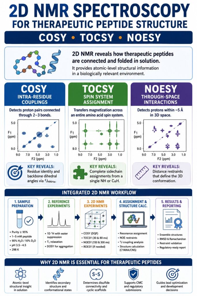
- Advanced 2D NMR techniques for peptide analysis — including COSY, TOCSY, and NOESY — deliver detailed atomic-level structural insights that cannot be fully achieved through conventional 1D NMR or mass spectrometry alone, making them indispensable in therapeutic peptide research.
- COSY experiments identify short-range scalar couplings between neighboring protons within amino acid residues, TOCSY propagates these correlations across complete amino acid spin networks, and NOESY detects spatial proton-proton interactions that reveal the peptide’s three-dimensional architecture in solution.
- Structural assignment of peptides typically follows the classical Wüthrich methodology, beginning with amino acid spin system recognition using TOCSY, followed by sequential residue connectivity analysis through NOESY, and ultimately progressing to structure generation using experimentally derived distance restraints.
- In pharmaceutical peptide development, 2D NMR plays a major role in determining disulfide bond arrangements, evaluating cyclic peptide conformations, characterizing helical or beta-sheet secondary structures during lead optimization, and identifying aggregation-sensitive regions important for formulation studies.
- Experimental conditions such as solvent composition (for example H₂O/D₂O mixtures or DMSO-d₆), peptide concentration, solution pH, and acquisition temperature significantly influence spectral quality as well as NOE intensity and polarity in NOESY measurements.
- Among all NOESY acquisition variables, the choice of mixing time (τm) is especially critical because accurate interproton distance determination depends on confirming linear NOE buildup behavior under carefully optimized conditions.
- Computational structure-calculation platforms such as CYANA, ARIA, and Rosetta integrate experimentally derived NMR restraints to generate statistically validated ensemble-based three-dimensional peptide models with measurable structural precision.
- Regulatory authorities increasingly expect comprehensive solution-state conformational characterization for innovative peptide therapeutics, and high-quality 2D NMR datasets are commonly included as core structural evidence within IND and NDA regulatory submissions.
The Spectral Overlap Problem in Therapeutic Peptides: Why Two-Dimensional NMR Is Required
A 20-residue peptide typically contains approximately 20 amide NH protons, 20 alpha-protons, and numerous sidechain protons distributed within a crowded 1D ¹H NMR spectral range between 0.5 and 9.5 ppm. The resulting resonance overlap is not merely inconvenient; it makes definitive resonance assignment impossible using 1D spectroscopy alone.
The addition of a second frequency dimension separates resonances according to their coupling partners rather than relying exclusively on chemical shift dispersion. This provides several major analytical advantages:
- Amide NH region (6.5–9.5 ppm): Even in peptides containing more than 30 residues, TOCSY cross-peaks resolve individual amino acid spin systems because the second dimension separates signals according to alpha-proton chemical shifts.
- Aliphatic region (0.5–4.5 ppm): COSY experiments separate overlapping sidechain coupling patterns that are indistinguishable in conventional 1D spectra.
- Critical importance for cyclic peptides: N-methylated and backbone-cyclized peptides frequently lack amide NH protons on modified residues. Two-dimensional experiments restore connectivity information that cannot be recovered from 1D data.
The enhanced chemical shift dispersion achievable with 600–800 MHz NMR spectrometers, which represent the standard instrumentation range for advanced peptide NMR analysis, provides sufficient resolution for assigning peptides containing up to approximately 50 residues as single structural entities.
Understand the technical distinctions between mapping workflows and complete sequencing protocols by reading our guide on peptide mapping vs. peptide sequencing key differences.
COSY (COrrelation SpectroscopY): Resolving Intra-Residue Scalar Couplings
COSY cross-peaks identify proton pairs connected through two-bond and three-bond scalar couplings (²J and ³J interactions). In peptide systems, these couplings primarily correspond to direct NH–CαH and CαH–CβH interactions within individual amino acid spin systems.
What COSY Reveals in a Therapeutic Peptide
In a standard DQF-COSY (Double Quantum Filtered COSY) experiment, diagonal peaks are selectively suppressed relative to cross-peaks, significantly improving the signal-to-noise ratio for coupled proton pairs. For peptides dissolved in H₂O/D₂O (90:10), the following correlations are particularly informative:
| Correlation Type | Chemical Shift Range | Diagnostic Value |
|---|---|---|
| NH–CαH | NH: 7.5–9.5 ppm; CαH: 3.5–5.5 ppm | Confirms residue identity; ³JHNHα reports φ dihedral angle |
| CαH–CβH | 3.5–5.5 ppm / 1.5–3.5 ppm | Initiates spin system assignment for Ala, Ser, Cys, Asp, and Asn |
| CβH–CγH | 1.5–3.5 ppm / 0.8–2.5 ppm | Extends spin systems for Leu, Ile, Val, Lys, and Arg |
| CαH–CαH (Gly) | 3.8–4.2 ppm | Distinguishes glycine, the only amino acid containing two CαH protons |
The ³JHNHα Coupling Constant: A COSY-Derived Reporter of Backbone Geometry
The vicinal coupling constant extracted from NH–CαH COSY cross-peak line shapes provides direct information about the peptide backbone φ torsion angle through the Karplus relationship:
3JHNHα=Acos2ϕ−Bcosϕ+C{}^{3}J_{HN H\alpha}=A\cos^{2}\phi-B\cos\phi+C3JHNHα=Acos2ϕ−Bcosϕ+C
Interpretation of ³JHNHα values provides immediate conformational insight:
- ³JHNHα < 5 Hz: Indicates φ angles near −60°, consistent with helical conformations.
- ³JHNHα > 8 Hz: Indicates φ angles between approximately −120° and −150°, characteristic of extended or beta-sheet conformations.
- ³JHNHα ≈ 6–7 Hz: Suggests conformational averaging and the coexistence of multiple structural states.
For therapeutic peptide candidates in early-stage development, this parameter alone can provide valuable conformational screening information before full structural calculations are performed.
DQF-COSY vs. E.COSY in Peptide Analysis
- DQF-COSY: The standard choice for peptide characterization because it suppresses water and diagonal signals while producing cleanly phased cross-peaks.
- E.COSY: Preferred when highly accurate coupling constant measurements are required, particularly for φ dihedral angle determination in cyclic peptides and disulfide-rich scaffolds. E.COSY spectra produce characteristic L-shaped multiplet patterns that enable sub-Hz coupling measurements.
TOCSY (TOtal Correlation SpectroscopY): Complete Spin System Assignment
TOCSY experiments propagate magnetization through all scalar-coupled protons within a single amino acid spin system. As a result, complete sidechain assignments can be obtained from a single NH or CαH cross-peak row, even when protons are separated by multiple covalent bonds.
TOCSY Mixing Time Optimization for Therapeutic Peptides
The TOCSY spin-lock mixing time (τmix) determines the number of covalent bonds over which coherence transfer occurs.
| τmix (ms) | Magnetization Transfer Depth | Optimal For |
|---|---|---|
| 20–30 ms | 2–3 bonds | Short spin systems: Ala, Ser, Cys, Gly |
| 50–60 ms | 3–5 bonds | Medium spin systems: Asp, Asn, Leu, Val, Thr |
| 80–100 ms | 5–8 bonds | Long spin systems: Lys, Arg, Ile |
For therapeutic peptides containing more than 15 residues, running two TOCSY experiments at τmix = 30 ms and τmix = 80 ms is standard analytical practice. The shorter mixing-time experiment identifies spin system classes, while the longer mixing-time experiment extends connectivity across complete sidechain networks.
TOCSY in H₂O vs. DMSO-d₆: Solvent Selection and Structural Relevance
The solvent system selected for TOCSY experiments has major implications for the biological relevance of the resulting structural data.
H₂O/D₂O (90:10), 298 K
- NH protons remain observable without excessive exchange.
- Temperature-dependent amide chemical shift coefficients (-Δδ/ΔT) provide hydrogen bonding information.
- The solvent environment closely resembles physiological conditions.
- Water suppression methods such as WATERGATE or excitation sculpting are required.
DMSO-d₆, 298–310 K
- NH resonances are sharp and fully visible due to slower exchange.
- Particularly useful for poorly water-soluble therapeutic peptides.
- ³JHNHα values may differ substantially from aqueous measurements.
- Not ideal for final biologically relevant conformational characterization.
TFE-d₃/H₂O Mixtures
- Commonly used to intentionally stabilize helical conformations.
- Appropriate for evaluating helix-forming propensity rather than native-state structural behavior.
For regulatory-quality structural characterization workflows at ResolveMass Laboratories Inc., aqueous conditions are always prioritized for primary datasets, while DMSO measurements are treated as supplementary data for poorly soluble compounds.
TOCSY Spin System Signatures for Common Amino Acids
| Amino Acid | TOCSY Signature | Distinguishing Feature |
|---|---|---|
| Glycine | CαH₂ doublet only | Two CαH protons; lacks NH–CβH correlation |
| Alanine | NH, CαH, CβH | Three-proton spin system; characteristic CβH near 1.4 ppm |
| Valine | NH, CαH, CβH, two CγH₃ | Branched sidechain; methyl resonances at 0.8–1.0 ppm |
| Leucine | NH, CαH, CβH, CγH, two CδH₃ | Extended sidechain with terminal methyl doublets |
| Isoleucine | NH, CαH, CβH, CγH₂, CδH₃ | Asymmetric branching and complex CβH multiplet |
| Lysine | NH through CεH₂ | Longest aliphatic spin system; CεH₂ near water resonance |
| Threonine | NH, CαH, CβH, CγH₃ | CβH near 4.2 ppm; methyl doublet near 1.2 ppm |
| Proline | CαH, CβH, CγH, CδH | No NH proton due to tertiary amine structure |
NOESY (Nuclear Overhauser Effect SpectroscopY): Determining 3D Conformation Through Through-Space Interactions
NOESY cross-peaks arise between proton pairs separated by approximately 5 Å or less in three-dimensional space, regardless of whether the protons are covalently connected. These through-space interactions provide the primary distance restraints used in peptide 3D structure determination.
The NOE Distance Relationship and Its Practical Implications
The cross-relaxation rate (σ) between two protons is proportional to the inverse sixth power of the internuclear distance:
σ∝r−6×f(τc)\sigma\propto r^{-6}\times f(\tau_c)σ∝r−6×f(τc)
Because of the strong r⁻⁶ dependence:
- Cross-peaks at 1.8–2.5 Å are strong and commonly correspond to sequential backbone HN–HN contacts in helices.
- Cross-peaks at 3.0–4.0 Å are medium intensity and frequently define turn geometries.
- Cross-peaks at 4.0–5.0 Å are weak and often represent long-range tertiary contacts.
Accurate quantitative distance extraction requires:
- Calibration of NOE volumes using a known reference distance, such as glycine geminal CαH₂ protons at 1.78 Å.
- Verification of linear NOE buildup using multiple mixing times (τm = 50, 100, 200, and 300 ms).
- Correction for spin diffusion effects at longer mixing times.
NOESY Mixing Time Selection for Therapeutic Peptides
| Peptide MW (Da) | τc (ns, ~298 K) | Recommended τm | Spin Diffusion Risk |
|---|---|---|---|
| < 500 | < 0.1 | 300–500 ms | Low (positive NOE regime) |
| 500–2,000 | 0.1–1.0 | 200–300 ms | Moderate (near zero-crossing) |
| 2,000–5,000 | 1–5 | 100–200 ms | Moderate–High |
| 5,000–10,000 | 5–15 | 50–150 ms | High (negative NOE, fast relay) |
The Zero-Crossing Challenge
Small peptides with molecular weights below approximately 1,000 Da in aqueous solution at 298 K operate near the condition ωτc ≈ 1, where NOE intensities approach zero. Several strategies are commonly used to overcome this issue:
- Lowering the temperature to increase τc, for example by measuring at 278 K.
- Increasing solvent viscosity using 10–30% d₆-glycerol.
- Using ROESY instead of NOESY, since rotating-frame NOEs remain negative for small molecules.
Sequential NOE Assignment: The Wüthrich Strategy Applied to Therapeutic Peptides
The classical Wüthrich sequential assignment strategy remains the foundation of peptide NMR structure determination:
- TOCSY: Identify all amino acid spin systems and assign tentative residue types.
- NOESY dNN(i,i+1): Establish backbone connectivity through NH(i)–NH(i+1) cross-peaks, particularly strong in helices.
- NOESY dαN(i,i+1): Detect alpha-H(i)–NH(i+1) contacts, commonly strong in extended conformations.
- NOESY dβN(i,i+1): Use beta-H(i)–NH(i+1) contacts as secondary confirmatory restraints.
- Medium-range NOEs: dNN(i,i+2), dαN(i,i+3), and dαβ(i,i+3) identify helix geometry and turn structures.
- Long-range NOEs: dαα(i,j) interactions where |i−j| > 4 identify beta-sheet contacts and tertiary organization.
NOE Patterns Diagnostic for Secondary Structure
| Secondary Structure | Characteristic NOE Pattern |
|---|---|
| α-Helix | Strong dNN(i,i+1), medium dαN(i,i+3), medium dαβ(i,i+3) |
| 3₁₀-Helix | Strong dNN(i,i+1), medium dαN(i,i+2), absent dαN(i,i+3) |
| β-Sheet (parallel) | Strong dαα(i,j), absent dNN(i,i+1) |
| β-Sheet (antiparallel) | Strong dαα(i,j), weak dNN(i,j±1) |
| β-Turn Type I | dαN(i,i+2), dNN(i+2,i+3), dαN(i,i+3) |
| β-Turn Type II | dαN(i,i+2), dNN(i+1,i+2), absent dNα(i,i+1) |
| Random Coil | Strong dαN(i,i+1) only; lacks medium- and long-range NOEs |
Integrated 2D NMR Workflow for Therapeutic Peptide Structure Determination
A complete solution-state structural characterization workflow at ResolveMass Laboratories Inc. follows a standardized analytical sequence.
Phase 1 — Sample Preparation and Quality Control
- Peptide purity ≥ 95% by HPLC-MS prior to NMR analysis
- Concentration: 1–5 mM in 90% H₂O / 10% D₂O
- pH range: 3.5–4.5 to minimize rapid NH exchange
- Standard temperature: 298 K, with additional temperatures as needed
- Internal reference: DSS at 0.00 ppm
Phase 2 — 1D ¹H and Reference Experiments
- 1D ¹H spectroscopy with water suppression
- T₁ relaxation measurements for recycle delay optimization
- DOSY experiments for aggregation screening and hydrodynamic radius analysis
Phase 3 — 2D NMR Experiments
- DQF-COSY for scalar coupling assignment
- TOCSY at τmix = 30 and 80 ms
- NOESY at τmix = 150 and 300 ms
- ROESY for small peptides when appropriate
- Optional ¹H-¹³C HSQC and HMBC for modified peptide characterization
Phase 4 — Assignment and Structure Calculation
- Manual assignment using Sparky, NMRFAM-Sparky, or CcpNmr Analysis
- NOE volume integration and restraint generation
- Dihedral angle restraints derived from ³JHNHα coupling constants
- Structure calculation using CYANA or CNS
- Ensemble refinement to obtain the lowest-energy structural family
- Validation using PROCHECK-NMR and AQUA
Phase 5 — Reporting
- Backbone and heavy atom RMSD values
- Ramachandran plot statistics
- NOE restraint violation analysis
- NMR-derived structural descriptors for CMC documentation
For teams developing metabolic therapies, review our specialised glp-1 analog peptide sequencing workflow to see how sequencing parameters are customized for blockbusters in this class.
Application-Specific Considerations in Therapeutic Peptide NMR
Disulfide-Bridged Peptides
For disulfide-rich peptides such as defensins, conotoxins, and insulin analogs, 2D NMR remains the definitive method for assigning disulfide connectivity when multiple cysteine residues are present. Long-range NOESY cross-peaks between cysteine CβH protons forming disulfide bonds are commonly observed in the 2.9–3.3 ppm region when the corresponding protons lie within 4–5 Å.
While partial reduction followed by mass spectrometry can suggest possible disulfide arrangements, NMR provides direct conformational information and clarifies whether the disulfide network stabilizes specific secondary structural motifs.
Learn how impurities and modifications are tracked during structural assessments by reading about glp-1 peptide impurity characterization.
Cyclic Peptides and N-Methylated Scaffolds
Backbone cyclization removes conventional N- and C-terminal assignment references used in linear peptide analysis. Therefore, sequential assignment strategies must be modified:
- Begin assignment from the most downfield amide NH resonance.
- Confirm ring closure using NOESY through-space connectivities.
- Identify N-methylated residues through missing NH resonances and characteristic alpha-proton chemical shifts.
Amphipathic Helical Peptides
Temperature-dependent amide chemical shift coefficients (-Δδ/ΔT) distinguish solvent-exposed residues from hydrogen-bonded residues:
- -Δδ/ΔT > 4 ppb/K: Solvent exposed and not hydrogen bonded
- -Δδ/ΔT < 2 ppb/K: Internally hydrogen bonded and consistent with helical organization
When combined with NOESY-derived helical patterns, these measurements provide residue-specific hydrogen bond maps without requiring crystallographic analysis.
Examine practical analytical findings from a real-world compound setup in our peptide characterization of ganirelix generic project report.
From 2D NMR Distance Restraints to Final 3D Structure
Transforming raw NMR data into a validated three-dimensional peptide structure requires multiple computational and analytical stages:
- NOE volume integration: Integrated peak volumes, rather than peak heights, must be measured accurately.
- Distance calibration: Reference distances establish the r⁻⁶ calibration relationship.
- Restraint classification: Distance restraints are categorized as strong, medium, or weak.
- Pseudoatom corrections: Additional upper-bound corrections are applied for unresolved methyl and methylene groups.
- Structure calculation: CYANA commonly performs torsion-angle dynamics followed by refinement using AMBER or OPLS force fields.
- Quality assessment: Backbone RMSD values below 1.0 Å generally indicate well-defined structures, whereas RMSD values above 2.0 Å may indicate insufficient restraints or conformational heterogeneity.
If your program requires high-throughput sequencing support, evaluate our specialized glp-1 peptide sequencing cro services.
Regulatory and CMC Importance of 2D NMR Data for Therapeutic Peptides
ICH Q6B guidance, together with evolving FDA and EMA expectations for peptide therapeutics and complex biologics, increasingly recognizes solution-state structural characterization as a key component of physicochemical characterization packages.
Regulatory submissions involving 2D NMR peptide characterization commonly include:
- Complete proton resonance assignment tables
- COSY-derived coupling constant analyses
- NOESY-derived distance restraint lists
- Ensembles of calculated structures with RMSD and Ramachandran statistics
- Spectral comparability studies between drug substance and reference standards
2D NMR characterization is especially valuable in IND submissions involving first-in-class peptide therapeutics where crystallographic analysis is impractical and where biologically relevant solution-state conformations are more informative than crystal packing arrangements.
Ensure your filings align with global health agency metrics by checking out fda requirements for peptide characterization.
Conclusion: The Essential Role of 2D NMR in Modern Therapeutic Peptide Development
The structural insight provided by COSY, TOCSY, and NOESY experiments, ranging from local backbone torsion angles to complete three-dimensional conformational ensembles, makes 2D NMR for peptide characterization indispensable in modern therapeutic peptide research and pharmaceutical development. No other analytical platform simultaneously delivers residue-specific conformational information in solution, hydrogen bond mapping, disulfide connectivity assignment, cyclic scaffold characterization, and regulatory-grade structural validation from a single sample preparation.
When advanced 2D NMR methodologies are combined with optimized sample preparation, carefully selected experimental parameters, and validated computational structure calculation workflows, the resulting structural characterization packages provide scientifically rigorous and regulatorily defensible data that support peptide therapeutic programs from lead optimization through full CMC documentation.
For therapeutic peptide programs requiring advanced 2D NMR structural characterization, ResolveMass Laboratories Inc. provides comprehensive end-to-end analytical support, including sample preparation consultation, spectral acquisition, resonance assignment, structural calculation, and regulatory-ready reporting.
Explore our dedicated regional offerings for a peptide sameness study for anda
or connect with our teams offering peptide sameness study services in canada.
Frequently Asked Questions (FAQs)
The amount of peptide required for 2D NMR characterization depends on instrument sensitivity, probe type, and peptide solubility. In most cases, a standard 600 MHz NMR system requires approximately 0.5–2 µmol of peptide dissolved in about 550 µL of solvent to achieve concentrations within the 1–5 mM range. When cryogenic probes are available, significantly lower quantities, sometimes as little as 0.1–0.5 µmol, may still provide usable TOCSY or NOESY spectra. However, lower sample concentrations generally require longer acquisition times to maintain acceptable signal quality and spectral resolution.
Yes, 2D NMR can identify peptide aggregation, although severe aggregation often compromises spectral quality and limits structural interpretation. Aggregation is commonly detected through broadened resonances in 1D spectra, abnormal diffusion behavior in DOSY experiments, and reduced TOCSY or NOESY cross-peak intensity. Several strategies can help reduce aggregation, including lowering peptide concentration, adjusting pH, modifying temperature, or adding mild denaturants such as low concentrations of urea or guanidinium chloride. If aggregation remains unresolved, alternative structural techniques such as solid-state NMR or cryo-electron microscopy may be necessary.
NOESY and ROESY both provide through-space structural information, but they operate through different relaxation mechanisms. NOESY relies on longitudinal cross-relaxation and works best for medium-sized molecules, whereas very small peptides often produce weak or near-zero NOE intensities under standard aqueous conditions. ROESY overcomes this limitation by using a spin-lock field that generates observable cross-peaks regardless of molecular size, making it particularly useful for peptides below approximately 1,000–1,500 Da. Although ROESY is highly effective for small therapeutic peptides, careful interpretation is required because relay artifacts resembling TOCSY correlations can sometimes appear in the spectra.
A well-defined therapeutic peptide structure generally requires approximately 10–15 NOE-derived distance restraints per residue to achieve strong structural convergence. For example, a 20-residue helical peptide may generate 200–300 clearly assignable NOEs that provide accurate backbone and sidechain positioning. Lower restraint densities may still reveal secondary structural features such as helices or turns, but the overall three-dimensional precision becomes less reliable. The quality of restraint distribution across the sequence is equally important because uneven restraint coverage can weaken structural confidence in flexible regions.
Peptides containing proline residues frequently exist as mixtures of cis and trans conformers that exchange slowly on the NMR timescale. This behavior typically produces two separate resonance sets corresponding to the two conformational states. TOCSY experiments often reveal duplicate spin systems for residues adjacent to proline, while NOESY or EXSY cross-peaks can demonstrate chemical exchange between the isomers. In addition, the alpha-proton chemical shift of the residue immediately following proline often provides a useful indicator, with cis conformers generally appearing further downfield than trans conformers.
As peptide size increases beyond approximately 40–50 residues, several technical challenges begin to reduce the effectiveness of conventional 2D ¹H NMR analysis. Larger peptides generate extensive spectral overlap, making resonance assignment increasingly difficult even in high-field instruments. In addition, faster transverse relaxation leads to broader peaks and reduced sensitivity, while spin diffusion complicates quantitative NOE interpretation. To overcome these limitations, researchers often use isotope labeling with ¹³C and ¹⁵N, multidimensional heteronuclear experiments, perdeuteration strategies, or ultrahigh-field spectrometers operating in the 800–1,000 MHz rang
In comparability assessments, 2D NMR acts as a highly sensitive structural fingerprinting tool for evaluating consistency between peptide batches or biosimilar products. Complete proton resonance assignments obtained from TOCSY and NOESY spectra are compared directly with reference standards to identify even subtle conformational differences. Small chemical shift changes in backbone amide or alpha-proton resonances may indicate structural alterations caused by manufacturing changes, degradation, or incorrect folding. This approach is particularly important for disulfide-rich peptides, where altered connectivity can significantly affect the overall conformational ensemble.
Water suppression is essential in peptide NMR because the water resonance is extremely intense and can obscure nearby amide proton signals that are critical for structural analysis. Techniques such as WATERGATE and excitation sculpting effectively suppress the water signal while preserving neighboring NH resonances. In contrast, presaturation methods may unintentionally reduce the intensity of exchangeable amide protons through saturation transfer effects, especially at physiological pH where proton exchange is rapid. Proper water suppression therefore plays a major role in maintaining accurate TOCSY and NOESY cross-peak detection and ensuring reliable resonance assignments.
Reference:
- International Conference on Harmonisation of Technical Requirements for Registration of Pharmaceuticals for Human Use. (1999). Q6B specifications: Test procedures and acceptance criteria for biotechnological/biological products. https://database.ich.org/sites/default/files/Q6B%20Guideline.pdf
- Boutin, J. A., Tartar, A. L., van Dorsselaer, A., & Vaudry, H. (2019). General lack of structural characterization of chemically synthesized long peptides. Protein Science, 28(5), 857–867. https://doi.org/10.1002/pro.3601
- Patel, A., & Patel, R. (2024). Analytical techniques for peptide-based drug development: Characterization, stability and quality control. International Journal of Science and Research Archive, 12(1). https://doi.org/10.30574/ijsra.2024.12.1.1108
- European Medicines Agency. (2023). Draft guideline on the development and manufacture of synthetic peptides (EMA/CHMP/CVMP/QWP/387541/2023). European Medicines Agency. https://www.ema.europa.eu/en/documents/scientific-guideline/draft-guideline-development-manufacture-synthetic-peptides_en.pdf

