The Strategic Role of Biosimilar Characterization Services in Biopharmaceutical Approvals
Biosimilar Characterization Services play a critical role in establishing analytical similarity by delivering comprehensive structural and functional comparisons between a biosimilar candidate and its reference medicinal product. The objective is to demonstrate that there are no clinically meaningful differences between the two products. This extensive analytical evaluation forms the cornerstone of the “Totality of Evidence” framework required by global regulatory authorities for marketing authorization.
To ensure that subsequent-entry biologics meet established standards of safety and efficacy, developers must rely on robust Biosimilar Characterization Services that satisfy the stringent comparability requirements imposed by international regulatory agencies. The process of demonstrating biosimilarity is governed by the “Totality of Evidence” framework established by the US Food and Drug Administration (FDA) and the European Medicines Agency (EMA). Unlike conventional small-molecule generic drugs, which typically establish equivalence through relatively simple chemical quantification, complex biotherapeutic macromolecules are produced in living expression systems and exhibit inherent microheterogeneity. This intrinsic complexity necessitates multidimensional structural fingerprinting.
Under international regulatory standards, including the International Council for Harmonisation (ICH) Topic Q6B guideline, developers are required to conduct comprehensive structural and physicochemical comparability studies. Standardization of these analytical activities is essential because even subtle changes in manufacturing processes can introduce structural variations that may affect clinical efficacy, product stability, and patient safety through immunogenic responses. By integrating advanced analytical characterization early in biotherapeutic development programs, sponsors can significantly reduce the risk of downstream clinical failures, lower overall development costs, and accelerate time to market. High-resolution mass spectrometry has emerged as the analytical gold standard for these comparative assessments, offering sub-Dalton accuracy and deep molecular characterization throughout the therapeutic product lifecycle.
Discover how our expert analytical workflows establish definitive structural identity. Read our comprehensive guide on Biosimilar Characterization Using Mass Spectrometry.
Article Summary
- Biosimilar Characterization Services are essential for regulatory approval, providing detailed structural and functional comparisons between biosimilars and reference biologics to demonstrate analytical similarity and support the totality-of-evidence approach.
- High-resolution mass spectrometry is the cornerstone of biosimilar characterization, enabling precise evaluation of protein structure, sequence integrity, post-translational modifications, and product heterogeneity.
- Peptide mapping using LC-MS/MS verifies primary amino acid sequences, helping detect sequence variants, degradation products, and terminal modifications that could impact biosimilarity claims.
- Intact mass, top-down, and middle-up analyses provide complementary structural insights, allowing scientists to assess molecular weight, proteoform distributions, glycosylation patterns, and large-scale structural differences.
- Comprehensive glycan profiling is critical because glycosylation influences efficacy and safety, with HILIC-MS workflows used to characterize glycan composition, site occupancy, and biological functionality.
- Disulfide bond mapping and conformational studies ensure correct protein folding, helping identify structural abnormalities that may affect stability, biological activity, or immunogenicity.
- Mass spectrometry-based impurity profiling offers advantages over traditional ELISA methods, enabling direct identification and quantification of host cell proteins and other process-related impurities that may pose safety risks.
- Early implementation of advanced characterization strategies reduces development risk, supports regulatory compliance, accelerates biosimilar development timelines, and helps demonstrate that any observed differences are not clinically meaningful.
Primary Sequence Verification Workflows in Biosimilar Characterization Services
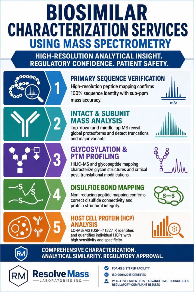
Primary sequence verification within Biosimilar Characterization Services confirms that the amino acid sequence of the biosimilar is fully identical to that of the reference product. This verification is achieved through high-resolution peptide mapping, which delivers sub-parts-per-million mass accuracy while enabling site-specific identification of sequence variations.
Definitive confirmation of the primary amino acid sequence is a fundamental regulatory requirement. Any transcriptional error or amino acid substitution introduced during cell line development can generate sequence variants that undermine biosimilarity claims. The accepted analytical standard for sequence confirmation is peptide mapping, a bottom-up proteomic strategy involving enzymatic digestion of the therapeutic protein into individual peptide fragments, followed by liquid chromatography coupled with online tandem mass spectrometry (LC-MS/MS).
Learn how to systematically structure your comparative data packages for global agencies. View our framework on how to Prove Biosimilarity Using LC-MS.
Advanced Peptide Mapping Protocols for Biosimilar Characterization Services
Advanced peptide mapping protocols employed in Biosimilar Characterization Services combine enzymatic digestion with high-resolution liquid chromatography-tandem mass spectrometry to confirm sequence coverage. This bottom-up methodology enables direct, site-specific identification of terminal modifications, chemical degradation products, and single-amino-acid substitutions.
To achieve complete sequence coverage, the target protein is generally digested using highly specific proteases such as sequencing-grade trypsin. The resulting peptide fragments are separated through reversed-phase liquid chromatography (RPLC) utilizing advanced column chemistries, including ACQUITY Premier Peptide CSH and bioZen WidePore columns. These columns incorporate bio-inert hardware surface technologies designed to eliminate non-specific metal-ion interactions.
The separated peptides are subsequently ionized through electrospray ionization (ESI) and analyzed using high-resolution mass spectrometers operating in either Data-Dependent Acquisition (DDA) mode or Data-Independent Acquisition (DIA) mode, including MS^E. By comparing experimentally observed mass-to-charge (m/z) ratios of precursor ions and their corresponding collision-induced dissociation (CID) or higher-energy collisional dissociation (HCD) product ions (b and y ions) with theoretical peptide sequences, researchers routinely achieve sequence coverage exceeding 95%. This degree of analytical resolution enables precise localization of sequence variants, terminal clipping events such as C-terminal lysine truncation, and chemical degradation processes including deamidation and oxidation.
The complexity of these workflows requires careful optimization of chromatographic conditions. A representative optimized RPLC-MS/MS peptide mapping method is summarized below:
| Method Parameter | Target Specification / Condition | Analytical Significance |
|---|---|---|
| Chromatography System | ACQUITY Premier UPLC or Vanquish UPLC | Eliminates metal-adsorptive losses |
| Stationary Phase | ACQUITY Premier Peptide CSH C18 (1.7 μm, 2.1 × 100 mm) | Maximizes resolution of polar peptides |
| Mobile Phase A | 0.1% Formic Acid in Water | Promotes efficient peptide protonation and ionization |
| Mobile Phase B | 0.1% Formic Acid in 95% Acetonitrile / 5% Water | Provides optimal elution strength and volatility |
| Column Temperature | 60 °C | Minimizes secondary interactions and structural artifacts |
| MS Acquisition Mode | Data-Independent Acquisition (DIA / MS^E) | Ensures comprehensive and unbiased fragment collection |
| Mass Accuracy | Sub-5 ppm (1 ppm with lock-mass calibrant) | Confirms precise peptide chemical formulas |
During biosimilar development, sponsors are expected to conduct extensive structural characterization across multiple representative lots, ideally comparing at least 10 reference product batches with 10 biosimilar batches. This approach allows developers to assess and understand manufacturing-related lot-to-lot variability.
Review specific enzymatic digestion setups and high-resolution MS conditions. Read about Peptide Mapping in Biosimilars.
Comparative Intact and Subunit Mass Spectrometry Analysis
Intact and subunit mass spectrometry analyses determine the exact molecular weight of therapeutic proteins without requiring enzymatic digestion. These techniques rapidly identify global proteoform distributions and characterize macro-level heterogeneity. Both top-down and middle-up approaches provide a comprehensive assessment of major post-translational modifications and detect truncations at the intact molecular level.
Although peptide mapping delivers highly detailed site-specific information, it may overlook broader structural characteristics due to sample preparation artifacts or incomplete peptide recovery. To complement bottom-up analyses, biopharmaceutical developers routinely employ intact mass spectrometry (top-down) and subunit-level mass spectrometry (middle-up). Intact mass analysis measures the molecular weight of the entire undigested protein under either denaturing or native conditions. This technique provides a rapid overview of major proteoforms and serves as a highly sensitive identity test capable of revealing macro-level differences between the biosimilar and its reference product.
Understand how global structural variants are identified without prior digestion. Explore our technical article on Intact Mass Analysis Biosimilars.
Top-Down and Middle-Up Structural Characterization Techniques
Top-down and middle-up methodologies analyze either intact proteins or large protein fragments generated by highly specific enzymes such as IdeS. These approaches reduce spectral complexity while preserving critical regional structural information. Consequently, they enable high-resolution mass measurements of protein subunits and facilitate the detection of localized variations in glycosylation patterns and terminal modifications.
For monoclonal antibodies (mAbs) and other highly complex fusion proteins, intact mass spectra frequently become congested due to multiple charge states and overlapping glycoforms. To overcome these challenges, middle-up and middle-down workflows are employed. These workflows utilize highly specific enzymes such as the immunoglobulin-degrading enzyme of Streptococcus pyogenes (IdeS), which cleaves monoclonal antibodies at a defined location below the hinge region. Following online reduction of disulfide bonds using reagents such as TCEP or DTT, the antibody is separated into three distinct fragments of approximately 25 kDa each: the light chain (LC), the Fc/2 fragment, and the Fd’ fragment.
These fragments are subsequently separated using reversed-phase chromatography or hydrophilic interaction chromatography (HILIC) and analyzed using high-resolution mass spectrometry platforms such as Orbitrap and Q-TOF instruments. This strategy significantly reduces spectral complexity while enabling detailed profiling of regional post-translational modifications and terminal truncations.
Learn how native conditions preserve non-covalent interactions during mass analysis. Read our insights on Native Mass Spectrometry for Biosimilars.
Resolving Glycan Heterogeneity and Post-Translational Modifications
Glycan profiling is essential for characterizing carbohydrate structures attached to biotherapeutic molecules because alterations in glycosylation can directly influence pharmacokinetics, serum clearance, and immune-mediated effector functions. This analysis evaluates critical quality attributes including galactosylation, sialylation, and core fucosylation to confirm biological similarity.
Monoclonal antibodies and other therapeutic glycoproteins exhibit considerable heterogeneity in their carbohydrate structures, which are typically attached at defined consensus sequences, such as N-linked glycosylation at Asn-297 within the Fc region. The composition of these glycans strongly influences pharmacokinetics, serum half-life, and effector mechanisms including antibody-dependent cellular cytotoxicity (ADCC) and complement-dependent cytotoxicity (CDC). For example, the absence of core fucosylation (afucosylation) significantly increases binding affinity to the FcγRIIIa receptor on natural killer cells, resulting in enhanced ADCC activity. In contrast, terminal sialylation can contribute to the suppression of inflammatory signaling pathways.
Discover site-specific localization of modifications across product lifecycles. View our technical breakdown of Post-Translational Modifications (PTMs) in Biosimilars.
HILIC-MS Workflows for Glycoprotein Fingerprinting
HILIC-MS workflows characterize glycoproteins through enzymatic glycan release, fluorescent labeling, chromatographic separation, and high-resolution mass spectrometric analysis. This approach combines superior chromatographic separation with orthogonal mass spectrometric confirmation to identify and quantify isomeric glycan structures.
Therapeutic Antibody
↓
Enzymatic Cleavage: PNGase F
↓
Released N-Glycans + Deglycosylated Antibody
↓
Fluorescent Labeling: 2-AB / RapiFluor-MS
↓
Fluorophore-Tagged Glycans
↓
Chromatographic Separation on Amide-300 Phase
↓
HILIC Column (High Resolution)
↓
Fluorescence & HRMS Co-Detection
↓
Isotopic Peak Matching & Composition Verification
The standard analytical workflow for comprehensive glycosylation profiling begins with enzymatic release of N-linked glycans using Peptide-N-Glycosidase F (PNGase F). The released glycans are subsequently derivatized with fluorescent labels such as 2-aminobenzamide (2-AB) or RapiFluor-MS to improve ionization efficiency and analytical sensitivity. Following labeling, glycans are separated using HILIC, which effectively resolves isomeric glycan species through hydrophilic interactions. High-resolution mass spectrometry then provides orthogonal confirmation of glycan composition, linkage patterns, and site occupancy.
For proteins containing multiple glycosylation sites, glycopeptide mapping by RP-MS/MS is performed. This technique enables analytical scientists to assign specific glycan structures to individual amino acid residues within the protein sequence, resulting in a detailed profile of glycoform microheterogeneity.
Learn how we separate and resolve complex, co-eluting isomeric structures. Read about Glycosylation Analysis of Biosimilars.
Disulfide Bond Mapping and Conformational Analysis
Disulfide bond mapping confirms the correct covalent connectivity of biotherapeutics to ensure proper three-dimensional folding, structural stability, and biological safety. This analysis identifies both native disulfide arrangements and potentially harmful scrambled or shuffled configurations that may trigger immunogenic responses.
Correct disulfide bond pairing is essential for maintaining the tertiary and quaternary structures of highly disulfide-bonded proteins such as IgG1 and IgG4 monoclonal antibodies. During manufacturing or storage under stress conditions, proteins may undergo disulfide scrambling or shuffling, resulting in misfolding, loss of biological function, and increased immunogenicity risk. Traditional proteomic workflows employ reducing agents that cleave disulfide bonds prior to digestion, thereby obscuring native connectivity patterns. To preserve native disulfide structures, analytical laboratories utilize non-reducing peptide mapping methodologies.
See how stress testing reveals degradation pathways and structural vulnerabilities. Read more on Forced Degradation of Biosimilars.
Non-Reducing Peptide Mapping Protocols to Prevent Shuffling
Non-reducing peptide mapping protocols prevent artificial disulfide shuffling by alkylating free cysteine residues at carefully controlled neutral-to-acidic pH conditions before enzymatic digestion. This approach blocks reactive thiol groups while avoiding spontaneous rearrangements that commonly occur under alkaline digestion conditions.
A significant challenge associated with non-reducing workflows is the potential introduction of artificial disulfide shuffling during sample denaturation and digestion. This process is highly pH-dependent. Conventional trypsin digestion is typically performed at pH 8.0–8.5, conditions that promote formation of reactive thiolate anions capable of inducing spontaneous disulfide rearrangement. To prevent this phenomenon, alkylating agents such as N-ethylmaleimide (NEM) are introduced under strictly controlled pH conditions between 6.0 and 7.0 to irreversibly block free sulfhydryl groups before digestion.
The resulting mixture of disulfide-linked peptides is analyzed using high-resolution LC-MS/MS. Deconvolution of complex fragmentation spectra is achieved using advanced computational tools such as Byonic and DiSulFinder in combination with specialized fragmentation techniques including electron transfer dissociation (ETD) and ultraviolet photodissociation (UVPD), which selectively cleave either disulfide bonds or peptide backbones to identify linked peptide partners.
Evaluate specialized Orbitrap workflows for monitoring chemical modifications. Read about Charge Variant Analysis in Biosimilars (Mass Spectrometry Approaches for Heterogeneity).
Orthogonal Impurity Analysis: Mass Spectrometry Aligned with USP <1132.1>
Mass spectrometry-based impurity analysis aligned with USP General Chapter <1132.1> identifies and quantifies individual host cell proteins that co-purify with therapeutic products. This orthogonal analytical strategy complements traditional ELISA methodologies by providing protein-specific sequence identification and quantitative assessment of high-risk impurities.
Process-related impurities, particularly Host Cell Proteins (HCPs) originating from expression systems such as Chinese Hamster Ovary (CHO) cells or E. coli, present substantial risks to biopharmaceutical safety. Residual HCPs may function as immunogens or possess enzymatic activities, including protease and lipase activity, capable of degrading the active pharmaceutical ingredient and reducing product stability and shelf life.
Although sandwich ELISAs remain the historical industry standard for total HCP quantification, these assays possess inherent limitations. ELISAs rely on polyclonal antibodies raised against mock harvest materials, making them vulnerable to incomplete antibody coverage. Consequently, weakly immunogenic HCPs, low-abundance high-risk proteins, and hitchhiker proteins that co-purify with therapeutic products may escape detection.
Establish comprehensive contaminant identification profiles. Learn more about Impurity Profiling of Biosimilars.
Overcoming ELISA Limitations via LC-MS/MS Proteomics
LC-MS/MS proteomics addresses ELISA limitations by eliminating dependence on antibody coverage. This enables de novo detection of weakly immunogenic, low-abundance, and co-eluting host cell proteins. Sequence-specific identification provides developers with a comprehensive risk profile of impurities capable of affecting product stability and patient safety.
Recognizing these advantages, the United States Pharmacopeia implemented General Chapter <1132.1>, titled Residual Host Cell Protein Measurement in Biopharmaceuticals by Mass Spectrometry, which became official on May 1, 2025. This important regulatory framework outlines the use of LC-MS/MS as a highly specific complement or alternative to ELISA. Through high-resolution Data-Independent Acquisition (DIA) workflows, laboratories can directly detect and identify individual HCPs at sequence level down to sub-parts-per-million concentrations.
| Analytical Parameter | Traditional HCP ELISA | LC-MS/MS Mass Spectrometry (USP <1132.1>) |
|---|---|---|
| Primary Output | Total collective HCP concentration (ng HCP/mg DS) | Absolute identification and quantitation of individual protein species |
| Reagent Dependency | Dependent on polyclonal antibody coverage | Antibody-independent de novo detection utilizing host proteome databases |
| Detection Limitations | Limited detection of weakly immunogenic or low-abundance proteins | Detects all ionizable peptides and can overcome product masking |
| Specificity & Risk Assessment | Cannot identify high-risk enzymatic impurities | Directly identifies biologically active high-risk impurities |
| Validation Framework | Established for routine QC and batch release | Aligned with USP <1132.1> for advanced risk assessment and development |
Integrating mass spectrometry with ELISA provides a highly effective complementary strategy. While ELISA remains valuable for high-throughput batch release testing, mass spectrometry delivers the detailed molecular information required for process development, purification optimization, and biosimilar comparability assessments.
Build comparative portfolios across multiple representative production lots. Read our blueprint for Biosimilar Comparability Studies.
High-Resolution Mass Spectrometry Instrumentation and Chromatographic Platforms
High-resolution mass spectrometry platforms provide the sub-parts-per-million mass accuracy and exceptional resolving power required for characterization of complex biotherapeutic variants. These systems employ advanced mass analyzers and bio-inert chromatographic technologies to resolve overlapping isotopic charge states and trace-level molecular modifications.
To perform advanced characterization studies, contract research organizations must utilize cutting-edge instrumentation capable of delivering high resolving power, superior mass accuracy, and broad linear dynamic range. These characteristics are essential for resolving isotopic distributions of large biomolecular subunits and differentiating isobaric post-translational modifications such as oxidation and deamidation within complex analytical matrices.
Performance Metrics of Orbitrap and Q-TOF Mass Analyzers
Orbitrap and Q-TOF mass analyzers represent the leading high-performance instrument geometries for biotherapeutic characterization. Each platform offers unique strengths in resolving power, scan speed, dynamic range, native-state analysis, and advanced fragmentation capabilities.
The Thermo Scientific Orbitrap Exploris 480 mass spectrometer combines ultra-high resolving power of up to 480,000 FWHM at m/z 200 with a compact instrument footprint and outstanding long-term stability. Equipped with the BioPharma option, the system supports native mass spectrometry analysis of intact therapeutic proteins and large non-covalent complexes while maintaining efficient ion transmission up to m/z 8,000. An integrated lock-mass calibration source continuously refines calibration accuracy, enabling sub-1 ppm mass accuracy during extended automated analytical sequences.
For enhanced analytical flexibility, the Orbitrap Eclipse Tribrid mass spectrometer integrates a segmented quadrupole, an ultra-high-field Orbitrap analyzer, and a dual-pressure linear ion trap. This architecture supports advanced fragmentation techniques including ETD and UVPD. The system also incorporates Proton Transfer Charge Reduction (PTCR), which uses gas-phase ion-ion reactions to reduce charge states of highly charged ions and simplify deconvolution of complex isotopic envelopes during top-down sequencing.
The Waters Xevo G3 QTof represents a distinct hybrid quadrupole-time-of-flight platform designed for both detailed characterization and quantitative monitoring. Equipped with StepWave XS ion optics, the system enhances transmission efficiency for low-mass labile species while maintaining excellent performance for large biomolecules.
The Xevo G3 QTof delivers resolution exceeding 40,000 FWHM and supports up to five orders of linear dynamic range. These characteristics are particularly valuable for targeted Multi-Attribute Method (MAM) workflows in which selected peptide attributes must be monitored quantitatively across multiple manufacturing batches.
Learn how defining key attributes guides the optimization of chromatographic platforms. Read about Critical Quality Attributes (CQAs) in Biosimilars.
Strategic Biosimilar Characterization Services at ResolveMass Laboratories Inc.
ResolveMass Laboratories Inc. provides advanced analytical testing and research and development services designed to meet the highest international regulatory standards for biosimilar development. Operating from Montreal, Canada, the company combines state-of-the-art Orbitrap high-resolution mass spectrometry systems with extensive scientific expertise to deliver reproducible, regulatory-compliant analytical data.
Establishing analytical comparability remains the most important factor in achieving regulatory approval for subsequent-entry biologics. ResolveMass Laboratories Inc. functions as a specialized contract research organization (CRO) dedicated to delivering mass spectrometry-based analytical and research services that align with the most demanding international regulatory requirements.
The company operates within an FDA-registered facility (FDA Establishment Identifier No. 3042696771) and maintains ISO 9001:2015 certification, ensuring stringent quality management practices, traceable workflows, and comprehensive data integrity standards.
At ResolveMass Laboratories Inc., Ph.D.-level scientists employ advanced Orbitrap and Q-TOF high-resolution mass spectrometry technologies to characterize recombinant proteins, peptides, and advanced drug delivery polymers. The laboratory offers a comprehensive portfolio of services that includes:
- Accurate intact mass and subunit molecular weight determination.
- Ultra-high sequence coverage peptide mapping.
- Site-specific glycoform and PTM profiling.
- Comprehensive impurity profiling using LC-MS.
- Direct infusion unknown compound identification.
- High-sensitivity biomarker quantification in complex biological matrices.
By collaborating with ResolveMass Laboratories Inc., biopharmaceutical developers gain access to robust analytical datasets and customized scientific workflows that help accelerate development programs and streamline IND and ANDA submission pathways.
Conclusion: Minimizing Downstream Clinical Risks through Biosimilar Characterization Services
Minimizing downstream clinical risk begins with investment in comprehensive, state-of-the-art Biosimilar Characterization Services that establish analytical comparability early in product development. This analytical certainty reduces clinical trial expenses while providing regulatory agencies with a scientifically robust and well-justified product quality package.
Realizing the full commercial potential of subsequent-entry biotherapeutics requires a scientific and regulatory strategy founded on advanced analytical similarity assessment. The inherent structural complexity of large recombinant biotherapeutics necessitates sophisticated mass spectrometry workflows capable of characterizing primary sequences, post-translational modifications, glycosylation patterns, and host cell impurities.
By implementing highly sensitive top-down, middle-up, and bottom-up characterization strategies, biopharmaceutical developers can confidently demonstrate that any observed differences relative to the reference product are not clinically meaningful.
ResolveMass Laboratories Inc. supports biosimilar developers and biopharmaceutical innovators through fully validated, high-resolution mass spectrometry services. Operating from an FDA-registered and ISO 9001:2015 certified laboratory, its team of Ph.D.-level scientists provides the structural insight, scientific rigor, and regulatory alignment necessary to accelerate biotherapeutic development and support successful regulatory submissions.
Organizations seeking to collaborate with experienced mass spectrometry specialists on customized biosimilar comparability programs can contact the analytical team at ResolveMass Laboratories Inc. through the Contact Us page.
Frequently Asked Questions on Biosimilar Characterization Services
Peptide mapping and peptide sequencing serve different purposes in protein characterization. Peptide mapping compares experimentally generated peptide fragments with a known reference sequence to verify protein identity, sequence integrity, and post-translational modifications. Peptide sequencing, in contrast, determines the amino acid order directly without depending on a predefined database. In biosimilar development, peptide mapping is commonly used for comparability assessments, whereas sequencing is particularly valuable when analyzing unknown proteins or confirming novel sequence variants.
USP Chapter <1132.1> recognizes mass spectrometry as a powerful complementary technique for host cell protein analysis because it provides direct identification of individual protein impurities at the sequence level. Unlike ELISA-based methods, mass spectrometry does not depend on antibody recognition or reagent coverage. This enables the detection of trace-level contaminants, weakly immunogenic proteins, and potentially harmful enzymatic impurities that may remain undetected using conventional assays. As a result, it offers a more detailed understanding of product purity and process-related risks.
Surfactants such as Tween are commonly included in biopharmaceutical formulations to improve product stability, but they can create significant challenges during mass spectrometry analysis. These compounds often suppress ionization efficiency, contaminate ion sources, and reduce chromatographic performance, leading to lower analytical sensitivity. To obtain reliable data, samples typically require additional cleanup procedures such as solid-phase extraction, protein precipitation, or affinity purification. Removing these excipients before analysis helps ensure accurate characterization of the therapeutic protein.
Native mass spectrometry is selected when preserving the natural structure of a protein is essential for the analysis. This technique allows researchers to examine intact protein complexes, non-covalent interactions, and higher-order structural features under conditions that closely resemble the biological environment. Denaturing LC-MS, on the other hand, intentionally unfolds proteins to expose their amino acid sequences and subunits for detailed characterization. The choice between these approaches depends on whether structural integrity or sequence-level information is the primary analytical objective.
Hydrogen-Deuterium Exchange Mass Spectrometry (HDX-MS) is a highly sensitive technique used to evaluate protein conformation and structural dynamics. The method measures the exchange of hydrogen atoms with deuterium within the protein backbone, providing insight into solvent accessibility and hydrogen-bonding patterns. Regions that are more exposed exchange faster, while structurally protected regions exchange more slowly. This information helps scientists compare higher-order structures between a biosimilar and its reference product and identify subtle conformational differences.
The Drug-to-Antibody Ratio (DAR) is one of the most critical quality attributes for antibody-drug conjugates because it directly influences therapeutic performance, toxicity, and pharmacokinetics. DAR represents the average number of drug molecules attached to each antibody molecule. Variations in DAR distribution can alter clinical behavior and product consistency. High-resolution mass spectrometry is widely used to evaluate DAR profiles, ensuring that the biosimilar closely matches the reference product in both conjugation level and molecular distribution.
“Hitchhiker” host cell proteins are process-related impurities that remain associated with the therapeutic protein during purification because of non-covalent interactions. These proteins can persist through multiple downstream manufacturing steps and may affect product stability, efficacy, or safety. Mass spectrometry enables scientists to separate and identify these impurities by analyzing their unique peptide signatures. The resulting sequence information provides a detailed impurity profile that supports risk assessment and process optimization.
Collision Induced Unfolding (CIU) is an ion mobility-mass spectrometry technique that evaluates protein stability by monitoring structural changes under increasing collision energy. As protein ions unfold in the gas phase, unique unfolding patterns are generated that serve as conformational fingerprints. These fingerprints provide detailed information about structural stability and folding behavior. When used alongside traditional biophysical methods such as differential scanning calorimetry (DSC) and circular dichroism (CD), CIU offers an additional layer of sensitivity for detecting subtle structural differences.
Reference:
- Mouapi, K. N., Duka, I., Chiu, M. L., & Parasrampuria, D. A. (2022). Analytical similarity assessment of biosimilars: Global regulatory landscape, recent studies and major advancements in orthogonal platforms. Frontiers in Bioengineering and Biotechnology, 10, 832059. https://doi.org/10.3389/fbioe.2022.832059
- Switzar, L., Nicolardi, S., Rutten, J. W., Lesnik Oberstein, S. A. J., Aartsma-Rus, A., & van der Burgt, Y. E. M. (2016). In-depth characterization of protein disulfide bonds by online liquid chromatography–electrochemistry–mass spectrometry. Journal of the American Society for Mass Spectrometry, 27(1), 50–58. https://doi.org/10.1007/s13361-015-1258-z
- United States Pharmacopeia. (n.d.). Host cell protein contaminants in mAb and protein therapy manufacturing. USP. https://www.usp.org/biologics/mabs/host-cell-proteins
- Mouapi, K. N., Duka, I., Chiu, M. L., & Parasrampuria, D. A. (2022). Analytical similarity assessment of biosimilars: Global regulatory landscape, recent studies and major advancements in orthogonal platforms. Frontiers in Bioengineering and Biotechnology, 10, 832059. https://doi.org/10.3389/fbioe.2022.832059
- Pisupati, K., Tian, Y., Okbazghi, S., Benet, A., Ackermann, R., Ford, M., Saveliev, S., Hosfield, C. M., Urh, M., Carlson, E., Becker, C., Tolbert, T. J., Schwendeman, S. P., Ruotolo, B. T., & Schwendeman, A. (2017). A multidimensional analytical comparison of Remicade and the biosimilar Remsima. Analytical Chemistry, 89(9), 4838–4846. https://doi.org/10.1021/acs.analchem.6b04436
- Liu, H. F., Ma, J., Winter, C., & Bayer, R. (2023). Recovery and purification process development for monoclonal antibody production. mAbs, 15(1), 2265026. https://doi.org/10.1080/19420862.2023.2265026

