Introduction to Mass Spectrometry Characterization of Peptide-Oligonucleotide Conjugates
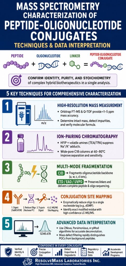
Mass spectrometry characterization of peptide-oligonucleotide conjugates is a sophisticated, multi-dimensional analytical approach used to verify the identity, purity, and stoichiometry of hybrid biotherapeutics. This process confirms both the peptide sequence and the nucleic acid structure within a single molecular entity. It addresses the inherent synthetic and structural incompatibilities between peptides and oligonucleotides by resolving complex charge-state distributions and validating covalent linkage sites. As a result, mass spectrometry characterization of peptide-oligonucleotide conjugates has become the principal analytical tool for supporting drug discovery programs, regulatory CMC submissions, and batch-to-batch quality assurance.
Peptide-oligonucleotide conjugates (POCs) represent a rapidly expanding therapeutic class that combines the targeting specificity and tissue-localization capabilities of synthetic peptides with the sequence-directed gene-silencing or gene-modulating functions of oligonucleotides. These hybrid molecules generally consist of three key components: a functional peptide sequence, such as a cell-penetrating peptide or tissue-targeting ligand; a chemical linker; and a nucleic acid payload, typically comprising siRNA, antisense oligonucleotides, microRNA, or aptamers. Through the covalent integration of these distinct biomolecular entities, researchers can overcome longstanding therapeutic challenges, including limited cellular membrane permeability, endosomal trapping, and rapid degradation by nucleases in vivo.
Understand how these molecular mechanisms translate into targeted therapeutics by reading our comprehensive guide on peptide oligonucleotide conjugates mechanism of action.
Despite their therapeutic advantages, the combination of peptide chemistry with phosphoramidite-based nucleic acid synthesis introduces significant structural complexity. The production of POCs typically follows one of two primary synthetic pathways: linear stepwise solid-phase synthesis on a common support or convergent post-synthetic ligation of separately synthesized and purified fragments, either in solution or on a solid phase. Stepwise solid-phase synthesis requires carefully selected orthogonal protecting groups capable of withstanding both acidic peptide deprotection conditions, which can cause nucleic acid depurination, and basic oligonucleotide deprotection conditions, which may damage sensitive amino acid residues. In contrast, post-synthetic ligation employs highly chemoselective reactions such as maleimide-thiol coupling, disulfide bond formation, oxime ligation, and copper-catalyzed or copper-free click chemistry. Although effective, these fragment-based assembly methods frequently introduce product heterogeneity, including unconjugated starting materials, truncated sequences, hydrolyzed linker derivatives, and over-modified conjugate species.
Delve deeper into the structural building blocks and synthesis chemistry by exploring our technical overview of peptide oligonucleotide conjugate linker chemistry
and discover the operational details behind peptide oligonucleotide conjugate synthesis methods.
Conventional quality control techniques developed for isolated peptides or purified nucleic acids are often inadequate for evaluating these hybrid constructs. The physicochemical properties of peptide-oligonucleotide conjugates are influenced simultaneously by the highly polyanionic and hydrophilic oligonucleotide region and the potentially hydrophobic or highly cationic peptide segment. To overcome these analytical challenges, specialized laboratories such as ResolveMass Laboratories Inc. have developed comprehensive validation workflows that combine high-resolution mass spectrometry with optimized chromatographic techniques to confirm intact molecular mass, sequence fidelity, and conjugation stoichiometry.
Ensure the safety and efficacy of your hybrid therapeutics with our robust framework for qc testing for peptide oligonucleotide conjugates.
Share via:
Article Summary:
- Peptide–oligonucleotide conjugates (POCs) are advanced therapeutic molecules that combine the targeting abilities of peptides with the gene-regulating functions of oligonucleotides, improving cellular delivery and biological effectiveness.
- Mass spectrometry is the primary analytical technique for characterizing POCs, enabling scientists to confirm molecular identity, purity, sequence integrity, conjugation sites, and overall product quality throughout drug development.
- High-resolution platforms such as Orbitrap FT-MS, Q-TOF, FT-ICR, and MALDI-TOF provide the accuracy and resolving power needed to analyze complex conjugates, detect impurities, and differentiate closely related molecular species.
- Optimized LC-MS workflows using ion-pairing reagents such as HFIP, TEA, and TPA improve chromatographic separation, reduce alkali metal adduct formation, and enhance sensitivity when analyzing oligonucleotide-containing conjugates.
- Advanced fragmentation techniques including ETD, EAD, and UVPD enable detailed sequencing of both peptide and oligonucleotide components while preserving sensitive linker structures and modification sites.
- Precise conjugation site mapping is achieved through enzymatic digestion, chromatographic separation, and tandem mass spectrometry, allowing researchers to identify attachment locations and distinguish structurally similar isomers.
- Modern data analysis tools, including advanced deconvolution algorithms and mass defect filtering, improve interpretation of complex spectra, support impurity detection, and provide reliable characterization for regulatory, quality control, and biotherapeutic development applications.
Advanced High-Resolution Platforms for Mass Spectrometry Characterization of Peptide-Oligonucleotide Conjugates
High-resolution mass spectrometry platforms, particularly Orbitrap FT-MS and Quadrupole Time-of-Flight (Q-TOF) instruments, provide the sub-parts-per-million mass accuracy required to resolve complex isotopic charge envelopes and verify the molecular formulas of intact conjugates. These advanced systems facilitate direct mass measurement of complete conjugates, synthetic intermediates, and degradation products, enabling comprehensive quality assessment throughout the development process. Their exceptional mass accuracy allows analysts to distinguish isobaric species and detect subtle post-translational or synthetic modifications with remarkable precision.
Discover how our high-resolution platforms provide absolute structural clarity through specialized peptide oligonucleotide conjugate analysis.
Successful mass spectrometry characterization of peptide-oligonucleotide conjugates requires instruments with substantial resolving power (>30,000 FWHM for Q-TOF platforms and >100,000 FWHM for Orbitrap systems) as well as broad mass-to-charge (m/z) transmission capabilities. Electrospray ionization (ESI) of these hybrid biomolecules generates extensive charge-state distributions. Consequently, intact mass analysis of large oligonucleotide conjugates and antibody-oligonucleotide conjugates (AOCs) requires instruments capable of transmitting and detecting ions across high m/z ranges, typically extending from 1,000 to 6,000 m/z.
Mass Analyzer Platform Comparison
Orbitrap FT-MS
- Typical Resolving Power: 100,000 to 500,000
- Mass Accuracy Range: <1 to 3 ppm
- Optimal Ionization Target: Intact conjugates, isotopic resolution, trace impurities
- Analytical Limitations: Slower scan speeds during ultra-high-resolution acquisition and higher instrument cost.
Q-TOF MS
- Typical Resolving Power: 30,000 to 60,000
- Mass Accuracy Range: 2 to 5 ppm
- Optimal Ionization Target: Online LC-MS/MS workflows, peptide mapping, subunit analysis
- Analytical Limitations: Reduced resolving capability for highly charged, high-mass analytes.
FT-ICR MS
- Typical Resolving Power: >1,000,000
- Mass Accuracy Range: <0.5 ppm
- Optimal Ionization Target: Isotopic fine structure analysis and direct-infusion screening
- Analytical Limitations: Highly susceptible to salt contamination and requires specialized superconducting magnet systems.
MALDI-TOF MS
- Typical Resolving Power: 10,000 to 25,000
- Mass Accuracy Range: 10 to 50 ppm
- Optimal Ionization Target: Rapid reaction monitoring and crude mixture screening
- Analytical Limitations: Lower mass accuracy, inability to perform online LC coupling, and susceptibility to laser-induced fragmentation.
In ESI mass spectrometry, gas-phase behavior is strongly influenced by ionization polarity. Due to the abundance of acidic phosphate groups within nucleic acids, negative-mode ESI is generally preferred for analyzing intact oligonucleotides and full-length conjugates. This approach produces highly resolved charge-state distributions while minimizing unwanted fragmentation.
For protein-based carriers such as monoclonal antibodies used in AOCs, the positive charge contribution of the antibody frequently dominates the overall ionization behavior of the conjugate. Consequently, positive-mode ESI remains highly effective for intact mass measurements of these species. Nevertheless, analysts must carefully optimize ionization conditions to avoid source-induced cleavage of chemically sensitive linker regions.
Evaluate how alternative delivery modalities stack up against each other by reading our structural breakdown on peptide vs antibody oligonucleotide conjugates.
Mobile Phase Optimizations for Liquid Chromatography-Mass Spectrometry Analysis
Ion-pairing reversed-phase liquid chromatography (IP-RPLC) coupled with mass spectrometry utilizes volatile fluoroalcohol buffers, including hexafluoroisopropanol (HFIP), together with organic amines to achieve effective chromatographic separation while minimizing alkali metal adduction. Controlled titration of HFIP with triethylamine (TEA) or tripropylamine (TPA) creates dynamic pH changes during droplet desolvation, significantly improving mass spectrometric sensitivity. This strategy mitigates signal splitting caused by sodium and potassium adduct formation, one of the most significant obstacles in obtaining high-quality ESI-MS spectra of nucleic acids.
Oligonucleotides are strongly polyanionic molecules that readily interact with alkali metal ions such as Na+ and K+, which are commonly introduced through reagents, laboratory glassware, and biological samples. Without suitable ion-pairing systems, negative-mode electrospray ionization produces extensive adduct formation, dispersing the analyte signal among multiple cation-associated species ([M − zH + nNa]z−) and reducing both sensitivity and mass accuracy.
To suppress this phenomenon, mobile phases typically contain a volatile organic amine and a fluoroalcohol. The amine, such as TEA, DIEA, or hexylamine, acts as an ion-pairing reagent by interacting with negatively charged phosphate groups, thereby increasing hydrophobicity and enhancing retention on reversed-phase columns. HFIP functions as both a volatile counter-ion and a buffering agent, maintaining a near-neutral pH environment.
Droplet Generation (pH ~7.0) → HFIP Evaporation → pH Increase → Amine-Mediated Na+/K+ Displacement → Formation of High-Sensitivity Deprotonated Ions
This process is governed by a dynamic pH-shift mechanism within the electrospray plume. During desolvation, HFIP, which has a boiling point of approximately 58°C, evaporates more rapidly than water and the accompanying amine. As HFIP is depleted, the pH of the shrinking droplet increases substantially, creating strongly basic conditions. Under these circumstances, the amine efficiently displaces sodium and potassium ions from the oligonucleotide through competitive interactions, allowing the analyte to enter the mass spectrometer as clean, deprotonated ions with minimal adduction.
Selection of the optimal amine-fluoroalcohol combination depends on the size and composition of the peptide-oligonucleotide conjugate under investigation.
Optimized IP-RPLC Conditions
Short POCs (10-mer to 15-mer)
- Column: Wide-pore C18, 1.7 μm, 300 Å
- Temperature: 60°C to 80°C
- Mobile Phase A: 15 mM Hexylamine, 50 mM HFIP in H₂O
- Mobile Phase B: 15 mM Hexylamine, 50 mM HFIP in Acetonitrile
- Flow Rate: 0.5 mL/min
Large POCs (>25-mer and Phosphorothioates)
- Column: Wide-pore C18, 3 μm, 300 Å
- Temperature: 80°C
- Mobile Phase A: 15 mM TPA, 100 mM HFIP in H₂O
- Mobile Phase B: 15 mM TPA, 100 mM HFIP in Methanol/Acetonitrile
- Flow Rate: 0.8 mL/min
Complex Mixtures and Standard QC Applications
- Column: YMC Accura Triart Bio C18, 1.9 μm
- Temperature: 50°C to 70°C
- Mobile Phase A: 400 mM HFIP adjusted to pH 7.0 with TEA (~8.6 mM TEA)
- Mobile Phase B: 400 mM HFIP, 8.6 mM TEA in 50% Methanol / 50% Water
- Flow Rate: 0.5 to 1.0 mL/min
Elevated column temperatures ranging from 60°C to 80°C are particularly important when analyzing conjugates containing highly cationic arginine-rich peptides or phosphorothioate-modified oligonucleotides. Increased thermal energy reduces non-specific electrostatic and hydrophobic interactions, minimizes aggregation, disrupts secondary structures, and produces sharper, more symmetrical chromatographic peaks.
Gas-Phase Fragmentation Chemistry for Sequence Verification and Structural Mapping
Comprehensive sequencing of peptide-oligonucleotide conjugates relies on complementary gas-phase fragmentation approaches that target different regions of the hybrid molecule. Collision-induced dissociation (CID) preferentially fragments the oligonucleotide backbone, whereas Electron Activated Dissociation (EAD) and Electron Transfer Dissociation (ETD) preserve linker structures while generating sequence-specific peptide fragments. This complementary behavior allows independent structural characterization of both molecular domains.
When subjected to conventional CID, peptide-oligonucleotide conjugates display highly asymmetric fragmentation patterns. The phosphodiester backbone and nucleobases require less energy for cleavage than peptide amide bonds. Consequently, tandem mass spectra are dominated by oligonucleotide fragment ions, including a-B, w, c, and d series ions, together with liberated nucleobases. In peptide-dT6 conjugates, for example, fragmentation primarily occurs within thymidylic acid residues while peptide backbone cleavage remains limited.
To achieve complete peptide sequence characterization and accurately identify modification sites, analysts frequently employ electron-based activation techniques such as ETD and EAD. These methods utilize low-energy electrons to induce rapid, non-ergodic fragmentation. Electron capture by protonated amino acid residues generates radical intermediates that rapidly cleave the peptide backbone, producing characteristic c and z ions.
Because electron transfer occurs on an extremely short timescale, vibrational energy does not have sufficient time to redistribute throughout the molecule. As a result, fragile modifications including phosphorylation, methylation, PEG linkers, alkyl spacers, disulfide bonds, maleimide-thiol linkages, and ester-based conjugation sites remain intact. This preservation enables direct identification of modified amino acid residues through retention of the linker on specific peptide fragments.
CID: Vibrational activation leading primarily to phosphodiester bond cleavage and nucleobase loss, resulting in limited peptide sequencing.
ETD/EAD: Radical-mediated activation causing peptide backbone cleavage while preserving conjugation linkers and labile modifications.
Ultraviolet Photodissociation (UVPD), particularly at 193 nm, offers an additional powerful approach for characterizing intact conjugates. UVPD generates extensive fragmentation across both peptide and oligonucleotide backbones simultaneously, producing a, b, c, x, y, z peptide ions alongside w, x, y and a, b, c, d oligonucleotide ions. This capability enables complete dual-backbone sequencing without the need for prior chemical or enzymatic cleavage.
Complete Conjugation Site Determination and Isomer Characterization
Accurate determination of conjugation sites often requires enzymatic reduction of the oligonucleotide component before peptide-level mapping can be performed. By digesting the nucleic acid segment to a single nucleoside monophosphate, analysts can employ conventional positive-mode ESI-MS/MS methods to identify the modified amino acid residue with high confidence. This workflow is particularly valuable for identifying positional isomers generated during convergent synthesis, as these species may exhibit significantly different biological activities.
For large bioconjugates such as antibody-oligonucleotide conjugates (AOCs), where monoclonal antibodies are linked to therapeutic siRNA molecules through MCC or SMCC linkers, site localization presents a substantial analytical challenge. The highly negative charge associated with the siRNA payload suppresses positive-mode ionization of antibody-derived fragments, limiting the effectiveness of standard protein mapping techniques.
To overcome this challenge, analysts employ a hierarchical localization strategy. Initial localization occurs at the antibody subunit level through digestion with highly specific proteases such as FabRICATOR, which cleaves antibodies below the hinge region. This process generates Fc fragments and heavy-chain/light-chain F(ab’)₂ fragments for subsequent analysis.
Size Exclusion Chromatography-Mass Spectrometry (SEC-MS): Separates antibody subunits according to hydrodynamic size, enabling rapid assessment of the oligonucleotide-to-antibody ratio (OAR) and providing insight into whether conjugation occurs within Fab or Fc regions.
Strong Cation Exchange-Mass Spectrometry (SCX-MS): Separates subunits based on charge properties, allowing differentiation of conjugation states due to the substantial charge contribution of the attached oligonucleotide.
Step 1: FabRICATOR Digestion
Step 2: SEC-MS and SCX-MS Screening
Step 3: Nuclease P1 Oligonucleotide Reduction
Step 4: Trypsin Digestion
Step 5: LC-MS/MS Site Mapping
For complete site localization at single-residue resolution, the oligonucleotide payload is enzymatically degraded using nucleases such as Nuclease P1 or Benzonase. These enzymes cleave phosphodiester bonds while preserving both the peptide backbone and conjugation linker. Following digestion, the large oligonucleotide is reduced to a single nucleoside monophosphate tag, such as deoxyguanosine monophosphate (329.2 Da), which remains covalently attached to the linker.
Subsequent reduction, alkylation, and tryptic digestion generate peptide fragments bearing a low-mass nucleotide tag. These peptides behave similarly to conventional tryptic peptides during reversed-phase chromatography and ionize efficiently in positive-mode ESI. Tandem mass spectrometry then identifies the exact modified residue through characteristic mass shifts observed on cysteine, lysine, or aspartic acid residues.
This level of site mapping is essential for distinguishing positional isomers that share identical intact masses but differ in conjugation location. Since many positional isomers co-elute during conventional LC-MS analysis, emerging technologies such as engineered aerolysin nanopores are gaining attention. These systems can distinguish peptides with identical masses based solely on differences in molecular shape and conformational behavior within a controlled electric field.
Overcome downstream translation and synthesis anomalies by anchoring your project with specialized peptide oligonucleotide conjugates preclinical services.
Computational Deconvolution Algorithms for Complex Data Interpretation
Computational deconvolution converts complex multiply charged ESI spectra into zero-charge molecular mass profiles, enabling confirmation of conjugate identity and detection of trace impurities. Selection of deconvolution algorithms capable of handling asymmetric isotope distributions characteristic of peptide-oligonucleotide conjugates is essential for achieving accurate results and avoiding computational artifacts. These advanced workflows support automated high-throughput analysis for purity testing and stability assessments.
Electrospray ionization generates highly complex charge-state distributions that require computational reconstruction to determine true molecular masses. The performance of this reconstruction depends heavily on the selected algorithm.
Maximum Entropy (MaxEnt) has historically served as the standard deconvolution approach for proteins, glycoproteins, and antibody-drug conjugates. This probabilistic method assumes Gaussian isotopic distributions and optimizes spectral fitting accordingly. Although highly effective for proteins, this assumption is often invalid for peptide-oligonucleotide conjugates. Their phosphorus-rich and oxygen-rich compositions produce asymmetric, non-Gaussian isotope envelopes. When MaxEnt attempts to fit these distributions, artificial peaks frequently appear at one-half or one-third of the actual molecular mass.
To address these limitations, several advanced algorithms have been developed:
ZNova Algorithm: Implemented in software platforms such as ProMass for MassLynx, ZNova utilizes charge-spacing calculations rather than probabilistic modeling. By measuring precise m/z spacing between adjacent charge states, it accurately determines charge values and molecular masses while minimizing artifacts. Typical mass accuracy ranges from −0.1 to +0.5 Da.
Parsimonious Charge Deconvolution: This method incorporates an objective function that penalizes unnecessary complexity and favors solutions containing the smallest possible number of spectral components. As a result, it effectively resolves overlapping charge envelopes and improves impurity characterization.
pMod (Peak Modeling): This enhanced deconvolution approach models actual isotopic distributions of biopolymers, enabling accurate processing of asymmetric isotope envelopes and heavily modified or phosphorothioated conjugates without introducing false spectral features.
Resolving Mass Defect Variations during Mass Spectrometry Characterization of Peptide-Oligonucleotide Conjugates
Mass defect analysis provides a powerful means of differentiating peptide-oligonucleotide conjugates from non-crosslinked peptide species by exploiting differences in the fractional components of exact monoisotopic masses. Oligonucleotides contain large amounts of oxygen and phosphorus, resulting in lower fractional masses than peptide-only species of equivalent nominal mass. This property enables rapid identification of hybrid molecules within complex proteolytic mixtures.
With the exception of carbon-12, which has a defined mass defect of zero (0.00000 Da), all elements exhibit either positive or negative mass defects. Hydrogen has a positive mass excess of +0.00783 Da, whereas oxygen, phosphorus, and sulfur possess negative mass defects of −0.00509 Da, −0.02624 Da, and −0.02793 Da, respectively. Because peptides contain substantial amounts of hydrogen and nitrogen, they typically exhibit positive fractional masses. Oligonucleotides, by contrast, contain highly oxygenated and phosphorylated sugar-phosphate backbones that contribute negative fractional mass characteristics.
Fractional Mass = Exact Mass − Nominal Mass
By plotting nominal mass against fractional mass for all ions detected during an LC-MS experiment, software can rapidly separate peptide-oligonucleotide conjugates from background peptides and unrelated protein digest products. For example, a peptide linked to three nucleotides exhibits a distinctive shift in fractional mass that clearly differentiates it from conventional tryptic peptides with the same nominal mass. This mass defect filtering strategy provides a rapid, label-free approach for profiling conjugation mixtures and nucleic acid-protein crosslinked species.
Analytical Guidelines and Conclusion for Mass Spectrometry Characterization of Peptide-Oligonucleotide Conjugates
The establishment of a robust, multi-dimensional analytical framework is essential for validating the stability, sequence integrity, and purity of peptide-oligonucleotide conjugates intended for regulatory submission. The integration of high-resolution ESI mass spectrometry with advanced chromatographic separation and computational analysis provides comprehensive confidence in biotherapeutic identity and quality. Achieving this level of analytical performance requires meticulous optimization of chromatography, fragmentation methodologies, and data interpretation workflows.
To facilitate regulatory compliance and accelerate development programs, analytical strategies should incorporate the following core principles:
Implement Ion-Pairing Optimization
Employ mobile phases containing volatile fluoroalcohols such as HFIP in combination with TEA or TPA on wide-pore C18 columns operated at elevated temperatures ranging from 60°C to 80°C. This approach suppresses alkali metal adduction and maximizes chromatographic resolution.
Utilize Soft, Electron-Based Fragmentation
Apply EAD, ETD, or UVPD activation methods during tandem mass spectrometry to preserve chemically sensitive linkers while achieving extensive sequence coverage across both peptide and oligonucleotide domains.
Execute Precise Site Mapping through Enzymatic Pre-Treatment
Digest the oligonucleotide payload to a single nucleoside monophosphate tag using Nuclease P1, enabling highly accurate positive-mode tryptic mapping and single-residue conjugation site localization.
Employ Advanced Deconvolution Algorithms
Replace traditional MaxEnt workflows with ZNova, Parsimonious Charge Deconvolution, or pMod to eliminate computational artifacts and improve the detection of trace impurities and degradation products.
Integrate Mass Defect Filtering
Utilize mass defect analysis to selectively identify peptide-oligonucleotide conjugates within complex proteomic backgrounds based on characteristic fractional mass signatures.
ResolveMass Laboratories Inc. is recognized as a leader in bioconjugate characterization, combining high-resolution Orbitrap FT-MS and Q-TOF platforms with customized chromatographic methodologies to generate comprehensive, regulatory-compliant analytical datasets. These validated workflows support every stage of biotherapeutic development, including discovery research, custom synthesis projects, CMC validation programs, and biosimilar comparability studies.
Transition your candidate from design to development by leveraging our specialized cmc services for peptide oligonucleotide conjugates
and scale your operations fluidly using our expert framework for the scale up of peptide oligonucleotide conjugates.
For specialized bioconjugate characterization, analytical method development, and regulatory documentation support, contact the scientific experts directly through the ResolveMass Laboratories Contact Page.
Frequently Asked Questions
Accurate chromatographic analysis of peptide-oligonucleotide conjugates requires the use of wide-pore C18 columns, such as 300 Å stationary phases or specialized bioinert columns designed for large biomolecules. Mobile phases typically contain volatile fluoroalcohols like HFIP combined with ion-pairing amines such as TEA, TPA, or hexylamine to improve retention and ionization efficiency. Elevated column temperatures between 60°C and 80°C help reduce secondary structure formation and minimize intermolecular interactions. Appropriate flow rates, generally ranging from 0.5 to 1.0 mL/min, further enhance peak shape and separation performance.
HFIP is preferred in LC-MS workflows because it is highly volatile and supports efficient ionization during electrospray mass spectrometry. While TEAA performs well as a chromatographic ion-pairing reagent, it often suppresses ion signals and reduces analytical sensitivity, particularly in negative-ion mode. During desolvation, HFIP rapidly evaporates, creating a favorable pH shift that promotes complete deprotonation of oligonucleotides. This process also reduces sodium and potassium adduct formation, resulting in cleaner spectra and improved mass accuracy.
Electron Activated Dissociation (EAD) and Electron Transfer Dissociation (ETD) provide more informative peptide sequencing compared to conventional CID methods. Instead of primarily fragmenting the oligonucleotide backbone, these techniques induce radical-driven cleavage along peptide amide bonds. Because the fragmentation occurs extremely rapidly, fragile linkers and chemical modifications remain intact throughout the process. This preservation enables detailed peptide sequence determination and allows precise identification of conjugation sites within the hybrid molecule.
Nuclease P1 is widely used to simplify structural analysis by selectively digesting the oligonucleotide portion of a conjugate. The enzyme breaks down the nucleic acid chain into a single nucleoside monophosphate that remains attached to the linker region. This significantly reduces both molecular size and overall negative charge, allowing the modified molecule to behave more like a conventional peptide during analysis. As a result, positive-mode ESI-MS/MS peptide mapping can be performed with greater sensitivity and accuracy.
Positional isomers arise when a linker attaches to different reactive amino acid residues within a peptide, such as lysine side chains or engineered cysteine residues. Although these species possess the same overall molecular weight, their attachment sites differ, which may influence biological activity and pharmacological performance. Advanced chromatographic methods can often separate these isomers based on subtle structural differences. Definitive identification is typically achieved through enzymatic digestion followed by high-resolution MS/MS mapping of the modified residue.
The Maximum Entropy (MaxEnt) algorithm was originally developed for proteins and assumes that isotopic distributions follow a symmetrical Gaussian pattern. Oligonucleotides and peptide-oligonucleotide conjugates often exhibit asymmetric isotope envelopes because of their phosphorus-rich composition. When MaxEnt attempts to fit these non-Gaussian patterns, it may generate artificial peaks that do not correspond to real molecular species. These artifacts frequently appear at one-half or one-third of the actual molecular mass, potentially complicating data interpretation.
Mass defect filtering takes advantage of the unique elemental composition of oligonucleotides and peptides. Oligonucleotides contain large amounts of oxygen and phosphorus, giving them different fractional mass characteristics than peptide-only fragments. By plotting nominal mass against fractional mass, analytical software can rapidly distinguish hybrid conjugates from background peptides and unrelated digest products. This approach provides a fast and effective method for detecting conjugated species within highly complex biological samples.
Reference:
- Flett, F. J., Walton, J. G. A., Mackay, C. L., & Interthal, H. (2015). Click chemistry generated model DNA-peptide heteroconjugates as tools for mass spectrometry. Analytical Chemistry, 87(19), 9595–9599. https://doi.org/10.1021/acs.analchem.5b02047
- Moser, M., & Gilar, M. (2025). High-resolution HPLC for separating peptide–oligonucleotide conjugates. Molecules, 30(11), 2378. https://doi.org/10.3390/molecules30112378
- Gait, M. J. (2003). Peptide-mediated cellular delivery of antisense oligonucleotides and their analogues. Cellular and Molecular Life Sciences, 60(5), 844–853. https://doi.org/10.1007/s00018-003-3044-5
- Pourshahian, S., & Limbach, P. A. (2008). Application of fractional mass for the identification of peptide:oligonucleotide cross-links by mass spectrometry. Journal of Mass Spectrometry, 43(8), 1081–1088. https://doi.org/10.1002/jms.1391
- Jensen, O. N., Kulkarni, S., Aldrich, J. V., & Barofsky, D. F. (1996). Characterization of peptide-oligonucleotide heteroconjugates by mass spectrometry. Nucleic Acids Research, 24(19), 3866–3872. https://doi.org/10.1093/nar/24.19.3866

