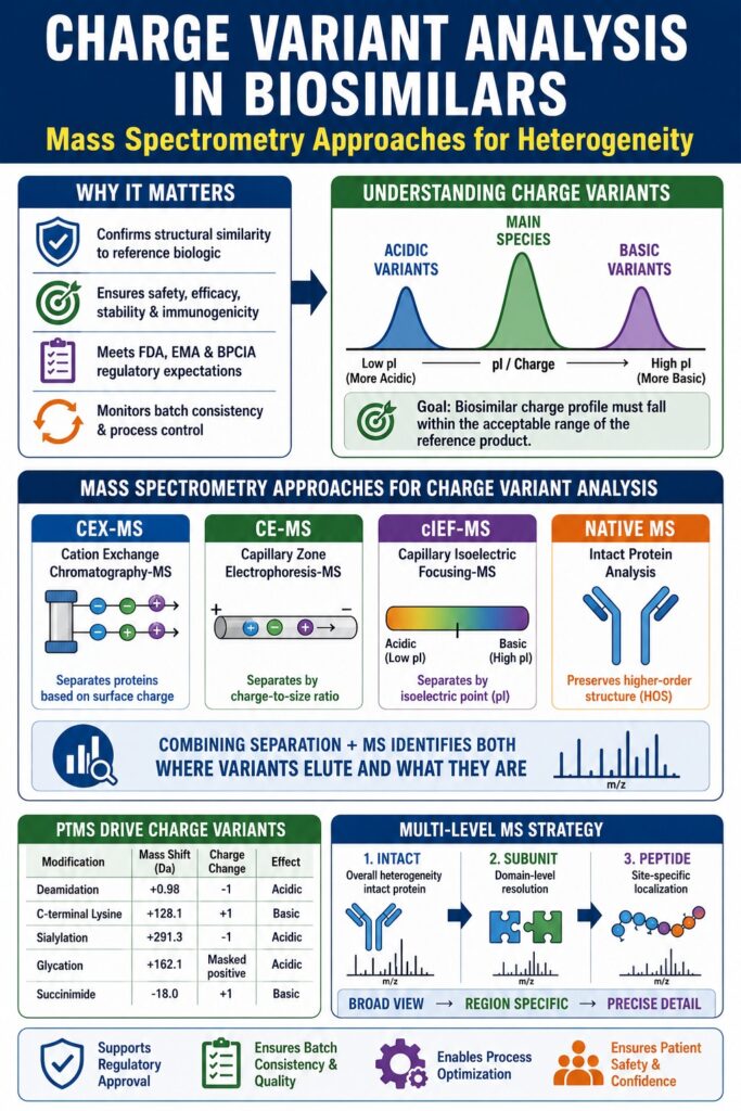
Why is Charge Variant Analysis in Biosimilars Important for Regulatory Approval?
Charge Variant Analysis in Biosimilars is an essential part of biosimilar development because it helps confirm that a biosimilar product is highly similar to its reference biologic. Regulatory agencies across the world expect detailed analytical evidence showing that the biosimilar matches the original product in terms of acidic, basic, and main protein variants. This comparison helps ensure that small molecular differences do not affect safety, efficacy, stability, or immunogenicity.
Since biologics are produced in living cells, some level of protein variability naturally occurs during manufacturing. Because of this, Charge Variant Analysis in Biosimilars plays a major role in monitoring protein heterogeneity and maintaining product consistency throughout development and commercialization.
Explore our specialized services: Regulatory Support for Generic Drug Development
Regulatory guidelines such as the Biologics Price Competition and Innovation Act (BPCIA) in the United States and European Medicines Agency (EMA) biosimilar guidance require detailed structural and functional characterization. Unlike traditional small-molecule drugs, biologics contain complex post-translational modifications and micro-heterogeneity that may change during manufacturing, purification, or storage.
Even different batches of the same reference biologic can show slight charge distribution changes. Therefore, biosimilar manufacturers must demonstrate that their products remain within the acceptable variability range observed in multiple innovator lots.
| Regulatory Concept | Significance in Charge Variant Analysis |
|---|---|
| Fingerprint-like Similarity | Achieving a highly comparable chromatographic and mass profile to the reference biologic. |
| Critical Quality Attributes (CQAs) | Charge variants are monitored because they directly affect potency, stability, and safety. |
| Totality of Evidence | Comprehensive analytical evidence reduces dependence on extensive clinical trials. |
| Clinically Meaningful Differences | Any variation in charge distribution must be shown to have no clinical consequence. |
| Interchangeability | Confirms that switching between products does not increase patient risk. |
Charge variants are much more than simple purity markers. Acidic variants caused by deamidation in complementarity-determining regions (CDRs) can reduce target binding and impact therapeutic activity. Basic variants, such as retained C-terminal lysine residues, may indicate process changes or upstream manufacturing differences.
A strong Charge Variant Analysis in Biosimilars program supports regulatory compliance, process control, batch consistency, and patient safety. Regulatory authorities increasingly expect developers to connect charge variant findings with stability studies and functional testing to support biosimilarity claims.
Need assistance with analytical evidence? Analytical Services for Generic Drug Development
Share via:
How Mass Spectrometry Improves Charge Variant Analysis in Biosimilars
Mass spectrometry provides high analytical sensitivity and molecular-level information for Charge Variant Analysis in Biosimilars. Unlike traditional UV-based techniques, mass spectrometry directly measures molecular mass and identifies the exact chemical modifications responsible for charge differences.
High-resolution accurate mass (HRAM) instruments can detect even very small changes in protein mass. This allows scientists to identify modifications such as deamidation, glycation, glycosylation shifts, oxidation, and lysine clipping with excellent precision.
Advanced characterization solutions:Biosimilar Characterization Using Mass Spectrometry
Traditional ion-exchange chromatography (IEX) methods can separate acidic and basic species, but they cannot always identify the root cause of each peak. Scientists often need additional offline testing to determine the modification behind the separation profile.
Native mass spectrometry (nMS) combined with online separation methods solves this limitation by providing both separation and molecular identification in a single workflow.
| Feature | UV-Based CVA | MS-Based CVA |
|---|---|---|
| Identification | Retention-time based interpretation | Direct molecular mass confirmation |
| Resolution | Limited to chromatographic separation | Enhanced with m/z dimension |
| Throughput | Requires secondary identification steps | Identification achieved in a single run |
| Sensitivity | Moderate | Excellent for low-abundance variants |
| Structural Insight | Minimal | Provides PTM and glycan characterization |
Modern workflows also use volatile mobile phases such as ammonium acetate and ammonium formate, which are compatible with electrospray ionization and preserve native protein structure during analysis.
Maintaining proteins under native conditions helps scientists study molecules in a form that closely resembles their natural biological state. This combination of structural preservation and detailed molecular analysis makes mass spectrometry a key technology for biosimilar characterization.
Confirm similarity with precision: Prove Biosimilarity Using LC-MS
Primary Separation Methods for Charge Variant Analysis in Biosimilars
Several advanced analytical techniques are commonly used in Charge Variant Analysis in Biosimilars, including:
- Cation-Exchange Chromatography (CEX-MS)
- Capillary Zone Electrophoresis (CZE-MS)
- Capillary Isoelectric Focusing (cIEF-MS)
Each method separates proteins based on different physicochemical properties such as ionic interaction, electrophoretic mobility, or isoelectric point.
Cation-Exchange Chromatography (CEX-MS)
CEX-MS is one of the most widely used methods for monoclonal antibody analysis. Most therapeutic antibodies have relatively basic isoelectric points, making CEX highly effective for separating acidic and basic variants.
Modern pH-gradient methods using volatile buffers improve mass spectrometry compatibility and enhance separation quality. CEX-MS is especially useful for resolving small changes caused by deamidation or lysine retention.
This method also improves sensitivity through on-column analyte concentration and provides excellent reproducibility for quality control applications.
Capillary Zone Electrophoresis (CZE-MS)
CZE-MS separates proteins based on charge-to-size ratio and electrophoretic mobility. It offers very high separation efficiency and can resolve variants that may overlap in chromatographic workflows.
Microchip CE-MS systems such as ZipChip have improved analysis speed and simplified workflows, making them useful during clone selection and early biosimilar development.
Capillary Isoelectric Focusing (cIEF-MS)
cIEF-MS separates proteins according to isoelectric point (pI). Modern automated cIEF-MS systems allow direct transfer of focused analytes into the mass spectrometer for accurate molecular analysis.
This technique provides valuable orthogonal confirmation of charge heterogeneity.
| Modality | Basis of Separation | Key Strength | MS Compatibility |
|---|---|---|---|
| CEX-MS | Surface charge / ionic interaction | Excellent for lysine variants | High with volatile pH gradients |
| CZE-MS | Charge-to-size ratio | Rapid separation and glycan selectivity | High with specialized interfaces |
| cIEF-MS | Isoelectric point | Direct pI determination | Moderate with ampholyte control |
| AEX-MS | Anionic surface charge | Useful for acidic proteins | High with volatile buffers |
Using multiple analytical methods together provides stronger confidence in biosimilar characterization and helps identify unresolved heterogeneity.
How Post-Translational Modifications Affect Charge Variant Analysis in Biosimilars
Post-translational modifications (PTMs) are major contributors to heterogeneity in Charge Variant Analysis in Biosimilars. These modifications can shift proteins toward acidic or basic regions by changing their surface charge.
PTMs may occur during:
- Cell culture
- Purification
- Formulation
- Long-term storage
Some modifications have little biological impact, while others may affect stability, potency, or pharmacokinetics.
Detailed PTM analysis: Post-Translational Modifications (PTMs) in Biosimilars
Acidic Variants
Acidic variants usually have lower isoelectric points than the main species. One of the most common causes is deamidation, which converts asparagine into aspartic acid or isoaspartic acid.
Other causes include:
- Glycation
- Fragmentation
- Sialylation
These changes can reduce antigen binding and alter therapeutic performance.
Basic Variants
Basic variants are often caused by incomplete enzymatic processing or temporary degradation intermediates.
Common examples include:
- Retained C-terminal lysine residues
- Succinimide intermediates
- N-terminal pyroglutamate formation
| Modification | Type | Mass Shift (Da) | Charge Shift |
|---|---|---|---|
| Deamidation | Acidic | +0.98 | -1 |
| C-terminal Lysine | Basic | +128.1 | +1 per Lys |
| Sialylation | Acidic | +291.3 | -1 per residue |
| Pyroglutamate | Main/Basic | -17.0 | Neutral / -1 |
| Glycation | Acidic | +162.1 | Masked positive charge |
| Succinimide | Basic | -18.0 | +1 |
Detailed PTM characterization supports process optimization, stability studies, and regulatory submissions.
The Role of Native Mass Spectrometry in Intact Protein Characterization
Native mass spectrometry allows proteins to be analyzed in folded, biologically relevant forms. Unlike denaturing methods, native MS preserves higher-order structure and non-covalent interactions.
This technique offers several benefits:
- Cleaner spectra
- Reduced salt adducts
- Better proteoform identification
- Improved structural insight
Native MS is especially useful for:
- Monoclonal antibodies
- Bispecific antibodies
- Fc-fusion proteins
Key Features of Native-MS Workflows
- Volatile Buffer Systems: Ammonium acetate is widely regarded as the preferred buffer for native mass spectrometry because it offers effective buffering performance while remaining highly compatible with electrospray ionization processes.
- Spectral Deconvolution: Intact proteins typically generate multiple charge states that appear as a charge distribution in the spectrum. Advanced deconvolution software is therefore essential for converting these charge envelopes into accurate neutral mass measurements.
- Preservation of Higher-Order Structure (HOS): Native MS enables the observation of molecular interactions and structural conformations that are usually disrupted under denaturing conditions. This capability allows researchers to directly correlate charge heterogeneity with the structural integrity of the protein.
- Reduced Adduct Formation: Analyses performed under native conditions generally produce cleaner spectra with fewer interfering salt adducts than denatured protein workflows, improving the accuracy of detecting and quantifying low-level variants.
Analyze proteins in their native state: Intact Mass Analysis of Biosimilars
The integration of native MS with online CEX or CE separations allows each chromatographic fraction to be characterized according to its intact native mass. This capability is especially valuable for complex biologics such as bispecific antibodies and fusion proteins, which often exhibit substantially greater heterogeneity than conventional IgG1 antibodies.
With advanced instrumentation such as the Orbitrap UHMR (Ultra-High Mass Range) platform, scientists are now able to distinguish isoforms within megadalton-scale protein complexes, significantly expanding the analytical limits of intact-mass characterization.
Multi-Level MS Strategies for Charge Variant Analysis in Biosimilars
Multi-level mass spectrometry strategies strengthen Charge Variant Analysis in Biosimilars by combining intact protein analysis (top-down), subunit analysis (middle-down), and peptide-level characterization (bottom-up) into one complete analytical workflow. This layered approach provides both a broad overview and highly detailed insight into protein heterogeneity. Intact mass analysis offers an overall picture of the total charge variant profile, while subunit analysis and peptide mapping help identify the precise location and quantity of individual modifications such as deamidation, glycation, or oxidation.
1. Intact Mass Analysis
The first stage of analysis focuses on evaluating the entire antibody molecule in its intact form. This approach is widely used in Charge Variant Analysis in Biosimilars because it preserves the natural combination of post-translational modifications present on a single protein molecule.
For instance, one intact mass spectrum can simultaneously display the distribution of lysine variants such as 0K, 1K, and 2K forms while also revealing overall glycan patterns across the antibody population. This provides a comprehensive view of molecular heterogeneity without breaking the protein apart.
However, although intact analysis delivers valuable global information, it usually does not provide enough sequence-level detail to identify the exact amino acid residue responsible for a modification such as deamidation.
2. Subunit Analysis
Subunit analysis acts as an intermediate strategy between intact protein analysis and peptide mapping. In this workflow, enzymes such as IdeS (Immunoglobulin-degrading enzyme from S. pyogenes) selectively cleave the antibody below the hinge region.
After reduction, this process generates smaller fragments of approximately 25 kDa, including the Fc portion and two Fab fragments. Studying these smaller subunits reduces spectral complexity and improves analytical resolution.
As a result, scientists can more easily detect and localize modifications within specific regions of the antibody, such as distinguishing deamidation events occurring in the light chain versus the heavy chain. This method maintains partial structural context while offering greater analytical clarity compared with intact analysis alone.
3. Peptide Mapping
Peptide mapping is considered the industry standard for highly detailed, site-specific characterization. In this method, the protein is completely digested into smaller peptides using enzymes such as trypsin.
The resulting peptides are analyzed by LC-MS/MS to determine the exact amino acid sequence and precisely identify the location of modifications. This technique provides extremely high sensitivity and accurate localization of PTMs.
Although peptide mapping delivers detailed molecular information, it also breaks apart the intact protein structure. Because of this, relationships between coexisting modifications on the original molecule are lost. In addition, sample preparation steps may introduce artificial modifications, including unintended deamidation or oxidation, which can complicate interpretation and correlation with the original intact charge variant profile.
Deep dive into peptide analysis: Peptide Mapping in Biosimilars
| Level of Analysis | Key Advantage | Major Limitation |
|---|---|---|
| Intact (Native) | Preserves overall heterogeneity | Limited sequence localization |
| Subunit (Middle-Up) | Domain-specific resolution | Requires enzymatic digestion |
| Peptide Mapping | Precise residue localization | Loss of intact structural context |
Using all three analytical levels provides a complete structural understanding for biosimilar comparability.
Orbitrap vs Q-TOF for Charge Variant Analysis in Biosimilars
Orbitrap and Q-TOF systems are both widely used for Charge Variant Analysis in Biosimilars, but they offer different strengths.
Orbitrap Systems
Orbitrap mass spectrometers are widely recognized as one of the most advanced technologies for intact protein characterization and Charge Variant Analysis in Biosimilars. These instruments operate using Fourier Transform (FT) technology, where ions oscillate inside an electrostatic field and their frequencies are measured with extremely high precision.
This analytical design allows Orbitrap systems to achieve ultra-high resolution, often reaching 480,000 FWHM or even higher in advanced instruments such as the Orbitrap Astral platform. Such high resolving power is extremely important for analyzing large biomolecules like monoclonal antibodies, which typically have molecular weights around 150 kDa.
The exceptional resolution helps scientists clearly distinguish isotope patterns and separate closely related glycoforms that might otherwise appear as overlapping or merged peaks in lower-resolution systems.
- Mass Stability: Orbitrap systems are well known for their outstanding mass stability and reproducibility. In many laboratories, calibration may only be required once per week, whereas several TOF-based systems may need more frequent calibration to maintain consistent performance.
- Cleaner Baselines: The Fourier Transform detection process also improves signal clarity by reducing background noise during data processing. This produces cleaner deconvoluted spectra and makes it easier to detect very low-abundance variants, including species present at levels below 1%.
- Native MS Capabilities: Modern Orbitrap instruments include specialized operating modes such as High Mass and UHMR (Ultra-High Mass Range) configurations. These modes are specifically designed to improve ion transmission and desolvation for very large biomolecules at high mass-to-charge (m/z) ratios, where many conventional systems experience performance limitations. Because of these advantages, Orbitrap platforms are frequently used in advanced Charge Variant Analysis in Biosimilars workflows that require high sensitivity, accurate mass measurement, and detailed proteoform characterization.
Q-TOF Systems
Quadrupole Time-of-Flight (Q-TOF) mass spectrometers determine molecular mass by measuring the time ions take to travel through a flight tube under vacuum conditions.
Although Q-TOF instruments generally provide lower resolution than Orbitrap systems for intact protein analysis, typically in the range of 30,000 to 60,000 FWHM, they offer several important advantages for high-throughput applications.
One major strength of Q-TOF systems is their wide intra-scan dynamic range. This capability allows accurate analysis of ions present at very different concentration levels within the same scan, making these instruments highly effective for bottom-up proteomics, peptide analysis, and metabolomics studies.
- Scan Speed: Q-TOF platforms can collect data at extremely high acquisition rates, sometimes reaching up to 300 Hz. This rapid scan speed makes them particularly suitable for coupling with ultra-fast analytical techniques such as microchip capillary electrophoresis (CE-MS).
- Cost and Ease of Use: Historically, Q-TOF systems were considered more affordable and easier to operate compared with early-generation Orbitrap instruments. While modern Orbitrap systems have become more user-friendly and biopharma-focused, Q-TOF instruments still remain popular for laboratories prioritizing speed, workflow flexibility, and high sample throughput. Both Orbitrap and Q-TOF technologies play valuable roles in modern Charge Variant Analysis in Biosimilars, and the choice between them often depends on the analytical goals, development stage, and required level of structural detail.
| Specification | Orbitrap | Q-TOF |
|---|---|---|
| Resolution | Ultra-high | High |
| Mass Accuracy | <1–3 ppm | <1–5 ppm |
| Native CVA Suitability | Excellent for intact proteins | Good for average mass analysis |
| Throughput | High | Very high |
| Software Ecosystem | Extensive | Robust |
How Ion Mobility Mass Spectrometry Supports Structural Analysis
Ion Mobility Mass Spectrometry (IM-MS) adds another analytical dimension by separating ions based on collision cross-section (CCS).
This helps scientists distinguish proteins with identical mass but different structures.
Advantages of IM-MS
- Differentiates structural isomers
- Improves fragmentation quality
- Provides structural fingerprints
- Supports higher-order structure analysis
Collision-Induced Unfolding (CIU)
CIU studies protein unfolding patterns in the gas phase and helps evaluate molecular stability differences.
| IM-MS Application | Benefit for Biosimilars |
|---|---|
| CCS Mapping | Confirms structural equivalence |
| Conformer Resolution | Distinguishes structural isomers |
| CIU Fingerprinting | Evaluates molecular stability |
| Native Complex Studies | Assesses binding stoichiometry |
Advanced Fragmentation Techniques for Top-Down Analysis
Advanced fragmentation approaches used in Top-Down Charge Variant Analysis in Biosimilars include Higher-Energy Collision Dissociation (HCD), Electron-Transfer Dissociation (ETD), and Ultraviolet Photodissociation (UVPD). These technologies allow scientists to directly analyze intact proteins and identify the precise location of charge-related modifications on separated protein variants.
By applying these fragmentation methods to intact monoclonal antibodies in the gas phase, researchers can confirm amino acid sequences within important regions such as complementarity-determining regions (CDRs) and accurately determine where post-translational modifications (PTMs) are located. This direct analysis is especially valuable because it avoids sample preparation artifacts that may occur during enzymatic digestion workflows.
Complementary Fragmentation Methods in Charge Variant Analysis in Biosimilars
Each fragmentation technique provides different types of structural information. For this reason, scientists often combine multiple methods to improve sequence coverage and increase confidence in protein characterization.
- Higher-Energy Collision Dissociation (HCD): Higher-Energy Collision Dissociation (HCD) is a fragmentation technique that breaks protein ions apart through energetic collisions with inert gas molecules. This process is commonly described as a “slow-heating” method because energy gradually spreads through the protein before fragmentation occurs. HCD usually generates strong fragmentation near the N-terminal and C-terminal regions of the protein and is highly effective for routine top-down analysis. Its robustness and compatibility with high-throughput workflows make it one of the most commonly used fragmentation methods in mass spectrometry laboratories.
- Electron-Transfer Dissociation (ETD): Electron-Transfer Dissociation (ETD) is considered a non-ergodic fragmentation technique because it fragments proteins without extensive energy redistribution across the molecule. In ETD, electrons are transferred from radical anions to protein ions, causing direct backbone cleavage while preserving delicate modifications such as glycosylation and phosphorylation. This makes ETD particularly useful for studying fragile post-translational modifications that may be lost during collision-based fragmentation methods. Because ETD preserves labile PTMs, it is highly valuable in detailed Charge Variant Analysis in Biosimilars, especially when identifying modifications that influence therapeutic activity or molecular stability.
- Ultraviolet Photodissociation (UVPD): Ultraviolet Photodissociation (UVPD) uses high-energy ultraviolet laser pulses to fragment proteins throughout the entire backbone structure. This method often produces extremely high sequence coverage, even for large and structurally complex proteins such as monoclonal antibodies. UVPD can generate fragmentation across multiple regions of the molecule simultaneously, providing detailed structural information that may be difficult to obtain using other techniques. Due to its broad fragmentation capability, UVPD is becoming increasingly important for advanced intact protein characterization and comprehensive biosimilar analysis.
By combining HCD, ETD, and UVPD, researchers can achieve more complete structural characterization in Charge Variant Analysis in Biosimilars. These advanced fragmentation strategies improve confidence in PTM identification, support regulatory comparability studies, and help ensure the structural consistency of biosimilar products.
Proton-Transfer Charge Reduction (PTCR)
A major difficulty in top-down analysis of intact monoclonal antibodies (mAbs) is spectral congestion, where a very large number of fragment ion peaks overlap within the spectrum. Proton-Transfer Charge Reduction (PTCR) helps solve this issue by lowering the charge states of fragment ions in the gas phase. This spreads the ions across a wider m/z range, making peak identification and interpretation much easier. Using PTCR, researchers have successfully achieved full sequencing of the CDR3 region in intact trastuzumab variants without requiring enzymatic digestion.
| Technique | Mechanism | Primary Benefit |
|---|---|---|
| HCD | Collision-induced fragmentation | Strong terminal coverage |
| ETD | Electron transfer fragmentation | Preserves fragile PTMs |
| UVPD | UV laser activation | Extensive sequence coverage |
| PTCR | Gas-phase charge reduction | Reduces spectral crowding |
How Microchip CE-MS Accelerates Biosimilar Development
Microchip CE-MS improves early-stage biosimilar development by delivering much faster analytical results compared with traditional LC-MS workflows. This rapid approach allows scientists to quickly screen large numbers of cell clones and frequently monitor bioreactor conditions during process development. Technologies such as ZipChip combine separation and ionization within a compact microfluidic platform, enabling high-resolution Charge Variant Analysis in Biosimilars with minimal sample preparation. The simple “dilute-and-shoot” workflow supports fast and efficient data-driven decision-making in accelerated development programs.
Benefits of Microchip CE-MS
- Faster charge variant profiling
- Minimal sample consumption
- Simplified sample preparation
- Rapid glycosylation assessment
| Benefit | Development Impact |
|---|---|
| Faster Turnaround | Accelerates screening workflows |
| Salt/Detergent Compatibility | Simplifies sample preparation |
| Automated Operation | Improves reproducibility |
| HRAM Integration | Maintains high identification confidence |
This technology supports rapid decision-making during process development and clone screening.
Regulatory Expectations for Charge Variant Percentages
Regulatory agencies do not define universal limits for acidic or basic variants. Instead, developers establish acceptable ranges using multiple reference product batches.
Regulatory Considerations
Developers must demonstrate that observed differences do not create clinically meaningful changes.
Establishing the Range of Similarity
Biosimilar manufacturers usually analyze 10 or more batches of the reference biologic to understand normal lot-to-lot variability. For monoclonal antibodies such as trastuzumab, the reference product may typically show a main peak representing around 60–70% of the total charge profile, while acidic variants account for approximately 20–30% and basic variants range between 5–10%. The biosimilar product is expected to demonstrate a closely comparable distribution.
- Clinical Significance: If a biosimilar shows a higher percentage of acidic variants compared with the innovator product, the additional variants must be carefully characterized. For example, if the biosimilar contains 35% acidic variants while the reference product contains 25%, the source of that additional 10% difference must be identified. When the increase is linked to modifications such as lysine retention, which is rapidly processed in vivo, the clinical concern may be limited. However, if the additional variants are caused by deamidation within the complementarity-determining regions (CDRs), they could negatively affect binding activity and therapeutic performance, potentially requiring manufacturing or process optimization.
- Purity Standards: In many biosimilar products, the main peak is generally expected to represent at least 50% of the total chromatographic area. The combined percentage of acidic and basic variants is commonly maintained below 40–50%, although acceptable ranges may differ depending on the specific biologic product and regulatory expectations.
Case Study: Trastuzumab and Adalimumab Biosimilar Comparability
For trastuzumab biosimilars such as Kanjinti and Herzuma, regulatory authorities accepted small differences in charge variant profiles because clinical studies demonstrated equivalent therapeutic performance compared with the reference product. In these studies, the primary clinical endpoint, pathological complete response (pCR), showed results comparable to the innovator biologic, supporting biosimilarity despite minor analytical differences.
In the case of adalimumab biosimilars, pharmacokinetic (PK) comparability is commonly evaluated using parameters such as area under the curve (AUC) and maximum serum concentration ($C_{max}$). Regulatory guidelines generally consider biosimilarity acceptable when these PK values fall within an 80–125% equivalence range under a 90% confidence interval. This framework allows for normal micro-heterogeneity in charge profiles while still confirming comparable clinical performance and safety.
| Parameter | Innovator Example | Biosimilar Target | Regulatory Interpretation |
|---|---|---|---|
| Main Peak % | 65% ± 5% | 62% | Acceptable |
| Acidic Variants % | 22% ± 3% | 29% | Requires further characterization |
| Basic Variants % | 8% ± 2% | 9% | Generally acceptable |
| Lysine Clipping | Full clipping | Partial clipping | Justifiable with in vivo evidence |
Specific product development services: Liraglutide Generic Development Services
Automated Software and Deconvolution in Charge Variant Analysis in Biosimilars
Advanced software platforms simplify complex mass spectrometry data analysis.
Popular tools include:
- Byos
- BioPharma Finder
- UNIFI
- BioAccord
Key Functions
- Automated PTM identification
- Charge envelope deconvolution
- Comparative mirror plots
- Multi-Attribute Method (MAM) monitoring
| Software Platform | Key Feature | Application |
|---|---|---|
| Byos | High-sensitivity analysis | Minor variant detection |
| UNIFI / BioAccord | Compliance-focused workflows | QC and regulated environments |
| BioPharma Finder | Intact and subunit deconvolution | Orbitrap-based characterization |
| OpenLab CDS | Streamlined data review | High-throughput workflows |
Automation improves reproducibility, data integrity, and regulatory compliance.
Case Studies: Charge Variant Analysis in Biosimilar Development
Case studies involving approved biosimilars, including products developed for Remicade (Infliximab) and Herceptin (Trastuzumab), show that Charge Variant Analysis in Biosimilars is often one of the earliest analytical methods used to detect meaningful differences between a biosimilar and its reference product. These findings typically lead to additional structural and functional studies to confirm whether the observed differences have any clinical impact.
For example, comparative analysis between the innovator biologic Remicade and the biosimilar Renflexis identified measurable mass differences during characterization studies. Further investigation demonstrated that these differences were mainly related to variations in glycosylation patterns rather than changes in the core amino acid sequence of the antibody. This type of detailed analysis helps establish that the biosimilar remains structurally and functionally comparable to the reference product.
Cetuximab Biosimilars
Cetuximab is a structurally complex monoclonal antibody because it contains glycosylation sites in both the Fc and Fab regions. During comparative microchip CE-MS studies involving the innovator product and two biosimilars, researchers observed that most acidic variants were linked to glycan structures present within the Fab region.
The analysis also showed that basic variants, mainly associated with C-terminal lysine residues, remained relatively consistent across all products. However, the acidic charge variant patterns displayed noticeable differences between the innovator and biosimilars.
These findings highlighted the importance of site-specific characterization in Charge Variant Analysis in Biosimilars, particularly for understanding how glycosylation-related heterogeneity in the Fab region may influence target binding and overall biological activity.
Trastuzumab Biosimilars
A comparative analysis of five trastuzumab samples, including one reference product and four biosimilars, used native top-down mass spectrometry to evaluate structural comparability. Although small differences were observed in the overall charge variant distribution, the fragmentation profiles and sequence coverage, including the critical CDR3 region, remained highly consistent across all samples.
The study provided strong evidence that the key functional regions of the antibodies were structurally equivalent, even though minor variability existed in the relative distribution of acidic and basic proteoforms.
Key Differences Identified in Biosimilar Comparative Studies
| Biosimilar Study | Charge Profile Observation | Root Cause | Clinical Outcome |
|---|---|---|---|
| Infliximab (CT-P13) | Shifted pI values | Glycan heterogeneity | No meaningful difference |
| Trastuzumab (ABP 980) | Increased acidic variants | Deamidation and glycation | No effect on HER2 binding |
| Cetuximab Biosimilars | Altered acidic peaks | Fab glycan variation | Functional verification required |
| Rituximab Biosimilars | Minor mobility shifts | Pyroglutamate and succinimide | Structurally comparable |
These examples show how modern analytical workflows help explain molecular differences while supporting biosimilarity conclusions.
Conclusions and Technical Recommendations
Charge Variant Analysis in Biosimilars has become one of the most advanced analytical areas in modern biopharmaceutical development. High-resolution mass spectrometry technologies now provide detailed structural insight into protein heterogeneity, helping manufacturers ensure quality, consistency, and patient safety.
By combining techniques such as CEX-MS, CE-MS, native MS, ion mobility analysis, and advanced fragmentation methods, developers can generate strong analytical evidence that meets global regulatory expectations.
Start your development project today: Formulation Development for Generic Drug Development
Technical Recommendations
- Use native-MS workflows with volatile buffers for direct online analysis
- Implement microchip CE-MS for faster process monitoring
- Invest in high-resolution Orbitrap systems for intact protein studies
- Combine intact, subunit, and peptide-level workflows
- Use validated automated software for PTM mapping and deconvolution
As biosimilar development continues to grow worldwide, advanced Charge Variant Analysis in Biosimilars workflows will remain essential for delivering safe, effective, and highly comparable biologic therapies.
For specialized support in high-resolution Charge Variant Analysis in Biosimilars and advanced mass spectrometry services, contact ResolveMass Laboratories: Contact Us
Frequently Asked Questions in Charge Variant Analysis
Acidic variants are protein forms that have a lower isoelectric point (pI) than the main antibody peak. These variants are commonly caused by modifications such as deamidation, glycation, or sialylation. Basic variants have a higher pI and are often linked to retained C-terminal lysine residues or succinimide formation. In cation-exchange chromatography, acidic variants usually elute earlier, while basic variants elute later.
Native mass spectrometry keeps proteins in a folded and biologically relevant state during analysis by using mild aqueous buffers. This approach produces lower charge states and reduces spectral overlap, making large proteins easier to analyze. It also provides a more accurate picture of natural protein heterogeneity and variant distribution compared with denaturing MS methods.
Deamidation is a chemical modification where asparagine residues convert into aspartic acid or isoaspartic acid, introducing an additional negative charge. If this change occurs within important binding regions such as complementarity-determining regions (CDRs), it may weaken antigen binding. As a result, the therapeutic activity and overall potency of the biosimilar can decrease.
Microchip CE-MS systems can complete charge variant separations within only a few minutes, while conventional LC-MS methods may require 30 to 60 minutes per sample. This major reduction in analysis time allows rapid screening of large sample sets during biosimilar development. Faster workflows help improve productivity during clone selection and process optimization.
In most cases, retained C-terminal lysine residues are not considered clinically significant because they are quickly removed by enzymes in the bloodstream after administration. However, they are still monitored closely as critical quality attributes (CQAs). Their presence can provide useful information about manufacturing consistency and purification performance.
Orbitrap mass spectrometers provide extremely high resolution and accurate mass measurements, which are essential for analyzing large and complex biomolecules such as monoclonal antibodies. Their resolving power allows scientists to separate closely related glycoforms and detect very small mass differences. This improves confidence in intact protein characterization and charge variant identification.
Traditional charge variant analysis mainly measures differences in protein charge rather than protein shape. However, when combined with advanced techniques such as Ion Mobility Mass Spectrometry (IM-MS) or Collision-Induced Unfolding (CIU), researchers can evaluate conformational differences and protein stability. These methods provide additional insight into higher-order structural comparability.
The “Totality of Evidence” approach means that regulatory agencies evaluate all available analytical, functional, nonclinical, and clinical data together rather than relying on one individual test. Charge variant analysis is only one part of the complete biosimilarity assessment. Approval is based on the combined evidence demonstrating that the biosimilar is highly similar to the reference product.
Reference:
- European Medicines Agency. (n.d.). Biosimilar medicines: Overview. European Medicines Agency. https://www.ema.europa.eu/en/human-regulatory-overview/biosimilar-medicines-overview
- Kirchhoff, C. F., Wang, X. M., Conlon, H. D., Anderson, S., Ryan, A. M., & Bose, A. (2017). Biosimilars: Key regulatory considerations and similarity assessment tools. Biotechnology and Bioengineering, 114(12), 2696–2705. https://doi.org/10.1002/bit.26437
- Na, Y., Kinzer, J. L., Morrissette, L., Cho, Y. S., Shay, B., Ford, M., & Schwendeman, A. (2025). Multimodal mass spectrometric characterization of structural microheterogeneity in rituximab reference and biosimilars. International Journal of Biological Macromolecules, 334(Pt 2), 149062. https://doi.org/10.1016/j.ijbiomac.2025.149062
- Gherghescu, I., & Delgado-Charro, M. B. (2021). The biosimilar landscape: An overview of regulatory approvals by the EMA and FDA. Pharmaceutics, 13(1), 48. https://doi.org/10.3390/pharmaceutics13010048
- U.S. Food and Drug Administration. (2025, October 29). FDA moves to accelerate biosimilar development and lower drug costs. FDA. https://www.fda.gov/news-events/press-announcements/fda-moves-accelerate-biosimilar-development-and-lower-drug-costs
- Du, Y., Walsh, A., Ehrick, R., Xu, W., May, K., & Liu, H. (2012). Chromatographic analysis of the acidic and basic species of recombinant monoclonal antibodies. mAbs, 4(5), 578–585. https://doi.org/10.4161/mabs.21328

