Introduction: Why Tissue and CSF Bioanalysis Is the Defining Step in CNS Drug Development
One of the greatest challenges in CNS drug development is not simply creating a molecule capable of interacting with a biological target in vitro. The real challenge lies in ensuring that the molecule successfully reaches the appropriate compartment within the brain, achieves an adequate free concentration, and remains present long enough to generate a meaningful pharmacological response. This is exactly where Tissue and CSF Bioanalytical Services become critically important. Without precise and reliable quantification of drug concentrations in brain tissue and cerebrospinal fluid (CSF), pharmacokinetic models, dose-selection strategies, and efficacy assessments are based more on assumptions than on measurable scientific evidence.
CNS drug development programs continue to experience exceptionally high attrition rates, with estimates exceeding 90% from Phase I through regulatory approval. A substantial number of these failures are associated with poor characterization of CNS penetration and inadequate understanding of target-site exposure during preclinical development. Comprehensive tissue and CSF bioanalysis is therefore not merely a supportive analytical activity. It serves as a fundamental risk-reduction strategy that directly influences development decisions and long-term program success.
At ResolveMass Laboratories Inc., our specialized tissue and CSF bioanalytical platform has been specifically developed to meet the demanding requirements of CNS drug development programs. Our approach addresses the unique analytical challenges associated with CNS matrices, regulatory expectations, and integrated PK/PD interpretation that conventional plasma-only bioanalysis cannot adequately support.
Learn more about how to optimize your program with our strategic Bioanalytical Strategy for Drug Development.
Share via:
📋 Article Summary:
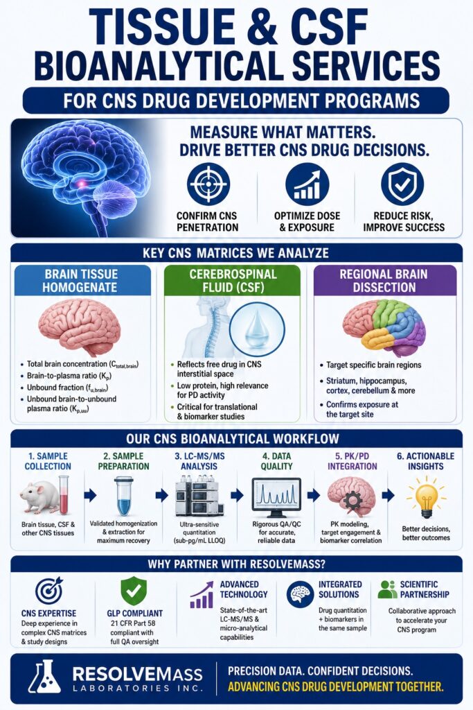
- Tissue and CSF Bioanalytical Services form the scientific foundation of CNS drug development by helping researchers confirm whether a drug candidate successfully penetrates the brain and interacts with its intended biological target.
- Commonly analyzed CNS matrices include brain tissue homogenate, cerebrospinal fluid (CSF), spinal cord tissue, and region-specific brain samples obtained through anatomical dissection.
- Key pharmacokinetic parameters generated through CNS bioanalysis include brain-to-plasma ratio (Kp), fraction unbound in brain tissue (fu,brain), and the unbound brain-to-unbound plasma ratio (Kp,uu), all of which are critical for evaluating CNS drug penetration and exposure.
- LC-MS/MS remains the industry-standard analytical platform for CNS bioanalysis because of its high selectivity, exceptional sensitivity, and ability to accurately quantify both total and free drug concentrations at ultra-low levels.
- CNS bioanalytical method development is significantly more complex than plasma-based analysis due to challenges such as phospholipid-rich brain matrices, extremely low CSF analyte concentrations, and non-specific binding during sample handling and processing.
- Regulatory studies supporting IND and NDA submissions require GLP-compliant bioanalytical validation, with assay development strategies designed in accordance with current FDA, EMA, and ICH bioanalytical method validation guidelines.
- ResolveMass Laboratories Inc. provides comprehensive tissue and CSF bioanalytical services, including assay development, method optimization, GLP validation, and regulated study sample analysis tailored specifically for CNS drug development programs.
The CNS Matrices That Drive Drug Development Decisions
Bioanalysis for CNS programs involves several anatomically and physiologically distinct matrices, each offering unique pharmacokinetic insights. The selection of matrices for analysis, along with the analytical strategy applied to them, directly influences go/no-go decisions throughout every stage of development.
Brain Tissue Homogenate
Brain tissue homogenate remains the most commonly used CNS matrix for quantifying total drug exposure within the brain. It provides the total brain concentration (Ctotal,brain), which is used to calculate the brain-to-plasma ratio (Kp). However, interpretation of total tissue concentration must always include evaluation of the unbound fraction. Since brain tissue is highly lipid-rich, containing approximately twenty times more lipid than plasma, highly lipophilic compounds can demonstrate artificially elevated total brain concentrations that may not accurately represent pharmacologically active free drug levels.
Critical parameters derived from brain homogenate analysis include:
- Total brain concentration (Ctotal,brain)
- Brain-to-plasma ratio (Kp = AUCbrain / AUCplasma)
- Fraction unbound in brain (fu,brain) determined through equilibrium dialysis
- Unbound brain-to-unbound plasma ratio (Kp,uu = Kp × fu,brain / fu,plasma)
Cerebrospinal Fluid (CSF)
CSF is widely considered the most informative individual matrix for estimating free drug exposure within the CNS interstitial space. Because CSF contains very low protein levels, approximately 0.4 mg/mL compared to roughly 70 mg/mL in plasma, drug concentrations measured in CSF closely reflect unbound interstitial fluid (ISF) concentrations. These concentrations are directly responsible for receptor occupancy and pharmacodynamic activity.
An important scientific consideration is that, for highly lipophilic compounds exhibiting substantial non-specific tissue binding in the brain, CSF drug concentrations frequently provide a more accurate estimate of pharmacologically active free drug than calculations derived indirectly from brain homogenate unbound fractions.
CSF bioanalysis is especially important in:
- Translational studies connecting preclinical findings with clinical development, including human lumbar CSF sampling during Phase I studies
- Intrathecal and intracerebral drug delivery programs
- CNS biomarker studies in which CSF functions as a liquid biopsy of the brain
- Programs focused on neuroinflammatory, neurodegenerative, or intracranial oncology indications
Secure expert compliance and data clarity for early-stage programs with our specialized Bioanalytical CRO for First-in-Human Studies.
Regional Brain Dissection Samples
For programs targeting specific anatomical structures within the brain, such as the striatum in Parkinson’s disease, the hippocampus in Alzheimer’s disease, or the prefrontal cortex in psychiatric disorders, whole-brain homogenate lacks sufficient spatial resolution. Regional brain dissection protocols enable quantification of drug concentrations within distinct anatomical areas, allowing confirmation of exposure directly at the target site rather than relying on averaged whole-brain values.
Commonly analyzed regions include:
- Cortex, hippocampus, striatum, and cerebellum
- Frontal lobe compared with parietal and occipital regions
- Brainstem, thalamus, and hypothalamus
- Spinal cord regions including cervical, thoracic, and lumbar segments
Spinal Cord and Additional CNS Tissues
Programs focused on diseases such as amyotrophic lateral sclerosis (ALS), spinal muscular atrophy (SMA), or neuropathic pain require detailed bioanalytical characterization of spinal cord tissue. Oligonucleotide therapeutics, gene therapies, and small-molecule compounds designed for intrathecal administration require extensive evaluation of spinal distribution in addition to brain penetration studies.
The Technical Architecture of Robust CNS Bioanalytical Methods
LC-MS/MS: The Essential Platform for CNS Matrices
LC-MS/MS (liquid chromatography-tandem mass spectrometry) is the validated analytical standard for quantifying drugs and metabolites in tissue and CSF matrices. Its unique combination of selectivity through MRM transitions, exceptional sensitivity capable of achieving sub-ng/mL or even sub-pg/mL lower limits of quantification (LLOQs), and high-throughput capability makes it ideally suited for the low-volume and analytically complex nature of CNS samples.
CNS matrix LC-MS/MS analysis is substantially more challenging than conventional plasma bioanalysis for several reasons:
| Challenge | Root Cause | Mitigation Strategy |
|---|---|---|
| High matrix effects from lipid-rich brain tissue | Co-eluting phospholipids suppress analyte ionization | Phospholipid-depleted SPE, optimized protein precipitation, and careful SIL-IS selection |
| Extremely low analyte concentrations in CSF | Minimal protein binding leads to rapid clearance and limited sample volume | Ultra-sensitive MRM acquisition and micro-extraction techniques |
| Non-specific adsorption | Analyte adhesion to collection tubes and pipette surfaces at low concentrations | Use of carrier proteins, low-bind plasticware, and matrix-matched calibrators |
| Variability in tissue homogenization | Inconsistent dilution factors influence back-calculated concentrations | Gravimetric homogenization procedures and strict SOP adherence |
| Limited rodent CSF volume | Mouse and rat CSF yields are typically only 5–10 µL per animal | Micro-LC methodologies, pooled sampling approaches, and IS normalization strategies |
Ensure the defensibility of your preclinical and clinical studies with our platform for Robust Bioanalytical Data.
Sample Preparation: The Most Underestimated Variable
Within CNS bioanalysis, sample preparation is frequently the stage at which analytical failures originate. Brain homogenate requires carefully controlled preparation conditions, typically involving a 1:3 (w/v) dilution in phosphate buffer at pH 7.4, to maintain consistent analyte recovery and minimize matrix variability. The choice of homogenization method, such as bead milling versus rotor-stator systems, together with post-homogenization storage conditions, can significantly affect assay performance and overall data quality.
For CSF samples, contamination with blood during collection is among the most common and serious analytical pitfalls. Even trace amounts of blood contamination can substantially elevate apparent analyte concentrations because of the major concentration differences between plasma and CSF. At ResolveMass Laboratories Inc., visual and hematological assessment of CSF clarity is treated as a mandatory quality checkpoint before any sample proceeds to bioanalytical testing.
Review our essential screening checklist to avoid Common Bioanalytical Mistakes during study execution.
Equilibrium Dialysis for fu,brain Determination
The unbound brain fraction (fu,brain) cannot be directly measured in vivo. Instead, it is determined in vitro through equilibrium dialysis of brain homogenate samples. In this assay, a spiked homogenate is equilibrated against a protein-free buffer across a semi-permeable membrane. The resulting concentration ratio between the buffer and homogenate compartments is then used to calculate fu,brain.
Important considerations include:
- Application of dilution corrections to convert diluted homogenate fu values into physiological fu,brain values
- Evaluation and correction for non-specific binding to the dialysis membrane
- Recognition of species-specific differences in fu,brain caused by unique tissue-binding proteins, making cross-species data essential for allometric scaling
- Awareness that target-mediated tissue binding, such as high-affinity interactions with CNS receptors, can artificially reduce apparent fu,brain in homogenate-based systems
Regulatory Strategy for Tissue and CSF Bioanalytical Method Validation
FDA and EMA Guidance Alignment
All CNS bioanalytical methods supporting IND or NDA/MAA submissions must comply with the following regulatory guidance documents:
- FDA Guidance for Industry: Bioanalytical Method Validation (2018)
- EMA Guideline on Bioanalytical Method Validation (2011, revised)
- ICH M10 Bioanalytical Method Validation and Study Sample Analysis (2022)
For non-plasma matrices such as brain tissue and CSF, these guidelines acknowledge that traditional plasma-based validation strategies often require modification. Surrogate matrices are frequently necessary when sufficient quantities of authentic blank tissue or CSF from the target species are unavailable.
Explore our complete verification frameworks designed to satisfy ICH M10 Bioanalytical Method Validation Guidelines.
Surrogate matrix approaches used at ResolveMass Laboratories Inc. include:
- Artificial CSF (aCSF) for CSF method development and validation
- Matching-species brain homogenate obtained from naïve animals for calibration curve generation
- Stability studies performed in the actual study matrix whenever sample volume allows
Navigate cross-border testing requirements smoothly by understanding the technical EMA vs. FDA Bioanalytical Method Validation Differences.
GLP vs. Non-GLP: Selecting the Appropriate Validation Tier
Not all tissue bioanalytical methods require full GLP validation. A tiered strategy is often scientifically and operationally appropriate.
| Development Stage | Recommended Assay Tier | Key Requirements |
|---|---|---|
| Lead optimization and early candidate selection | Qualified (fit-for-purpose) | Acceptable accuracy and precision, minimum three QC levels, basic stability assessment |
| IND-enabling investigations | GLP-compliant validated methods | Complete FDA/EMA/ICH M10 validation package |
| Clinical Phase I CSF studies | GLP-validated clinical-grade methods | Human matrix validation, parallelism assessment, and clinical chain-of-custody procedures |
| Regulatory submission support | GLP-validated with incurred sample reanalysis (ISR) | ISR of at least 30 samples with minimum two-thirds within ±20% of original values |
Ensure comprehensive data packages for regulatory submissions with our IND Bioanalytical Support services.
PK/PD Integration: Translating Tissue and CSF Data into Actionable Decisions
Tissue and CSF concentration data become most valuable when incorporated into a comprehensive quantitative PK/PD framework. Standalone exposure metrics, such as Kp = 3 or CSF/plasma = 0.4, may provide directional insight but remain strategically incomplete without broader pharmacological context.
The CNS drug exposure hierarchy is constructed from the following layers:
- Total plasma exposure (AUCplasma) obtained from standard PK studies
- Total brain exposure (AUCbrain, Kp) derived from brain homogenate analysis
- Free brain exposure (AUCu,brain, Kp,uu) calculated using homogenate analysis combined with fu,brain data
- Target-site occupancy assessed through receptor binding studies or SPECT/PET imaging
- Pharmacodynamic response measured through biomarkers or behavioral efficacy models
Successful CNS drug development requires reliable data across multiple levels of this hierarchy. Tissue and CSF bioanalytical services establish the foundational exposure data upon which downstream translational modeling and clinical decision-making depend.
Accelerate your early milestones using targeted Bioanalytical Services for Rapid Proof-of-Concept.
Multicompartment PK Modeling Using CNS Matrix Data
When plasma, CSF, and brain homogenate samples are collected at matched time points, they support the construction of multicompartment CNS pharmacokinetic models. These models can:
- Characterize both the rate and extent of BBB transport, including P-glycoprotein efflux and influx transporter contributions
- Estimate steady-state unbound brain exposure using single-dose preclinical data
- Support human dose projections through physiologically based pharmacokinetic (PBPK) modeling
- Guide dosing regimen optimization, including frequency, route of administration, and formulation strategy
Specialized Workflows at ResolveMass for CNS Programs
ResolveMass Laboratories Inc. has developed its CNS bioanalytical platform around three core operational principles: matrix fidelity, regulatory defensibility, and integrated PK/PD interpretation.
Key elements that distinguish our tissue and CSF bioanalytical workflows include:
- Dedicated CNS matrix processing suites featuring temperature-controlled tissue homogenization with gravimetric tracking systems designed to minimize dilution-related variability
- Sub-1 pg/mL LLOQ capability achieved through advanced micro-LC-MS/MS methodologies for low-volume rodent CSF samples
- Integrated fu,brain determination using in-house equilibrium dialysis performed concurrently with PK sample analysis to generate Kp,uu values at matching exposure time points
- Regional brain dissection support utilizing standardized protocols aligned with brain atlas coordinates for improved inter-study consistency
- Biomarker co-quantitation platforms capable of simultaneously measuring drug concentrations and CNS biomarkers such as Aβ42/40, tau, NFL, GFAP, and cytokines within the same CSF sample
- Fully GLP-compliant study execution under 21 CFR Part 58 with dedicated QA oversight and electronic data integrity systems
Streamline your timeline from discovery through synthesis and testing via our Integrated Chemistry and Bioanalytical CRO workflow.
Biomarker Analysis in CNS Tissues and CSF: Beyond Drug Quantitation
Modern CNS drug development programs increasingly require pharmacodynamic biomarker assessment in parallel with drug quantitation, often within the same biological matrix. CSF has emerged as the preferred liquid biopsy matrix for numerous CNS disorders, with commonly evaluated biomarkers including:
- Amyloid-β (Aβ42, Aβ40, and Aβ42/40 ratio) for Alzheimer’s disease target engagement
- Phospho-tau (p-tau181 and p-tau217) as markers of tauopathy progression
- Neurofilament light chain (NfL) as a broad neuronal injury biomarker
- GFAP (Glial Fibrillary Acidic Protein) for astrocyte activation assessment
- α-synuclein for Parkinson’s disease and Lewy body pathology evaluation
The simultaneous analysis of drug concentrations and biomarkers within limited CSF volumes, often ≤200 µL from rodent studies or limited aliquots from clinical lumbar punctures, creates substantial analytical challenges. ResolveMass Laboratories Inc. addresses these challenges using integrated hybrid LC-MS/MS and ligand-binding assay (LBA) workflows capable of delivering multiplexed, high-sensitivity analysis within a single sample volume.
Conclusion: Precision Tissue and CSF Bioanalytical Services Are Essential for CNS Drug Development Success
The history of CNS drug development contains many examples of programs that advanced based primarily on plasma pharmacokinetic data, only to fail because target-site exposure within the brain was never adequately characterized. Tissue and CSF Bioanalytical Services eliminate this critical information gap by transforming the question, “Does this drug reach the brain?” from a theoretical assumption into a measurable and scientifically defensible conclusion.
From GLP-validated brain homogenate assays and ultra-sensitive CSF quantitation methods to equilibrium dialysis for fu,brain determination and integrated PK/PD modeling, every aspect of CNS bioanalytical science requires specialized expertise, advanced instrumentation, and purpose-built infrastructure. At ResolveMass Laboratories Inc., these services are not peripheral capabilities. They represent the foundation of our scientific and operational focus.
Build a dependable framework for your upcoming development pipeline milestones by initiating a long-term Bioanalytical CRO Partnership.
If your CNS development program requires validated tissue and CSF bioanalytical services aligned with regulatory expectations and integrated into a comprehensive PK/PD strategy, we invite you to connect with our scientific team.
👉 Contact ResolveMass Laboratories Inc.
Frequently Asked Questions (FAQs)
CSF analysis is widely used in CNS research because it provides a closer representation of the unbound drug concentration present within the brain’s interstitial fluid. This free drug fraction is the portion responsible for receptor interaction and pharmacological activity. In contrast, plasma primarily reflects total circulating drug levels, much of which may be bound to proteins and therefore pharmacologically inactive. For highly lipophilic compounds or drugs with strong plasma protein binding, plasma concentrations may significantly overestimate the amount of active drug actually reaching CNS tissues.
Kp measures the ratio of total drug concentration in brain tissue relative to plasma concentration. However, total concentrations alone do not distinguish between bound and pharmacologically active drug. Kp,uu improves upon this by comparing the unbound drug concentration in the brain to the unbound concentration in plasma. This metric provides a more mechanistic understanding of CNS penetration and helps identify transporter-related limitations such as P-glycoprotein-mediated efflux, which can substantially reduce free drug exposure within the brain despite apparently favorable total brain concentrations.
The fraction unbound in brain tissue, known as fu,brain, is typically measured using equilibrium dialysis techniques performed on brain homogenate samples. In this process, homogenized brain tissue is placed opposite a protein-free buffer across a semi-permeable membrane and allowed to equilibrate under controlled conditions. After equilibrium is achieved, LC-MS/MS is used to quantify analyte concentrations in both compartments. The resulting concentration ratio is corrected for homogenate dilution to estimate the physiologically relevant in vivo unbound fraction within brain tissue.
CSF collection from rodents is highly restricted by the naturally limited fluid volume available in these species. In mice, cisterna magna puncture typically yields approximately 5–10 µL of CSF, while rats generally provide around 50–100 µL. Because these sample volumes are extremely small, highly sensitive analytical platforms such as micro-LC-MS/MS are required. In some studies, pooled sample strategies may also be necessary to achieve reliable quantification at very low analyte concentrations.
One of the primary causes of matrix effects in brain homogenate assays is the high concentration of phospholipids naturally present in brain tissue. These compounds can co-elute with the analyte during chromatography and interfere with ionization efficiency, leading to signal suppression or enhancement. To minimize these effects, laboratories often employ phospholipid-depleted solid-phase extraction (SPE), optimized chromatographic separation methods, and stable-isotope-labeled internal standards. These strategies help maintain assay accuracy, reproducibility, and overall analytical reliability.
Whole-brain homogenate analysis is generally appropriate when assessing overall CNS exposure for compounds that distribute broadly throughout the brain. However, regional brain dissection becomes essential when the therapeutic target is localized to a specific anatomical region, such as the striatum in Parkinson’s disease or the hippocampus in Alzheimer’s disease. Regional analysis allows researchers to confirm drug exposure directly at the intended target site rather than relying on averaged concentrations from the entire brain. This approach is particularly valuable when CNS distribution is heterogeneous or when precise target engagement must be demonstrated.
Bioanalytical validation for CNS tissue and CSF studies is primarily guided by FDA Bioanalytical Method Validation guidance, EMA bioanalytical validation recommendations, and the ICH M10 guideline. These regulatory frameworks establish requirements for assay selectivity, sensitivity, accuracy, precision, stability, and matrix effect evaluation. For non-plasma matrices such as brain tissue and CSF, additional considerations include the use of surrogate matrices and demonstration of assay performance within the actual study matrix whenever possible. GLP compliance under 21 CFR Part 58 is generally required for studies supporting IND submissions and regulatory filings.
Simultaneous analysis of drug concentrations and CNS biomarkers within a single CSF sample requires carefully planned sample allocation and highly sensitive analytical workflows. Typically, predefined aliquots are reserved for pharmacokinetic analysis using LC-MS/MS and biomarker analysis using ELISA, MSD, or multiplex platforms. In situations where sample volume is extremely limited, advanced multiplex LC-MS/MS methods may be used to quantify both small-molecule drugs and endogenous biomarkers from a single extract. This integrated approach helps preserve precious sample volume while maximizing the amount of scientific data generated.
Reference:
- FDA Guidance for Industry: Bioanalytical Method Validation. U.S. Food and Drug Administration. May 2018. https://www.fda.gov/media/70858/download
- EMA Guideline on Bioanalytical Method Validation. European Medicines Agency. July 2011. https://www.ema.europa.eu/en/documents/scientific-guideline/guideline-bioanalytical-method-validation_en.pdf
- ICH Harmonised Guideline: Bioanalytical Method Validation and Study Sample Analysis M10. International Council for Harmonisation. May 2022. https://www.ich.org/page/multidisciplinary-guidelines
- Central Nervous System Drug Development: An Integrative Biomarker Approach toward Individualized Medicine. PMC1201325. https://pmc.ncbi.nlm.nih.gov/articles/PMC1201325/

