

Introduction
Poly(lactic-co-glycolic acid) PLGA Nanoparticle Development offers an advanced biomaterial platform for the encapsulation, stabilization, and delivery of fragile subunit vaccine antigens to antigen-presenting cells, supporting the generation of durable immune responses. By transforming soluble recombinant proteins and synthetic peptides into particulate delivery systems, this approach addresses major barriers to immunogenicity while providing controlled antigen release and targeted tissue biodistribution.
Subunit vaccines provide important safety benefits compared with live-attenuated or inactivated whole-pathogen formulations because they have a defined chemical composition and do not possess infectious potential. However, their successful clinical translation is often limited by rapid enzymatic degradation, inefficient cellular internalization, and an inherently limited ability to stimulate cellular immunity on their own. Poly(lactic-co-glycolic acid) PLGA Nanoparticle Development addresses these pharmacokinetic and immunological limitations by providing steric protection for conformational epitopes within aliphatic polyester matrices, thereby reducing premature systemic degradation while allowing intracellular release profiles to be precisely tailored. In addition, particulate PLGA systems can act as physical adjuvants by improving dendritic cell recruitment, cellular uptake, and antigen processing. Research evaluated by ResolveMass Laboratories Inc. indicates that careful optimization of polymer molecular weight, lactide-to-glycolide ratios, surface charge, and adjuvant co-encapsulation is essential for promoting Major Histocompatibility Complex (MHC) Class I cross-presentation together with MHC Class II signaling pathways, providing a foundation for the development of next-generation subunit vaccines.
Explore our specialized PLGA long-acting injectable formulation services to accelerate clinical translation.
Share via:
Quick Summary:
- PLGA nanoparticles improve subunit vaccine performance by protecting fragile antigens, enabling controlled release, and enhancing delivery to antigen-presenting cells.
- Fabrication methods include double emulsion, microfluidic nanoprecipitation, active self-healing, and hydrophobic ion-pairing. Each offers different advantages in antigen loading, particle size, and protein stability.
- Physicochemical optimization of polymer composition, molecular weight, surface charge, and porosity helps control degradation and reduce premature burst release.
- Immune potentiation occurs through enhanced dendritic-cell uptake, endosomal escape, and MHC Class I cross-presentation, supporting both CD8+ cytotoxic T-cell and CD4+ helper T-cell responses.
- Co-delivery of adjuvants such as MPLA and CpG ODN can synchronize antigen and immune stimulation within the same antigen-presenting cell.
- Preclinical studies demonstrate improved antibody responses, cellular immunity, mucosal IgA, and protection against bacterial and parasitic pathogens.
- Clinical translation requires scalable manufacturing, particle-size and zeta-potential control, residual-solvent testing, and optimized freeze-drying to maintain vaccine quality and stability.
Methodologies in Poly(lactic-co-glycolic acid) PLGA Nanoparticle Development
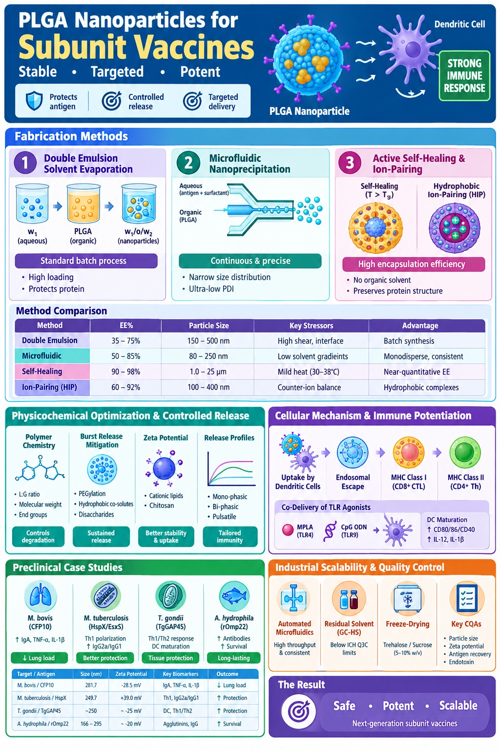
Poly(lactic-co-glycolic acid) PLGA Nanoparticle Development incorporates controlled fabrication approaches such as double emulsion solvent evaporation, microfluidic nanoprecipitation, and active self-healing microencapsulation to achieve high antigen loading while protecting the structural integrity of proteins. The appropriate synthesis strategy depends on several antigen-specific characteristics, including hydrophilicity, molecular weight, susceptibility to organic interfaces, and the desired final particle size.
Learn more about practical solutions for overcoming challenges in PLGA microsphere development during initial formulation design.
Double Emulsion Solvent Evaporation Mechanics
Water-in-oil-in-water (w₁/o/w₂) double emulsion solvent evaporation represents a primary batch-processing technique for incorporating hydrophilic subunit proteins into hydrophobic PLGA polymer matrices. The method creates an internal aqueous compartment containing the antigen within an organic polymer phase, after which solvent removal causes the polymer to solidify and form nanoparticles.
The primary emulsion (w₁/o) is initially produced by dispersing an aqueous antigen solution into an organic phase containing PLGA dissolved in volatile organic solvents, including dichloromethane or ethyl acetate. High-shear homogenization or ultrasonic agitation is commonly used to generate this primary emulsion. The resulting w₁/o emulsion is then introduced into a larger secondary aqueous phase (w₂) containing stabilizing surfactants, most commonly Poly(vinyl alcohol) (PVA) or Poloxamer 188, producing the final double emulsion (w₁/o/w₂). Controlled solvent evaporation or ambient solvent extraction subsequently removes the organic solvent, promoting polymer phase separation and solidification into rigid spherical nanoparticles. Important process parameters affecting mean particle diameter, polydispersity index (PDI), and entrapment efficiency include the phase volume ratios (w₁:o and o:w₂), polymer concentration, lactide-to-glycolide stoichiometry, applied shear energy, and surfactant concentration at the secondary interface. Nevertheless, high-energy sonication and contact with hydrophobic organic-aqueous interfaces may cause partial denaturation or aggregation of fragile tertiary protein structures. Protective excipients, including trehalose or recombinant serum albumin, may therefore be incorporated to help maintain protein stability.
Examine the specific role of surfactants and emulsifiers in PLGA microsphere fabrication for stability optimization.
Microfluidic Continuous Synthesis and Process Optimization
Microfluidic hydrodynamic focusing provides a continuous and highly reproducible method for producing PLGA nanoparticles with narrow size distributions and predictable scalability. Through precise control of fluid movement at the sub-millimeter level, microfluidic systems can minimize the batch-to-batch variability and elevated shear conditions commonly associated with conventional emulsification techniques.
Within microfluidic lab-on-a-chip architectures, an organic stream containing PLGA and lipophilic compounds is focused between converging aqueous sheath streams containing the subunit antigen and surfactant. Controlled diffusion across the laminar solvent-antisolvent boundary produces rapid, regulated supersaturation and nanoprecipitation. This process generates monodisperse nanoparticle populations with polydispersity indices routinely below 0.10. Parameters including total flow rate (TFR) and the flow rate ratio (FRR) between the aqueous and organic streams determine the relationship between mixing time and polymer aggregation time. As a result, these systems provide direct control over mean particle diameter, including particle sizes below 100 nanometers. Because the process avoids aggressive cavitation forces and extended exposure to organic solvents, antigen structural fidelity can be better preserved. This characteristic makes microfluidics an attractive manufacturing approach for clinical-grade subunit vaccine formulations.
Discover advanced techniques for controlling PLGA particle size and polydispersity index (PDI) in continuous processing.
Active Self-Healing and Hydrophobic Ion-Pairing Encapsulation
Active self-healing encapsulation and hydrophobic ion-pairing complexation are specialized approaches developed to achieve very high antigen encapsulation efficiency while minimizing or eliminating organic solvent-mediated protein degradation. These techniques address important limitations associated with conventional double emulsion systems, particularly when encapsulating highly soluble or structurally sensitive biomolecules.
Conventional double emulsion procedures may produce relatively low encapsulation efficiency for small or highly water-soluble antigens because these molecules can rapidly diffuse into the external continuous aqueous phase before the polymer matrix has sufficiently hardened. Active self-healing microencapsulation addresses this limitation by using preformed porous PLGA microspheres containing protein-trapping agents such as aluminum hydroxide (Al(OH)₃) gel. When these microspheres are incubated with low-concentration aqueous antigen solutions at temperatures above the hydrated polymer glass transition temperature (T > T_g), polymer chains can spontaneously rearrange and close the matrix pores. This mechanism can seal approximately 97% to 100% of the antigen within the polymer without exposing the protein to organic solvents or intense shear forces. In contrast, hydrophobic ion-pairing (HIP) complexation employs amphiphilic counter-ions, including dextran sulfate, to mask charged polar groups present on antigen surfaces. Reversible electrostatic complexation increases the apparent lipophilicity of the protein, promoting its partitioning into the organic phase during single-emulsion processing. This substantially improves entrapment efficiency and can also reduce the initial burst release.
Review effective strategies for encapsulating hydrophilic vs. hydrophobic APIs in PLGA to enhance entrapment efficiency.
| Fabrication Method | Typical Encapsulation Efficiency (EE%) | Mean Particle Diameter | Primary Processing Stressors | Key Technical Advantage |
|---|---|---|---|---|
| Double Emulsion (w₁/o/w₂) | 35% – 75% | 150 nm – 500 nm | High-shear cavitation, organic/water interface | Standardized batch synthesis for hydrophilic proteins |
| Microfluidic Nanoprecipitation | 50% – 85% | 80 nm – 250 nm | Minor local solvent concentration gradients | Continuous assembly, ultra-low PDI, high batch consistency |
| Active Self-Healing (T > T_g) | 90% – 98% | 1.0 μm – 25 μm | Mild thermal exposure (30°C – 38°C) | Zero organic solvent contact, near-quantitative entrapment |
| Hydrophobic Ion-Pairing (HIP) | 60% – 92% | 100 nm – 400 nm | Counter-ion stoichiometric balancing | Converts hydrophilic peptides into lipophilic complexes |
Physicochemical Optimization and Controlled Release Kinetics in Poly(lactic-co-glycolic acid) PLGA Nanoparticle Development
Physicochemical optimization in Poly(lactic-co-glycolic acid) PLGA Nanoparticle Development involves adjusting polymer backbone composition, surface charge, and matrix porosity to regulate hydrolysis kinetics and minimize premature antigen release. Strategic modification of these structural characteristics allows mono-phasic, bi-phasic, or pulsatile release profiles to be engineered according to specific immunological requirements.
Polymer Ester Chemistry and Matrix Hydrolysis
The degradation rate of the PLGA matrix and the corresponding antigen release profile are primarily influenced by the copolymer molar ratio of lactic acid to glycolic acid, overall molecular weight, and terminal end-group chemistry. Hydrolytic cleavage of ester bonds progressively breaks down the polymer matrix through bulk erosion, eventually producing non-toxic monomeric degradation products.
PLGA is a biodegradable aliphatic polyester that undergoes non-enzymatic bulk hydrolysis under physiological aqueous conditions and ultimately degrades into the endogenous metabolic products lactic acid and glycolic acid. Lactic acid contains a hydrophobic methyl side group that restricts water penetration into the polymer matrix, whereas glycolic acid does not contain this methyl group and is comparatively more hydrophilic. As a result, polymers containing higher proportions of lactide, such as PLGA 75:25, generally demonstrate slower degradation that can extend over several months. In comparison, 50:50 lactide-to-glycolide formulations undergo comparatively rapid hydrolysis, typically within weeks. Polymer molecular weight (M_w) also affects degradation behavior. Lower molecular weight chains, such as those in the 10–24 kDa range, tend to degrade more rapidly because they contain a greater relative density of hydrophilic end-groups and exhibit fewer chain entanglements. In addition, capping polymer termini with ester groups decreases initial water uptake compared with uncapped polymers containing carboxylic acid-terminated groups. This provides a direct chemical mechanism for modifying matrix degradation rates and establishing continuous release profiles.
Analyze the key physical and chemical differences between PLGA 50:50 and PLGA 75:25 for precise release control.
Strategies for Initial Burst Release Mitigation
Initial burst release, which refers to the rapid liberation of surface-adsorbed or pore-accessible antigen following administration, can be reduced through the incorporation of PEGylated diblock copolymers, hydrophobic small-molecule co-solutes, or disaccharide-based matrix stabilizers. Controlling this early release phase helps retain the antigen payload and supports prolonged exposure within immune tissues.
An uncontrolled initial burst can result in the release of as much as 50% of an encapsulated subunit antigen during the first few hours following administration. Such rapid antigen loss may waste valuable antigen, potentially contribute to systemic immune tolerance, and reduce the effectiveness of subsequent booster responses. Incorporating Poly(ethylene glycol) (PEG) into PLGA backbones to form PEG-PLGA modifies the surface energetics of the nanoparticles and establishes a hydrophilic steric barrier that decreases protein desorption and surface adherence. Co-encapsulation of hydrophobic immune potentiators or small molecules, including honokiol and capsaicin, can modify solvent exchange behavior during particle formation. This reduces internal pore connectivity and shifts the release pattern toward sustained Fickian diffusion. Disaccharides such as trehalose can also be incorporated into internal aqueous microdomains to reduce intra-particular protein aggregation and maintain native secondary structures during matrix erosion. These effects support more complete antigen release and help maintain a consistent antigen output over time.
Understand the structural factors governing bulk erosion vs. surface erosion in PLGA during matrix degradation.
Zeta Potential Modulation and Cationic Hybrid Platforms
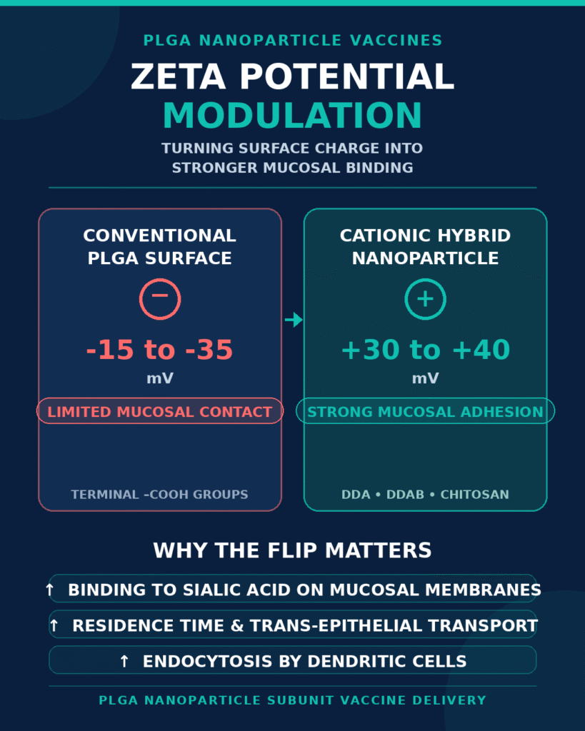
Modification of nanoparticle surface charge using cationic lipids or biopolymers can improve particle stability, increase adhesion to mucosal tissues, and facilitate efficient endocytosis by antigen-presenting cells. Adjusting the surface zeta potential can therefore transform conventionally repulsive particles into more strongly bioadhesive vaccine delivery systems.
Conventional PLGA nanoparticles generally exhibit negative surface charges (ζ = -15 mV to -35 mV), primarily because of terminal carboxylic acid groups. This negative charge may limit initial interactions with negatively charged mucosal surfaces and cellular membranes. Incorporating cationic components, including dimethyl dioctadecyl ammonium (DDA), didecyldimethylammonium bromide (DDAB), or chitosan, can produce hybrid nanoparticles with strongly positive zeta potentials (ζ = +30 mV to +40 mV). These cationic hybrid nanoparticles can establish electrostatic interactions with sialic acid residues on mucosal membranes, increasing local residence time and supporting trans-epithelial transport. Positive surface charge can also strengthen interactions with dendritic cell plasma membranes, promoting efficient endocytosis and macropinocytosis.
Cellular Mechanisms of Immune Potentiation and Cross-Presentation
Poly(lactic-co-glycolic acid) nanoparticles enhance subunit vaccine immunogenicity by promoting antigen uptake by dendritic cells, facilitating endosomal escape into the cytosol, and supporting Major Histocompatibility Complex (MHC) Class I cross-presentation to CD8+ T cells. This pathway allows purified protein antigens to generate cellular cytotoxic immune responses that are more commonly associated with live viral or bacterial vector systems.
Endosomal Escape and Proteasomal Processing
After uptake by professional antigen-presenting cells, PLGA nanoparticles undergo hydrolytic degradation within acidic phagolysosomal compartments. This process can promote localized membrane destabilization and facilitate the release of encapsulated antigen into the cytosol. Once in the cytosol, the subunit antigen can enter proteasomal processing pathways that contribute to MHC Class I presentation.
Professional antigen-presenting cells, including dendritic cells and macrophages, internalize PLGA nanoparticles through mechanisms such as clathrin-mediated endocytosis and macropinocytosis. As the particles progress through early and late endosomes, the acidic luminal environment, typically ranging from pH 4.5 to 5.5, accelerates PLGA ester hydrolysis and generates lactic acid and glycolic acid monomers. The accumulation of soluble degradation products can increase intra-endosomal osmotic pressure, promoting the movement of water and ions into the compartment until localized endosomal disruption occurs. This process can facilitate the transfer of encapsulated protein antigens into the cytoplasm, where they become accessible to cytosolic immunoproteasomes. Proteasomal degradation generates oligopeptides that are transported by Transporter associated with Antigen Processing (TAP) proteins into the endoplasmic reticulum, where they can be loaded onto newly synthesized MHC Class I molecules.
CD8+ Cytotoxic T Lymphocyte Activation via MHC Class I Presentation
Cross-presentation of PLGA-encapsulated subunit antigens through MHC Class I complexes promotes the differentiation of naive CD8+ T cells into active Cytotoxic T Lymphocytes (CTLs). At the same time, antigen that remains within endolysosomal compartments can enter MHC Class II processing pathways and provide support for CD4+ T helper cell responses.
Soluble subunit proteins that are not encapsulated are predominantly processed within endolysosomal compartments and presented through MHC Class II molecules to CD4+ T helper cells. As a result, they may have limited ability to generate the CD8+ CTL responses needed for eliminating intracellular pathogens. PLGA nanoparticles can redirect a portion of the antigen toward the cytosolic MHC Class I cross-presentation pathway, promoting antigen-specific CD8+ T-cell expansion and increased secretion of Interferon-gamma (IFN-γ), Tumor Necrosis Factor-alpha (TNF-α), and granzyme B. Simultaneously, antigen retained within endolysosomal compartments can undergo cathepsin-mediated cleavage and subsequent loading onto MHC Class II molecules, activating CD4+ Th1 and Th2 helper subsets. Activation of both presentation pathways provides an integrated immune response involving cellular and humoral protection.
Co-Delivery of Molecular Adjuvants for Synchronized APC Activation
Co-encapsulation of Toll-like Receptor (TLR) agonists together with subunit antigens within individual PLGA nanoparticles enables both signals to reach the same antigen-presenting cell at approximately the same time. This coordinated intracellular delivery can improve dendritic cell maturation while reducing unnecessary systemic inflammatory exposure.
When molecular adjuvants are administered separately from soluble antigens, the adjuvants may undergo rapid systemic clearance. This can result in off-target cytokine release while insufficiently stimulating the antigen-presenting cells responsible for processing the vaccine antigen. Incorporating hydrophobic TLR agonists such as Monophosphoryl Lipid A (MPLA, TLR4 agonist) or Unmethylated CpG Oligodeoxynucleotides (CpG ODN 1826, TLR9 agonist) within PLGA nanoparticles promotes co-uptake by individual dendritic cells. Once internalized, these dual-loaded nanoparticles can initiate intracellular TLR signaling cascades, resulting in nuclear translocation of NF-κB, increased expression of co-stimulatory markers such as CD80, CD86, and CD40, and local production of Interleukin-12 (IL-12) and Interleukin-1 beta (IL-1β). This coordinated co-delivery concentrates adjuvant activity within antigen-processing cells and can substantially improve the therapeutic index and safety profile.
Preclinical Case Studies and Targeted Delivery Efficacy
In vivo preclinical investigations indicate that PLGA nanoparticle-based subunit vaccines can provide enhanced protective efficacy, increased mucosal IgA production, and substantial reductions in pathogen burden compared with unencapsulated proteins or conventional alum-based formulations. These observations have been demonstrated across mucosal bacterial challenge models and complex parasitic infection models.
Pulmonary mucosal immunization using culture filtrate protein-10 (CFP10) encapsulated within PLGA nanoparticles approximately 280 nm in diameter, with a zeta potential of ζ = -28.5 mV, produced substantial protective immunity against Mycobacterium bovis in murine models. Intranasal administration of CFP10-PLGA nanoparticles produced elevated levels of secretory IgA in bronchoalveolar lavage fluid (BALF), increased serum concentrations of TNF-α and IL-1β, and enhanced antigen-specific IFN-γ production by splenocytes. Histopathological analysis after pathogen challenge demonstrated notable decreases in pulmonary inflammatory consolidation and bacterial burden. Similarly, in tuberculosis prime-boost regimens, PLGA:DDA hybrid nanoparticles co-encapsulating the multistage fusion protein HspX/EsxS and MPLA generated strong systemic Th1 polarization, increased IgG2a/IgG1 antibody ratios, and improved protection following BCG priming.
In parasitic disease models, PLGA nanospheres containing Toxoplasma gondii glidesome-associated protein 45 (TgGAP45) generated potent combined Th1/Th2 protective immunity and performed substantially better than commercial Montanide ISA 206/660 oil emulsions while avoiding inflammatory tissue damage at the injection sites. In aquaculture bacterial challenge models, PLGA nanoparticles containing recombinant outer membrane proteins (Omp22, OmpA, and maltoporin) provided sustained protein release over 40 days, increased agglutination antibody titers, and produced high survival rates following Aeromonas hydrophila challenge. Collectively, these findings illustrate the adaptability of PLGA nanoparticle carriers for different infectious disease targets and administration routes.
| Target Pathogen / Antigen | Formulation Matrix Metrics | Surface Zeta Potential | Key Immunological Biomarkers | Demonstrated Protective Outcome |
|---|---|---|---|---|
| M. bovis / CFP10 Protein | PLGA (w₁/o/w₂), 281.7 nm | -28.5 mV | Elevated BALF IgA, TNF-α, IL-1β, splenocyte IFN-γ | Reduced lung bacterial load & reduced inflammatory lesions |
| M. tuberculosis / HspX/EsxS | PLGA:DDA + MPLA, 249.7 nm | +39.0 mV | Dominant Th1 polarization, elevated IgG2a/IgG1 antibody ratio | Enhanced BCG prime-boost cellular protection |
| Toxoplasma gondii / TgGAP45 | PLGA Nanospheres | Negative (approximately -25 mV) | DC maturation, mixed Th1/Th2 cytokines, lymphocyte proliferation | Superior tissue protection vs. commercial oil emulsions |
| Aeromonas hydrophila / rOmp22 | PLGA (w₁/o/w₂), 166 nm – 295 nm | Negative (approximately -20 mV) | Elevated agglutination titers, persistent circulating IgG | Long-lasting survival enhancement post-challenge |
Industrial Scalability and Quality Control Standards in Poly(lactic-co-glycolic acid) PLGA Nanoparticle Development
Moving PLGA nanoparticle subunit vaccine formulations from laboratory-scale research into cGMP industrial manufacturing requires stringent control of critical quality attributes (CQAs). Key considerations include automated microfluidic processing, rigorous residual solvent analysis, and validated freeze-drying procedures. Robust and reproducible analytical methods are essential for maintaining batch-to-batch consistency and satisfying regulatory expectations.
Successful clinical translation of nanovaccine formulations requires tight specifications for particle size, size distribution, zeta potential, antigen recovery, and endotoxin content throughout manufacturing scale-up. Multi-channel microfluidic skid platforms can support high-throughput continuous production while maintaining consistent local channel dimensions, thereby preserving comparable fluid-mixing conditions during scale expansion. Gas Chromatography-Headspace (GC-HS) analysis is necessary to confirm that residual processing solvents, including dichloromethane and ethyl acetate, remain substantially below the toxicological thresholds established under International Council for Harmonisation (ICH) Q3C. Long-term liquid formulation stability can also require optimized freeze-drying (lyophilization) procedures using lyoprotectants such as trehalose or sucrose at concentrations of 5%–10% w/v. These excipients help immobilize particle structures within an amorphous glass matrix and reduce particle fusion, antigen leakage, and loss of tertiary protein structure during storage. Analytical capabilities deployed by ResolveMass Laboratories Inc. support comprehensive characterization of these critical quality parameters, helping facilitate the advancement of PLGA nanoparticle formulations toward clinical development.
Access our full range of PLGA characterization and testing services for IND and ANDA filings.
Conclusion
Poly(lactic-co-glycolic acid) PLGA Nanoparticle Development represents a versatile and clinically relevant platform for addressing fundamental immunogenicity and stability challenges associated with conventional subunit vaccines. Through controlled matrix erosion, targeted intracellular antigen delivery, and coordinated adjuvant presentation, this technology supports the rational development of safe and highly potent vaccine formulations.
Poly(lactic-co-glycolic acid) PLGA Nanoparticle Development addresses the critical gap between the safety characteristics of subunit antigens and the vector-like cellular immunogenicity required for effective protection against intracellular pathogens. By providing structural stabilization, limiting burst release, and enabling surface charge optimization, PLGA nanocarriers can facilitate both MHC Class I cross-presentation and MHC Class II processing pathways, supporting balanced T-cell and antibody-mediated immune responses. Emerging process intensification strategies, including continuous microfluidic assembly and active self-healing encapsulation, offer scalable and reproducible manufacturing pathways while supporting preservation of structural protein integrity. ResolveMass Laboratories Inc. continues to advance analytical and formulation strategies designed to support the progression of complex polymeric nanovaccine candidates from early discovery through preclinical optimization.
Learn more about evaluating PLGA polymer sameness for ANDA regulatory filings.
To consult with our advanced formulation scientists or explore custom analytical testing and process development, contact ResolveMass Laboratories Inc. at https://resolvemass.ca/contact/.
Frequently Asked Questions
The main advantage of double emulsion (w₁/o/w₂) solvent evaporation is its ability to encapsulate water-soluble proteins within protected internal aqueous compartments. The organic PLGA phase surrounds these compartments before polymer solidification, helping preserve the hydrophilic antigen during nanoparticle formation. This approach also enables sustained and controlled antigen release.
PLGA nanoparticles can promote CD8+ CTL activation through intracellular antigen delivery followed by MHC Class I cross-presentation. After cellular uptake, acidic phagolysosomal conditions promote PLGA degradation and facilitate antigen release into the cytosol. The antigen can then undergo proteasomal processing and TAP-mediated transport for loading onto MHC Class I molecules.
Initial burst release can rapidly deplete antigen located near the nanoparticle surface, reducing the duration of controlled delivery and potentially increasing unwanted inflammatory exposure. Excessive early release may also compromise the intended immunization profile. Strategies such as PEGylation, hydrophobic co-encapsulation, and active self-healing microencapsulation can help reduce premature antigen release.
The lactide-to-glycolide ratio influences PLGA hydrophobicity, water penetration, and the rate of ester bond hydrolysis within the polymer matrix. A higher lactide proportion, such as 75:25, generally produces a more hydrophobic matrix with slower degradation and prolonged antigen release. In comparison, a 50:50 composition typically undergoes faster hydrolysis and provides more rapid release.
Cationic surface modification with DDA or chitosan can shift the naturally negative zeta potential of PLGA nanoparticles toward positive values, often above +30 mV. This promotes electrostatic association with negatively charged mucin and cellular membranes. As a result, nanoparticles may exhibit improved mucosal retention and enhanced interaction with dendritic cells, supporting cellular uptake.
Co-encapsulation of TLR agonists such as CpG ODN or MPLA allows antigen and immune-stimulatory signals to reach the same antigen-presenting cells in a coordinated manner. This synchronized delivery can strengthen dendritic cell activation and increase expression of co-stimulatory molecules. It may also provide more localized immune stimulation while limiting unnecessary systemic inflammatory responses.
Active self-healing microencapsulation protects fragile proteins by introducing the antigen into preformed porous PLGA microspheres under mild aqueous conditions. Trapping agents such as Al(OH)₃ help retain the protein while exposure to temperatures above the polymer Tg promotes pore closure. This process minimizes contact with organic solvents and avoids high-shear sonication that could damage sensitive proteins.
Microfluidic continuous synthesis provides controlled hydrodynamic focusing and predictable mixing conditions during nanoparticle formation. This enables production of highly uniform particles with precise size control and low PDI, often below 0.10. The continuous process also supports scalable manufacturing while reducing the protein aggregation and batch variability associated with high-energy bulk sonication.
PLGA nanoparticle formulations can be stabilized through lyophilization using disaccharide cryoprotectants such as trehalose or sucrose, commonly at 5%–10% w/v. During freeze-drying, these excipients form an amorphous glass matrix that helps protect both the nanoparticle structure and encapsulated protein. This reduces aggregation, matrix collapse, leakage, and loss of protein secondary structure during storage.
Reference:
- Du, X., Xue, J., Jiang, M., Lin, S., Huang, Y., Deng, K., Shu, L., Xu, H., Li, Z., Yao, J., Chen, S., Shen, Z., & Feng, G. (2021). A multiepitope peptide, rOmp22, encapsulated in chitosan-PLGA nanoparticles as a candidate vaccine against Acinetobacter baumannii infection. International Journal of Nanomedicine, 16, 1819–1836. https://doi.org/10.2147/IJN.S296527
- Silva, A. L., Soema, P. C., Slütter, B., Ossendorp, F., & Jiskoot, W. (2016). PLGA particulate delivery systems for subunit vaccines: Linking particle properties to immunogenicity. Human Vaccines & Immunotherapeutics, 12(4), 1056–1069. https://doi.org/10.1080/21645515.2015.1117714
- Liang, Z., Li, M., Ni, J., Hussain, T., Yao, J., Song, Y., Liu, Y., Wang, H., & Zhou, X. (2022). CFP10-loaded PLGA nanoparticles as a booster vaccine confer protective immunity against Mycobacterium bovis. BioImpacts, 12(5), 395–404. https://doi.org/10.34172/bi.2022.23645
- Lee, J., Neustrup, M. A., Slütter, B., O’Mahony, C., Bouwstra, J. A., & van der Maaden, K. (2024). Intradermal vaccination with PLGA nanoparticles via dissolving microneedles and classical injection needles. Pharmaceutical Research, 41(2), 305–319. https://doi.org/10.1007/s11095-024-03665-7
- Rauta, P. R., & Nayak, B. (2015). Parenteral immunization of PLA/PLGA nanoparticle encapsulating outer membrane protein (Omp) from Aeromonas hydrophila: Evaluation of immunostimulatory action in Labeo rohita (rohu). Fish & Shellfish Immunology, 44(1), 287–294. https://doi.org/10.1016/j.fsi.2015.02.007
Get In Touch With Us
About The Author


