


Introduction
Delivering comprehensive Bioanalytical Support for an AAV Gene Therapy IND requires the validation of highly sensitive molecular and immunoassay platforms capable of performing reliably across complex tissue matrices. These validated approaches are essential for generating biodistribution, shedding, and immunogenicity profiles that satisfy regulatory expectations. Investigational New Drug (IND) submissions for adeno-associated virus (AAV)-based therapeutics require robust nonclinical data packages that provide sufficient scientific justification for initiating first-in-human clinical trials. Recombinant AAV vectors are designed to transport genetic payloads into target tissues; however, their non-replicating episomal characteristics, diverse tissue tropisms, and possibility of off-target transduction create substantial bioanalytical challenges during nonclinical development.
Moving a therapeutic program from preclinical candidate selection toward IND submission requires careful alignment with the expectations described in the International Council for Harmonisation (ICH) S12 guidance (Nonclinical Biodistribution Considerations for Gene Therapy Products) and the ICH M10 bioanalytical method validation framework. Bioanalytical testing programs must be capable of quantitatively determining vector genome copy numbers, characterizing vector shedding over longitudinal timepoints, and assessing both humoral and cellular host immune responses. Advanced molecular technologies, including Droplet Digital PCR (dPCR), quantitative PCR (qPCR), and hybrid liquid chromatography-tandem mass spectrometry (LC-MS/MS), can be applied to overcome challenges associated with matrix-induced suppression, very low vector copy numbers, and assay cross-reactivity.
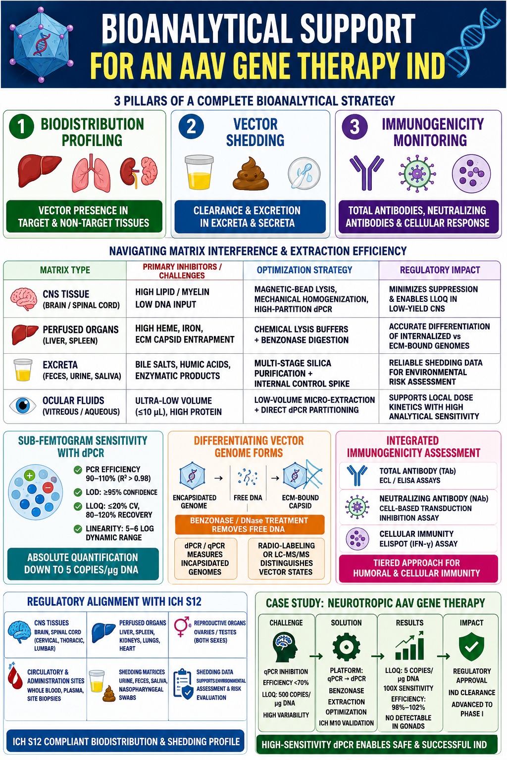
A complete bioanalytical strategy supporting an IND submission is built around three fundamental testing pillars:
- Biodistribution Profiling: Determining vector genome persistence, distribution, and clearance in target and non-target tissues across the required core organ systems.
- Vector Shedding Kinetics: Measuring vector clearance and excretion through excreta and secreta to support environmental risk assessments.
- Immunogenicity Monitoring: Conducting tiered assessment of total antibodies (TAb), neutralizing antibodies (NAb), and cellular immune responses (ELISpot).
Explore how tailored testing frameworks can accelerate your regulatory timeline: IND Bioanalytical Support Solutions
Share via:
Quick Summary:
- AAV IND bioanalytical support requires highly sensitive, validated molecular and immunoassay platforms to generate reliable biodistribution, shedding, and immunogenicity data.
- Three core testing pillars—biodistribution profiling, vector shedding kinetics, and immunogenicity monitoring—form the foundation of a complete bioanalytical strategy.
- Matrix-specific challenges in tissues such as the brain, liver, spleen, and ocular fluids can affect extraction and assay performance. Optimized workflows, magnetic-bead extraction, and internal controls improve data reliability.
- High-partition dPCR enables absolute quantification of low-copy vector genomes, with sensitivity reaching 5 copies/µg DNA in the case study. Validation covers efficiency, LOD, LLOQ, precision, accuracy, and linearity.
- Vector genome differentiation using nuclease digestion and orthogonal analytical techniques helps distinguish encapsidated genomes from free DNA and ECM-bound capsids, reducing the risk of misinterpreting biodistribution results.
- Tiered immunogenicity assessment combines Total Antibody (TAb), Neutralizing Antibody (NAb), and cellular ELISpot assays to evaluate humoral and cellular immune responses.
- ICH S12 and ICH M10 alignment supports regulatory-grade nonclinical data packages. In the neurotropic AAV case study, dPCR improved sensitivity 100-fold, reduced matrix-related variability, and supported successful IND clearance and Phase I advancement.
Navigating Novel Tissue Matrix Interference and Extraction Efficiency in Bioanalytical Support for an AAV Gene Therapy IND
Matrix interference associated with novel biological tissues can be controlled through optimization of nucleic acid extraction workflows and implementation of internal reference controls designed to maintain consistent and linear amplification across challenging organ homogenates. Nonclinical biodistribution investigations require evaluation of vector genome presence in a defined panel of biological matrices, including the brain, spinal cord, liver, kidneys, heart, lungs, spleen, injection sites, and gonads. Because each tissue possesses a different biochemical composition, analytical performance can vary considerably. For example, central nervous system (CNS) tissues may contain substantial lipid fractions, splenic and hepatic tissues can contain abundant heme, and ocular fluids may have high protein concentrations. These matrix characteristics can interfere with polymerase activity and contribute to inconsistent nucleic acid extraction efficiency.
Discover strategies to overcome biological matrix challenges in your nonclinical workflows: Navigating IND-Enabling Bioanalytical Studies
To achieve consistent linear amplification and recovery, bioanalytical workflows can combine automated magnetic bead-based extraction procedures with matrix-matched calibration controls. Incorporating exogenous internal control spikes, such as linearized non-target plasmids or synthetic DNA constructs, provides an effective mechanism for monitoring extraction efficiency and identifying polymerase inhibition during analysis. Establishing whether a negative result represents genuine target absence or matrix-associated signal suppression is particularly important when producing regulatory-grade data intended for an IND submission.
Learn how to mitigate critical analytical pitfalls in complex study designs: Common Bioanalytical Mistakes to Avoid
| Biological Matrix Type | Primary Matrix Inhibitors / Analytical Challenges | Extraction & Assay Optimization Strategy | Impact on Regulatory Compliance |
|---|---|---|---|
| CNS Tissue (Brain/Spinal Cord) | High lipid/myelin content and low total DNA input from micro-dissections. | Automated magnetic-bead lysis, mechanical homogenization, and high-partition dPCR. | Minimizes matrix suppression and enables LLOQ verification in low-yield CNS sub-regions. |
| Perfused Organs (Liver, Spleen) | High heme, iron ions, and extracellular matrix (ECM) capsid entrapment. | Chemical cell-lysis buffers combined with pre-analytical Benzonase digestion. | Enables accurate differentiation and quantification of internalized versus ECM-bound vector genomes. |
| Excreta (Feces, Urine, Saliva) | Bile salts, humic acids, and complex enzymatic degradation products. | Multi-stage silica column purification combined with internal control DNA spiking. | Improves the reliability of environmental safety data used for regulatory shedding risk analysis. |
| Ocular Fluids (Vitreous/Aqueous) | Ultra-low sample volume (≤10 μL) and high protein concentration. | Low-volume micro-extraction procedures coupled with direct dPCR partitioning. | Supports localized administration dose-kinetics while maintaining analytical sensitivity. |
Discover how specialized tissue parsing enhances vector quantification: Tissue and CSF Bioanalytical Services
Achieving Sub-Femtogram Sensitivity and Quantitation Limits for Low-Copy Vector Genomes
Sub-femtogram assay sensitivity and ultra-low limits of quantitation (LLOQ) can be achieved through Droplet Digital PCR (dPCR) partitioning, which separates target nucleic acids physically and reduces the impact of matrix-associated amplification suppression. In contrast to quantitative PCR (qPCR), which determines target quantities by comparing fluorescence signals with an external standard curve, dPCR divides the reaction mixture into thousands of discrete nanoliter-sized droplets. Absolute target quantification is subsequently calculated from the proportion of positive and negative droplets using Poisson statistics. This approach eliminates dependence on a conventional standard curve while increasing assay tolerance to residual matrix inhibitors.
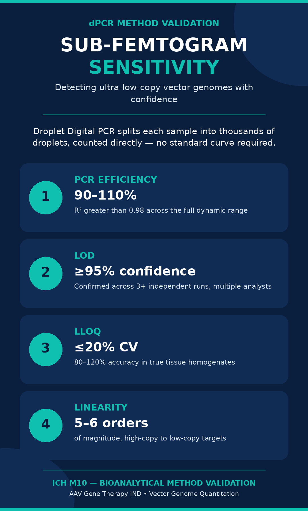
Method validation requirements, consistent with 2024 American Association of Pharmaceutical Scientists (AAPS) consensus recommendations and ICH M10 standards, require comprehensive assessment of multiple assay performance characteristics:
- PCR Efficiency: Amplification efficiency should remain within 90% to 110%, with a coefficient of determination (R²) greater than 0.98 across the established dynamic range.
- Limit of Detection (LOD): The lowest copy number that can be detected with ≥95% statistical confidence should be established across a minimum of three independent analytical runs conducted by multiple analysts over several days.
- Lower Limit of Quantitation (LLOQ): The LLOQ should be formally established as the lowest concentration of target vector genome that can be quantified with acceptable precision, typically ≤20% CV, and accuracy, generally within 80%–120% recovery, in the presence of target tissue homogenates.
- Linearity and Dynamic Range: Linearity should be demonstrated across approximately 5 to 6 orders of magnitude to adequately characterize both highly exposed target organs and tissues exhibiting low-copy non-target vector clearance.
See how high-precision assay design ensures regulatory compliance: Generating Robust Bioanalytical Data
Differentiating Encapsidated Vector Genomes, Free DNA, and Extracellular Matrix-Bound Capsids
Distinguishing encapsidated and potentially functional vector genomes from degraded, unencapsidated DNA and extracellularly associated capsids requires pre-analytical nuclease digestion strategies combined with orthogonal radiolabeling or hybrid mass spectrometry approaches. Conventional nucleic acid extraction procedures release DNA from multiple sources within a biological sample. Consequently, these methods may unintentionally co-purify unencapsidated DNA contaminants, degraded vector fragments, and intact viral particles. To preferentially measure vector genomes protected within intact capsids, tissue samples can be treated with endonuclease enzymes, including Benzonase or DNase I, before capsid lysis. This treatment enzymatically removes exposed and non-encapsidated nucleic acids, allowing subsequent dPCR/qPCR measurements to more specifically represent intact vector-encapsidated genomes.
Scientific investigations have also shown that AAV vectors can exhibit rapid post-administration clearance while viral capsids may temporarily localize within the extracellular matrix (ECM) of highly perfused organs, particularly the liver and spleen. This behavior can result in a quantitative difference between capsid protein concentrations measured using ELISA or hybrid LC-MS/MS and vector genome copy numbers determined using dPCR. Dual-isotope radiolabeling approaches, such as co-delivery of 125I non-residualizing and 111In residualizing probes, can help distinguish internalized and endocytosed AAV vectors from capsids that remain associated with extracellular tissue structures. Characterizing these different vector states is important because it reduces the possibility of incorrectly interpreting extracellular accumulation as evidence of off-target cellular transduction during nonclinical safety assessments.
Explore how integrated chemistry platforms solve complex structural assay challenges: Integrated Chemistry and Bioanalytical CRO Capabilities
Integrated Immunogenicity Assessment and Anti-AAV Antibody Screening in Bioanalytical Support for an AAV Gene Therapy IND
Integrated immunogenicity assessments included within IND packages generally employ tiered bioanalytical workflows that combine Total Antibody (TAb) ligand-binding assays, cell-based Neutralizing Antibody (NAb) transduction inhibition assays, and cellular ELISpot measurements. Pre-existing humoral immunity against naturally occurring AAV serotypes is reported in approximately 30% to 60% of the human population. Even relatively low concentrations of neutralizing antibodies may interfere with vector activity by neutralizing vector particles, modifying biodistribution, increasing systemic clearance, or contributing to inflammatory toxicities.
Bioanalytical method development for humoral immunogenicity therefore emphasizes the establishment of reliable screening and confirmatory assay cut-points:
- Total Antibody (TAb) Assays: These assays can be developed using electrochemiluminescence (ECL) or ELISA formats. One important technical challenge in TAb assay development is the limited availability of free primary amine groups on the external AAV capsid surface. This limitation can complicate chemical conjugation with biotin or ruthenium tags. Optimization of molar labeling ratios is therefore necessary to minimize capsid structural disruption while maintaining the conformational epitopes required for antibody recognition.
- Neutralizing Antibody (NAb) Assays: These assays are performed using cell-based transduction inhibition systems that measure vector-mediated reporter gene expression, such as luciferase or green fluorescent protein, in permissive cell lines. The resulting functional assay determines the dilution factor at which a serum sample inhibits viral entry and subsequent transgene expression.
- Cellular Immunogenicity: Cellular immune responses can be evaluated using Enzyme-Linked Immunospot (ELISpot) assays that measure Interferon-gamma (IFN-γ) secretion from isolated Peripheral Blood Mononuclear Cells (PBMCs) following stimulation with AAV capsid or transgene peptide pools.
Learn how to protect study validity with complete regulatory traceability: Ensuring Data Integrity in Bioanalytical Studies
Regulatory Alignment with ICH S12 Guidance and IND-Enabling Data Packages
Alignment with the ICH S12 guidance requires the application of appropriately validated bioanalytical methods across a defined core set of tissues and biological fluids to generate comprehensive nonclinical biodistribution and shedding profiles. The finalized ICH S12 framework establishes harmonized international expectations for the nonclinical development of gene therapy products and describes the scope, timing, and analytical rigor expected before first-in-human clinical trials can begin.
A regulatory-grade ICH S12 data package generally encompasses the following biological matrices:
- Central Nervous System Tissues: Brain micro-dissections, cervical spinal cord, thoracic spinal cord, and lumbar spinal cord.
- Visceral Organs and Perfused Tissues: Liver, spleen, kidneys, lungs, and heart.
- Reproductive Organs: Gonads, including ovaries or testes, assessed in both sexes to evaluate potential germline vector persistence.
- Circulatory and Administration Sites: Whole blood, blood plasma, and tissue biopsies collected from the site of administration.
- Shedding Matrices: Urine, feces, saliva, and nasopharyngeal swabs collected at longitudinal post-dose timepoints to evaluate potential environmental risk.
Alongside tissue biodistribution characterization, vector shedding studies determine the extent and persistence of viral vector excretion through biological fluids. Shedding information is important for supporting the Environmental Assessment (EA) required by regulatory authorities because it helps characterize potential transmission risks to untreated individuals and possible environmental exposure. Under ICH S12, shedding assessments should use quantitative and appropriately validated assays in the relevant biological matrices so that reliable vector clearance kinetics can be established following administration.
Stay ahead of evolving global validation standards for regulatory submissions: ICH M10 Bioanalytical Method Validation Guidelines
Case Study: Bioanalytical Support for an AAV Gene Therapy IND Submission in a Neurodegenerative Model
Transitioning from conventional qPCR to high-partition dPCR successfully addressed tissue matrix suppression and reduced the LLOQ by 100-fold, allowing sensitive detection of vector genomes required to support IND clearance for a novel neurotropic gene therapy.
Nonclinical Challenge
A biopharmaceutical sponsor had developed an engineered AAV vector intended to cross the blood-brain barrier for treatment of a rare neurodegenerative disorder. During the initial nonclinical toxicology program in non-human primates (NHPs), the sponsor encountered substantial bioanalytical difficulties while attempting to quantify vector genome copy numbers in micro-dissected spinal cord regions and specific brain sub-structures. The established qPCR assay demonstrated considerable matrix-associated inhibition, resulting in inconsistent PCR efficiencies below 70% and excessive inter-assay variability greater than 35% CV. In addition, the qPCR method had an LLOQ of 500 vector copies/μg DNA, which was not sufficiently sensitive for identifying low-level off-target transduction in peripheral tissues, including the gonads and heart. This analytical limitation represented a significant regulatory compliance challenge ahead of the planned IND submission.
Methodological Solution
A multi-stage bioanalytical strategy was implemented to address the identified analytical limitations:
- Platform Migration to dPCR: The analytical platform was changed from conventional qPCR to high-partition Droplet Digital PCR (dPCR), with the assay designed to target a conserved region within the transgene promoter.
- Extraction Workflow Engineering: A customized sample homogenization workflow based on bead-milling in specialized lysis buffers was developed. This was followed by automated magnetic bead purification optimized to reduce interference from myelin-associated lipids.
- Nuclease Integration: Pre-treatment with Benzonase digestion was incorporated into the workflow to remove non-encapsidated free vector DNA before thermal lysis of the viral capsid.
- Validation Parameters: The revised method was validated according to ICH M10 guidelines across the relevant target tissue matrices. Validation included assessment of dynamic range, precision, accuracy, and tolerance to matrix effects.
Learn how specialized testing capabilities expedite early safety milestones: Bioanalytical CRO Services for First-in-Human Studies
Quantifiable Results and Impact
The optimized dPCR bioanalytical method provided absolute target quantification to as low as 5 vector copies/μg of genomic DNA, representing a 100-fold improvement in sensitivity compared with the legacy qPCR platform. Across the evaluated brain homogenates, amplification efficiency remained consistently between 98% and 102%, without dependence on standard curve interpolation. At late post-dose timepoints, the assay demonstrated non-detectable vector levels in NHP gonad samples, supporting the regulatory safety assessment concerning potential germline transmission. The completed bioanalytical package successfully passed regulatory review, allowing the sponsor to obtain IND clearance and progress the therapeutic program into Phase I clinical trials.
Explore rapid decision-making frameworks for candidate selection: Bioanalytical Services for Rapid Proof-of-Concept
Conclusion: Advancing Bioanalytical Support for an AAV Gene Therapy IND
High-precision Bioanalytical Support for an AAV Gene Therapy IND serves as a critical link between preclinical candidate selection and clinical trial execution by producing validated biodistribution, shedding, and immunogenicity datasets. As the development of engineered AAV capsids and increasingly complex genetic payloads continues to expand, sponsors require analytical partners with the expertise and infrastructure to perform sophisticated molecular and immunoassay testing. Addressing matrix-specific extraction limitations, implementing high-sensitivity dPCR platforms, and developing comprehensive immunogenicity profiles can help therapeutic developers meet the analytical expectations associated with ICH S12 and ICH M10. ResolveMass Laboratories Inc. delivers scientific rigor, regulatory alignment, and customized method validation capabilities to support complex gene therapy programs throughout nonclinical development and toward clinical advancement.
Establish a strategic analytical partnership to streamline your pipeline: Strategic Bioanalytical CRO Partnership Solutions
For customized bioanalytical method development, validation, and IND-enabling regulatory support, visit ResolveMass Laboratories Contact Us.
Frequently Asked Questions
Droplet Digital PCR (dPCR) divides a reaction into thousands of individual partitions, allowing target molecules to be quantified directly through Poisson statistics. Unlike qPCR, it does not depend on an external standard curve for absolute quantification. This partitioning approach can also improve detection and quantitation of low-copy vector genomes in complex biological matrices.
Pre-analytical nuclease digestion using enzymes such as Benzonase or DNase I can remove exposed, unencapsidated vector DNA from tissue homogenates. DNA protected within intact AAV capsids remains available for subsequent capsid lysis and molecular analysis. This workflow helps ensure that dPCR or qPCR results more accurately represent encapsidated vector genomes rather than free DNA contamination.
Matrix inhibition is assessed by introducing known quantities of target DNA into non-transduced tissue homogenates and comparing the measured response with appropriate controls. Internal control DNA spikes can help identify extraction losses or amplification inhibition during analysis. Optimized purification procedures and partition-based platforms such as dPCR can further reduce the influence of matrix-related interference.
Lipid-rich brain tissue can interfere with nucleic acid extraction and amplification, requiring specialized sample-processing workflows. Mechanical bead-milling can be combined with optimized detergent-based lysis and magnetic-bead nucleic acid extraction to reduce lipid-associated interference. These approaches improve DNA recovery and help maintain reliable assay performance in CNS tissue samples.
Humoral immunogenicity assessment typically incorporates Total Antibody (TAb) assays, using formats such as ELISA or electrochemiluminescence, to detect binding antibodies against AAV capsids. Cell-based Neutralizing Antibody (NAb) assays are used to determine whether antibodies functionally inhibit AAV-mediated transduction. Together, these assays provide complementary information about the host antibody response.
Biodistribution studies determine where vector genomes are present within tissues and organs, including their persistence and clearance over time. Shedding studies instead evaluate the release and elimination of viral vectors through biological materials such as urine, feces, and saliva. Shedding data are particularly important for assessing potential environmental exposure and transmission risks.
Following AAV administration, viral capsids can temporarily associate with the extracellular matrix (ECM) of highly perfused organs, including the liver and spleen. These extracellular capsids may follow different clearance kinetics than vector genomes that have entered cells. Consequently, capsid protein measurements and dPCR-based vector genome measurements may show different patterns during early post-dose sampling.
Quantitative nucleic acid assays should be evaluated for critical characteristics such as specificity, selectivity, LLOQ, LOD, precision, accuracy, linearity, dynamic range, and matrix effects. Additional considerations include PCR efficiency, typically targeted within 90%–110%, and stability under relevant sample-storage conditions. These parameters establish whether the assay is sufficiently reliable for its intended bioanalytical application.
Pre-existing anti-AAV neutralizing antibodies can be evaluated by analyzing baseline serum samples with validated cell-based transduction inhibition assays. The resulting measurements establish NAb titers before administration of the vector. Characterizing baseline immunity is important because pre-existing NAbs may influence vector exposure, accelerate clearance, affect biodistribution, and potentially reduce therapeutic efficacy.
Reference:
- U.S. Food and Drug Administration. (2023, May). S12 nonclinical biodistribution considerations for gene therapy products: Guidance for industry. https://www.fda.gov/regulatory-information/search-fda-guidance-documents/s12-nonclinical-biodistribution-considerations-gene-therapy-products
- U.S. Food and Drug Administration. (2023, May). S12 nonclinical biodistribution considerations for gene therapy products: Guidance for industry. https://www.fda.gov/media/167605/download
- Wang, H., Li, R., Sadekar, S., Kamath, A. V., & Shen, B.-Q. (2024). A novel approach to quantitate biodistribution and transduction of adeno-associated virus gene therapy using radiolabeled AAV vectors in mice. Molecular Therapy—Methods & Clinical Development, 32(3), 101326. https://doi.org/10.1016/j.omtm.2024.101326
Get In Touch With Us
About The Author


