


Introduction: Why Characterization of Long-Acting Biologics in PLGA Demands a Dedicated Framework
The characterization of long-acting biologics encapsulated within poly(lactic-co-glycolic acid) (PLGA) matrices cannot be approached using the same analytical strategies applied to conventional biologic drug products. A monoclonal antibody (mAb) in aqueous solution and the identical mAb encapsulated within a 50:50 PLGA microsphere intended for monthly subcutaneous administration represent fundamentally different analytical challenges, despite containing the same active pharmaceutical ingredient.
PLGA systems, including microspheres, nanospheres, implants, and in situ forming depots, remain the leading biodegradable polymer platform for long-acting injectables (LAIs) across numerous therapeutic areas. These include oncology applications such as leuprolide acetate, psychiatric therapies such as risperidone, endocrinology products such as octreotide, and a growing number of biologic development programs targeting autoimmune diseases and chronic central nervous system disorders. The same properties that make PLGA an effective controlled-release platform, including tunable degradation profiles spanning days to months, an established FDA approval history, and biodegradation into lactic and glycolic acid, also create one of the most analytically challenging environments for therapeutic proteins.
Bulk hydrolytic erosion progressively acidifies the internal polymer microenvironment. Studies have demonstrated that local pH values within PLGA microspheres may decrease to between 1.5 and 4, meaning that an encapsulated protein can remain exposed for extended periods to conditions that would normally be classified as severe degradation stress in a conventional stability program.
This article examines the analytical methodologies, technical considerations, and regulatory expectations required to establish a scientifically robust characterization program for long-acting biologics formulated within PLGA delivery systems.
Learn More: Explore specialized PLGA Contract Manufacturing options for advanced drug delivery systems.
Share via:
Article Summary
- Characterization of long-acting biologics in PLGA systems requires a comprehensive analytical strategy that evaluates polymer properties, particle morphology, protein stability, encapsulation efficiency, and controlled drug release behavior.
- The acidic microenvironment formed during PLGA degradation, where internal pH may fall to nearly 1.5, is a major contributor to protein denaturation, aggregation, unfolding, and chemical instability within the formulation.
- Analytical techniques including SEC-HPLC, GPC/SEC-MALS, Dynamic Light Scattering (DLS), Scanning Electron Microscopy (SEM), FTIR spectroscopy, Differential Scanning Calorimetry (DSC), and Circular Dichroism (CD) spectroscopy are essential for complete characterization of PLGA-biologic systems.
- In vitro release testing (IVRT) for PLGA-based long-acting injectables is highly complex because release mechanisms involve both polymer degradation and protein diffusion, requiring carefully designed membrane-based or membrane-free testing approaches.
- Polymer molecular weight and polydispersity index (PDI) strongly influence release kinetics, and even modest variations in molecular weight can significantly alter burst release and sustained-release profiles.
- Regulatory agencies such as the FDA and EMA expect extensive higher-order structure (HOS) characterization of proteins recovered from PLGA matrices, rather than relying solely on drug loading or encapsulation data.
- Bioanalytical methods used for pharmacokinetic studies must differentiate between free released biologic and drug that remains associated with the PLGA carrier system, creating analytical challenges not typically encountered in conventional biologic formulations.
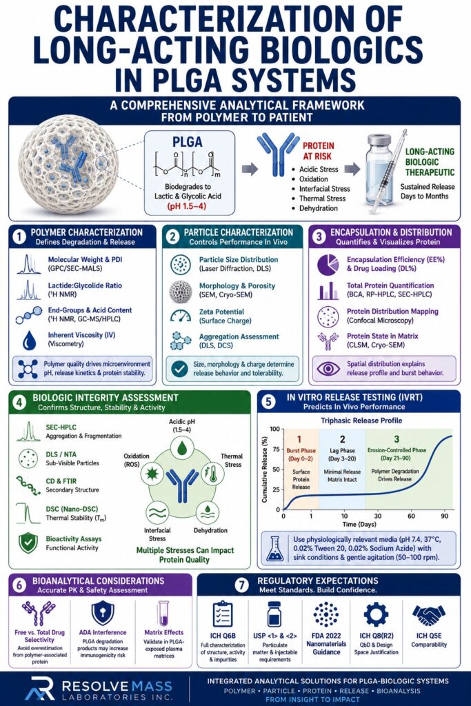
Polymer Characterization as a Prerequisite to Biologic Characterization
The polymer must be comprehensively characterized before any interpretation of encapsulated biologic data can be considered reliable. Variability in PLGA properties directly influences protein release kinetics, degradation-induced microenvironmental pH, encapsulation efficiency, and long-term biologic stability.
Biologic stability within PLGA systems is not determined solely by the protein itself. Instead, it is an emergent property of the combined polymer-protein system. Inadequate characterization of the polymer introduces significant uncertainty into downstream analytical findings.
Molecular Weight and Polydispersity Index (PDI)
Gel Permeation Chromatography (GPC), ideally coupled with Multi-Angle Light Scattering (MALS), is considered the gold standard for determining the absolute molecular weight of PLGA. The following parameters are critical for characterization:
| Parameter | Analytical Method | Regulatory Significance |
|---|---|---|
| Number-average Mw (Mn) | GPC/SEC-MALS | Controls onset of polymer degradation |
| Weight-average Mw (Mw) | GPC/SEC-MALS | Predicts protein release kinetics |
| Polydispersity Index (PDI = Mw/Mn) | GPC | Evaluates batch-to-batch consistency |
| Inherent Viscosity (IV) | Ubbelohde viscometry | Serves as a surrogate for Mw in QC environments |
Even a 10–15% variation in molecular weight between PLGA lots can significantly influence burst release behavior and lag-phase duration in encapsulated biologics. For 28-day or 90-day LAI products, such variability may directly translate into clinically meaningful pharmacokinetic differences.
Technical Resource: To understand how molecular mass variations affect performance, read about How to Determine the Molecular Weight of PLGA Polymers
and review the foundational principles of PLGA Polymer Molecular Weight and PDI.
Lactide:Glycolide (L:G) Ratio Confirmation
The lactide-to-glycolide ratio, commonly expressed as 50:50, 75:25, or 85:15 (L:G), determines polymer hydrophilicity and degradation kinetics. Proton Nuclear Magnetic Resonance (¹H NMR) spectroscopy in deuterated chloroform (CDCl₃) remains the primary analytical technique for confirming this ratio.
The methine proton resonance associated with the lactide repeat unit at approximately 5.15 ppm and the methylene proton resonance from glycolide at approximately 4.8 ppm enable direct integration-based determination of the copolymer composition.
Technical Resource: Dive deeper into advanced molecular quantification by reading about NMR Spectroscopy for Accurate Monomer Ratio verification.
End-Group Analysis and Acid Content
Acid-capped and ester-capped PLGA polymers exhibit fundamentally different degradation behaviors. Acid-capped PLGA undergoes more rapid internal acidification because free carboxylic acid end-groups accelerate autocatalytic hydrolysis.
End-group analysis using ¹H NMR, combined with quantification of residual monomer content such as lactide and glycolide by GC-MS or reverse-phase HPLC, is especially important for formulations containing pH-sensitive biologics, including growth factors, interleukins, and antibody fragments.
Particle Characterization: Size, Morphology, and Surface Properties
Particle size, morphology, and surface charge are critical physical parameters influencing injection-site tolerability, lymphatic transport, phagocytic clearance, and the overall in vivo release profile of the encapsulated biologic.
Particle Size Distribution
Laser diffraction and Dynamic Light Scattering (DLS) are complementary analytical approaches for particle sizing. Laser diffraction, typically reported using volume-based D10/D50/D90 values, is the preferred compendial method under USP <429> for microsphere formulations ranging from approximately 1–100 µm.
DLS, which reports intensity-weighted Z-average diameter, is more suitable for PLGA nanoparticle systems below 1 µm.
An important analytical limitation must be considered: DLS cannot reliably resolve multimodal particle populations in microsphere formulations. Differential centrifugal sedimentation (DCS) provides substantially higher resolution for particles between approximately 0.1 and 30 µm and should be considered when bimodal distributions are suspected.
Morphological Characterization by SEM
Scanning Electron Microscopy (SEM) provides critical structural information, including:
- Surface porosity, which directly influences burst release behavior
- Hollow versus solid microsphere architecture
- Particle aggregation resulting from manufacturing artifacts
- Internal protein distribution when combined with Cryo-SEM techniques
Cryo-SEM preserves hydrated structural features and can reveal protein localization within the polymer matrix.
A smooth, non-porous particle surface is generally associated with lower burst release of encapsulated proteins, whereas porous or wrinkled surfaces often indicate rapid initial release and possible protein adsorption during manufacturing.
SEM analysis should be conducted both before release testing and after incubation intervals such as 24 hours and 7 days to evaluate morphological evolution during polymer degradation.
Zeta Potential
Surface charge analysis using electrophoretic light scattering (ELS), typically expressed as zeta potential in millivolts, provides insight into suspension stability and protein adsorption behavior at the polymer interface.
PLGA microspheres commonly exhibit slightly negative zeta potentials ranging from approximately −10 to −30 mV in physiological media due to exposed carboxylate end-groups. These negatively charged surfaces may interact electrostatically with positively charged regions of proteins.
Encapsulation Efficiency, Drug Loading, and Protein Distribution
Encapsulation efficiency (EE%) and drug loading (DL%) are foundational quantitative descriptors of PLGA-biologic systems. However, for biologics, these values are meaningful only when combined with characterization of the protein’s physical state within the matrix.
Total Protein Quantification
The most widely used extraction method involves dissolving the PLGA matrix in an organic solvent such as DMSO, acetone, or methylene chloride, followed by phase separation and quantification of the aqueous protein fraction.
Common analytical methods include:
- Micro-BCA or Bradford assays: Suitable for total protein quantification, although residual organic solvents may interfere with assay performance.
- Reverse-phase HPLC: Appropriate for small peptide biologics below approximately 5 kDa with favorable chromatographic behavior.
- SEC-HPLC: Preferred for larger proteins because it simultaneously evaluates aggregation and fragmentation.
Protein Distribution Mapping
Bulk extraction methods provide no information regarding protein localization within the polymer matrix.
Confocal laser scanning microscopy (CLSM) using fluorescently labeled proteins enables visualization of:
- Core-shell versus homogeneous distribution
- Surface-associated versus matrix-entrapped protein fractions
- Protein clustering, which may indicate early-stage aggregation
Regulatory agencies increasingly expect spatial distribution data to explain biphasic or multiphasic release profiles observed during in vitro release testing.
Characterization of Biologic Integrity Inside PLGA: The Core Analytical Challenge
Demonstrating that proteins recovered from PLGA matrices retain their native higher-order structure, biological activity, and absence of degradation-related modifications represents the most scientifically demanding aspect of PLGA-biologic characterization.
The PLGA microenvironment introduces five primary categories of protein degradation risk:
| Stress Type | Origin | Common Protein Impact |
|---|---|---|
| Acid hydrolysis | Lactic/glycolic acid accumulation | Asp-Pro cleavage, deamidation |
| Thermal stress | Polymer glass transition during processing | Unfolding, aggregation |
| Oxidation | Residual peroxides and process-related artifacts | Met/Trp/Cys oxidation |
| Interfacial stress | Water-oil emulsion manufacturing | Surface-induced unfolding |
| Dehydration | Lyophilization during manufacturing | Misfolding and conformational collapse |
Size-Exclusion Chromatography (SEC-HPLC) for Aggregation and Fragmentation
SEC-HPLC remains the primary orthogonal analytical method for detecting high-molecular-weight species (HMWS) and low-molecular-weight species (LMWS) in proteins recovered from PLGA systems.
The mobile phase must be carefully optimized because residual organic solvents from polymer extraction can induce artificial protein unfolding during chromatography if not adequately removed through dilution or buffer exchange.
Critical reporting parameters include:
- Monomer percentage
- HMWS percentage, including dimers and larger aggregates
- LMWS percentage, including fragments and half-body species
- Identification of any novel species absent in the reference standard
Dynamic Light Scattering (DLS) and Nanoparticle Tracking Analysis (NTA) for Sub-Visible Particles
Protein aggregation below SEC detection limits and within the sub-visible particle range of approximately 0.1–10 µm can be evaluated using DLS and NTA.
For PLGA-derived samples, degradation products from the polymer itself may contribute particulate material in the same size range. Therefore, blank polymer extract controls are essential to differentiate protein-derived aggregates from polymer debris.
Circular Dichroism (CD) and FTIR for Secondary Structure
Far-UV Circular Dichroism spectroscopy between 190–260 nm provides information regarding α-helix and β-sheet secondary structural content.
Any spectral shift between PLGA-recovered protein and the non-encapsulated reference standard may indicate polymer-induced conformational changes. This is particularly important for monoclonal antibodies and other β-sheet-rich proteins that are highly susceptible to acid-induced unfolding.
Attenuated Total Reflectance Fourier Transform Infrared Spectroscopy (ATR-FTIR) offers a significant advantage because it enables secondary structure analysis directly within the solid PLGA matrix, minimizing extraction-induced artifacts.
Key spectral regions include:
- Amide I band: 1600–1700 cm⁻¹
- Amide II band: approximately 1550 cm⁻¹
- β-sheet structures: approximately 1630–1640 cm⁻¹
- α-helical structures: approximately 1650–1660 cm⁻¹
Differential Scanning Calorimetry (DSC) for Thermal Stability
Differential Scanning Calorimetry evaluates melting temperature (Tm) and unfolding enthalpy of recovered biologics.
A reduction in Tm relative to the reference standard indicates partial unfolding or destabilization caused by the PLGA microenvironment. Even relatively small Tm shifts of 2–3°C may have implications for long-term stability and immunogenicity risk.
Nano-DSC is generally preferred because PLGA extraction procedures often yield limited protein quantities.
In Vitro Release Testing (IVRT): Designing Methods That Reflect Reality
In vitro release testing for PLGA-based biologics is technically complex because release is driven primarily by polymer degradation rather than simple diffusion.
Protein release from PLGA systems is governed by several interconnected mechanisms:
- Diffusion of surface-associated protein
- Polymer swelling and water penetration
- Bulk erosion and pore formation
- Protein diffusion through the degrading matrix
This multiphasic release mechanism requires carefully designed IVRT methodologies.
Approved Method Formats
| IVRT Format | Applicability | Key Limitation |
|---|---|---|
| Sample-and-replace | Microspheres in buffered media | Labor-intensive; sampling removes material |
| Dialysis membrane (USP 4 flow-through) | Nanoparticles and hydrogel depots | Membrane fouling from aggregated protein |
| Membrane-free (immersion/centrifuge) | Microspheres >5 µm | Potential for incomplete separation |
| In situ forming depot-specific systems | Atrigel-type formulations | Matrix geometry changes after injection |
The FDA’s 2023 Draft Guidance on IVRT for Extended-Release Injectables recommends physiologically relevant media such as phosphate-buffered saline at pH 7.4 containing 0.02% Tween 20 and/or 0.02% sodium azide to prevent microbial growth. Testing should be conducted at 37°C with gentle agitation between approximately 50–100 rpm.
Maintaining sink conditions is essential but may be difficult for highly loaded biologic formulations where released protein concentrations approach solubility limits.
Learn More: Read about the tailored mechanics of sustained-release drug delivery by looking at PLGA for Depot Formulation designs
and explore the underlying physics of PLGA for Controlled Release.
The Burst Release Challenge
Most PLGA-biologic systems demonstrate a triphasic release profile:
- Initial burst phase (Day 0–2): Rapid release of surface-associated or near-surface protein
- Lag phase (Day 3–20): Minimal release while the polymer matrix remains largely intact
- Erosion-controlled phase (Day 21–90): Polymer degradation creates diffusion pathways for protein release
Burst release presents both formulation and analytical challenges. Characterization programs should quantify the burst fraction and verify that proteins released during this phase maintain structural integrity. Although burst-phase proteins experience minimal exposure to acidic polymer interiors, they are subjected to the greatest interfacial stress during manufacturing.
Technical Resource: For a detailed breakdown of how polymer structural mechanics govern the progression of these release phases, review the analysis on PLGA Ratio Release Kinetics.
Bioanalytical Considerations for In Vivo Pharmacokinetic Characterization
Bioanalytical characterization of PLGA-encapsulated biologics requires methods capable of distinguishing systemically released drug from polymer-retained payload, a distinction that is generally irrelevant for conventional biologic formulations but essential for long-acting injectables.
Validated ligand-binding assays (LBAs), including PK ELISA and electrochemiluminescence (ECL) immunoassays, are commonly used to quantify free and/or total drug in serum or plasma.
Critical bioanalytical considerations include:
- Free versus total drug selectivity: Partially polymer-associated proteins or protein corona complexes may produce artificially elevated free-drug measurements.
- Anti-drug antibody (ADA) interference: PLGA degradation products may exhibit adjuvant-like effects, potentially increasing ADA formation compared to non-encapsulated biologics. ADA assays must therefore demonstrate sufficient drug tolerance.
- Matrix effects from PLGA degradation products: Endogenous lactic acid and glycolic acid may suppress or enhance assay responses depending on assay chemistry. Matrix factor validation should therefore incorporate reference standards prepared in PLGA-exposed plasma matrices.
Learn More: Discover the clinical development pathway of extended-release delivery systems by viewing PLGA Long-Acting Injectable Formulation criteria.
Regulatory Expectations for the Characterization Package
Regulatory agencies generally evaluate PLGA-encapsulated biologics as combination products consisting of both a biodegradable device component and a biologic drug component. Consequently, characterization programs must satisfy expectations derived from both biologic and device regulatory frameworks.
Key regulatory expectations include:
- ICH Q6B compliance: Full characterization of primary structure, post-translational modifications, higher-order structure, biological activity, and impurity profiles.
- USP <1> and <2> requirements: Applicable to injectable microsphere suspensions and particulate matter evaluations.
- FDA Guidance for Industry: Drug Products, Including Biological Products, That Contain Nanomaterials (2022): Particularly relevant for PLGA nanoparticle systems.
- ICH Q8(R2) Pharmaceutical Development: Design-space justification for parameters such as polymer molecular weight, L:G ratio, emulsion speed, and solvent evaporation conditions.
- ICH Q5E Comparability: Required when changes occur in polymer suppliers or PLGA manufacturing processes during development.
Learn More: To ensure your testing satisfies comprehensive regulatory standards, review the criteria for comparative reference product testing under PLGA Characterization for RLD
and standard protocols for Q1/Q2 Polymer Equivalence Assessment.
Analytical Technology Spotlight: Emerging Methods Advancing PLGA-Biologic Characterization
Several emerging analytical technologies are becoming increasingly important for advanced characterization of PLGA-biologic systems.
Microfluidic Modulation Spectroscopy (MMS)
MMS is an automated infrared spectroscopy platform that offers improved sensitivity compared with conventional FTIR for detecting protein secondary structure changes at low concentrations within complex matrices.
Cryo-Electron Microscopy (Cryo-EM)
Cryo-EM is gaining traction for nanoparticle characterization because it enables visualization of protein organization within PLGA matrices at nanometer-scale resolution without staining artifacts.
Asymmetric Flow Field-Flow Fractionation (AF4) Coupled with MALS
AF4-MALS enables characterization of protein-PLGA nanoparticle complexes ranging from approximately 10–1000 nm without column interaction artifacts. This approach is particularly advantageous for hydrophobic or aggregated protein-polymer complexes that are difficult to analyze by SEC.
Hydrogen-Deuterium Exchange Mass Spectrometry (HDX-MS)
HDX-MS provides peptide-level higher-order structural characterization of extracted proteins, enabling identification of specific domains destabilized by the PLGA microenvironment.
Single-Particle ICP-MS
Single-particle ICP-MS is useful for PLGA nanoparticles containing metal-labeled proteins or nanotracers, enabling real-time release kinetics analysis in complex biological matrices.
Conclusion: Building a Fit-for-Purpose Characterization Strategy for Long-Acting Biologics
Characterization of long-acting biologics formulated within PLGA systems is not a single analytical exercise. It is a layered, orthogonal characterization strategy that begins with detailed polymer analysis and extends through bioanalytical evaluation supporting clinical pharmacokinetics.
Each analytical tier informs the next. Polymer molecular weight determines degradation kinetics. Degradation kinetics define the duration and severity of acidic stress exposure. Acidic stress influences protein structural stability. Structural stability ultimately affects immunogenicity risk assessment and regulatory evaluation.
Organizations that approach PLGA-biologic characterization as a conventional formulation analysis project frequently underestimate the scientific complexity involved, particularly the necessity for higher-order structural characterization of proteins recovered directly from the polymer matrix.
The analytical fingerprint of a biologic encapsulated in PLGA is fundamentally distinct from that of the same molecule formulated as a conventional biologic drug product.
ResolveMass Laboratories Inc. applies a comprehensive analytical framework to PLGA-biologic characterization programs, including polymer molecular weight analysis by GPC-MALS, particle morphology characterization by SEM and DLS, and higher-order protein structure assessment using SEC, CD, DSC, and FTIR. These analytical capabilities support both scientific understanding and regulatory submission requirements across microsphere, nanoparticle, implant, and in situ forming depot systems encompassing multiple biologic modalities.
Ready to advance your PLGA-biologic characterization program?
Contact the ResolveMass Laboratories team to discuss your project.
Frequently Asked Questions (FAQs)
The greatest analytical challenge lies in proving that the biologic maintains its native structural integrity after prolonged exposure to the acidic environment generated during PLGA degradation. Internal pH levels inside degrading PLGA microspheres may become extremely low, which can trigger protein unfolding, aggregation, deamidation, and hydrolytic cleavage. Because of this, analytical testing must confirm that the recovered protein remains stable and biologically active. Techniques such as SEC-HPLC, Circular Dichroism (CD), and Differential Scanning Calorimetry (DSC) are commonly used to evaluate these structural changes.
The molecular weight of PLGA strongly influences how quickly the polymer undergoes degradation and erosion. High molecular weight PLGA generally breaks down more slowly, which delays the onset of sustained drug release and extends the lag phase. Even relatively small variations between polymer batches can significantly alter release kinetics and ultimately affect pharmacokinetic performance in vivo. For this reason, each polymer lot used during development and manufacturing must be carefully characterized using GPC or SEC-MALS methods.
No, SEC-HPLC cannot be performed directly on intact PLGA microspheres because the encapsulated protein must first be extracted from the polymer matrix. Typically, the polymer is dissolved using an organic solvent, followed by recovery of the protein into an aqueous phase prior to analysis. The extraction process itself must be thoroughly validated to ensure that it does not artificially create aggregation or fragmentation artifacts. Control studies using spiked reference proteins are generally included to confirm extraction reliability and protein stability.
The FDA generally recommends a sample-and-replace release testing approach performed in phosphate-buffered saline at physiological pH under controlled temperature conditions near 37°C. Gentle agitation and maintenance of sink conditions are also considered essential for accurate release evaluation. Depending on the formulation type, dialysis membrane systems, flow-through systems, or membrane-free methods may be appropriate. Regardless of the method selected, developers must demonstrate that the procedure is discriminating and capable of detecting formulation-related differences in release behavior.
Distinguishing protein aggregates from polymer-derived particles requires carefully designed control experiments. Analysts typically run blank PLGA samples processed under identical extraction conditions to establish the background particle profile produced solely by polymer degradation. Once the baseline is defined, protein-related aggregates can be identified through comparative analysis or protein-selective detection methods. Advanced techniques such as fluorescence correlation spectroscopy or AF4 coupled with UV detection at 280 nm can further improve specificity for protein-containing particles.
ATR-FTIR is one of the most widely used techniques for examining protein secondary structure directly within intact PLGA systems. This method evaluates the amide I and amide II spectral regions, which provide structural information without requiring protein extraction from the polymer. More advanced approaches such as synchrotron FTIR microspectroscopy and Raman spectroscopy can offer higher spatial resolution and detailed mapping of protein distribution inside individual microspheres. These techniques are especially valuable for identifying structural perturbations caused by the PLGA microenvironment.
Zeta potential measurements provide useful information about electrostatic stability, but they do not offer a complete picture of nanoparticle behavior. Protein-loaded PLGA nanoparticles may still become unstable because of protein adsorption at the particle surface, charge neutralization, or osmotic effects associated with protein release. Therefore, zeta potential data should always be supported with additional stability studies. Techniques such as DLS under accelerated conditions and SEC analysis for monitoring protein integrity are commonly included in comprehensive stability programs.
Encapsulation efficiency for monoclonal antibodies can be measured using either indirect or direct analytical approaches. The indirect method involves quantifying the amount of unencapsulated antibody remaining in the supernatant after microsphere formation, while the direct method requires dissolving the PLGA matrix and measuring the total recovered protein. SEC-HPLC is generally preferred for monoclonal antibody analysis because it can simultaneously quantify intact monomer content and detect aggregation. This ensures that only structurally intact antibody molecules are counted as successfully encapsulated protein.
PLGA-encapsulated biologics are generally regulated as combination products because they incorporate both a biologic drug component and a polymer-based delivery device component. Regulatory oversight is typically assigned according to the product’s primary mode of action, which may involve either CDER or CBER within the FDA. The biologic component must comply with standards such as ICH Q6B, while the PLGA material must also satisfy applicable device biocompatibility requirements, including ISO 10993 guidelines. This dual regulatory framework creates additional characterization and documentation requirements compared with traditional biologic products.
Reference:
- ICH Q6B. Specifications: Test Procedures and Acceptance Criteria for Biotechnological/Biological Products. International Council for Harmonisation. 1999. https://www.ich.org/page/quality-guidelines
- ICH Q8(R2). Pharmaceutical Development. International Council for Harmonisation. 2009. https://www.ich.org/page/quality-guidelines
- U.S. Food and Drug Administration. (2022). Bioequivalence studies with pharmacokinetic endpoints for drugs submitted under an abbreviated new drug application. https://www.fda.gov/regulatory-information/search-fda-guidance-documents/bioequivalence-studies-pharmacokinetic-endpoints-drugs-submitted-under-abbreviated-new-drug
- FDA. Draft Guidance for Industry: Drug Products, Including Biological Products, That Contain Nanomaterials. US Food and Drug Administration. April 2022. https://www.fda.gov/media/157812/download
Get In Touch With Us
About The Author


