

Introduction
Neutralizing Antibody (NAb) Assays for Biosimilars evaluate the functional ability of anti-drug antibodies (ADAs) to inhibit the mechanism of action of a therapeutic biologic. They serve as a critical safety and analytical comparability measure in biosimilar clinical development. Detecting and characterizing these neutralizing responses requires sophisticated bioanalytical strategies that align with Food and Drug Administration (FDA) and European Medicines Agency (EMA) guidelines to demonstrate analytical parity and evaluate potential clinical risks.
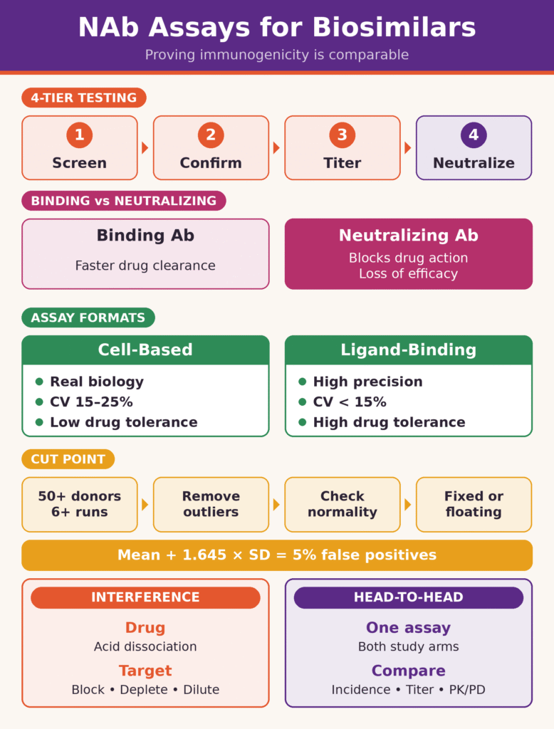
The regulatory framework for immunogenicity assessment follows a structured, four-tier testing hierarchy:
- Tier 1 (Screening): High-sensitivity immunoassays identify all potential binding anti-drug antibodies (BAbs) in serum.
- Tier 2 (Confirmatory): Competitive inhibition procedures eliminate false-positive results by establishing that the detected signal is specific to the therapeutic molecule.
- Tier 3 (Titer Quantification): Serial dilutions are used to characterize the magnitude of the confirmed antibody response.
- Tier 4 (Neutralization Assessment): Functional bioassays or competitive binding platforms determine whether confirmed ADAs actively neutralize biological activity.
Although binding anti-drug antibodies (BAbs) recognize non-active epitopes and primarily affect pharmacokinetics by accelerating immune-mediated clearance, neutralizing anti-drug antibodies (NAbs) bind directly to or near the active paratope, receptor-binding domain, or catalytic site. Consequently, NAbs interfere with the drug’s primary mechanism of action (MoA), potentially reducing therapeutic efficacy, causing complete treatment failure, or producing adverse cross-neutralization of endogenous protein counterparts.
The bioanalytical landscape for biosimilar development has undergone a substantial paradigm shift. Modernized regulatory pathways increasingly streamline or replace traditional Phase III comparative clinical efficacy trials with comprehensive pharmacokinetic (PK), pharmacodynamic (PD), and comparative immunogenicity evaluations. This shift increases the evidentiary requirements for Neutralizing Antibody (NAb) Assays for Biosimilars. In the absence of a Phase III clinical efficacy and safety assessment as a supporting component, NAb assays must demonstrate high analytical sensitivity, robust drug tolerance, statistically sound cut point determination, and unbiased head-to-head comparative interpretation between the biosimilar candidate and its reference product.
To understand how these immunogenicity assessments fit into the broader analytical evaluation of biosimilars, review the principles of biosimilar characterization services.
Shae via:
Quick Summary:
- What NAb assays do: They test whether anti-drug antibodies block a biologic’s mechanism of action. This is a key safety and comparability measure in biosimilar development under FDA and EMA guidance.
- Four-tier testing: Samples go through screening, confirmation, titer measurement, and finally a neutralization test. Binding antibodies mainly speed up drug clearance, while neutralizing antibodies can reduce efficacy or cause treatment failure.
- Higher stakes now: Regulators increasingly rely on PK, PD, and immunogenicity data instead of Phase III efficacy trials. As a result, NAb assays need strong sensitivity, drug tolerance, and statistically sound design.
- Two assay formats: Cell-based assays are biologically relevant and preferred for complex or agonistic mechanisms, but they vary more (15–25% CV). Competitive ligand-binding assays are more precise (below 10–15% CV) and drug-tolerant, and they suit receptor-blocking mAbs.
- Cut point determination: At least 50 drug-naïve samples are tested across 6 or more runs by 2 or more analysts. Outliers are removed using IQR rules and normality is checked. ANOVA decides between a fixed and a floating cut point, typically targeting a 5% false-positive rate (Mean + 1.645 × SD).
- Managing interference: Acid dissociation, SPEAD, and BEAD overcome drug interference. Blocking antibodies, immunoaffinity depletion, and optimized sample dilution (MRD) reduce interference from soluble targets.
- Head-to-head comparison: A single validated assay, using the biosimilar as the reagent, tests samples from both the biosimilar and reference arms. NAb incidence, titers, onset, and their impact on PK/PD are compared to show clinical equivalence.
Bioanalytical Formats for Neutralizing Antibody (NAb) Assays for Biosimilars
Bioanalytical formats for Neutralizing Antibody (NAb) Assays for Biosimilars include cell-based functional bioassays (CBAs) and non-cell-based competitive ligand-binding assays (CLBAs). The selection of an appropriate format depends on the target biologic’s cellular mechanism of action, the complexity of its signaling pathways, and the required analytical precision. Regulatory agencies recommend functional CBAs when a biotherapeutic acts through complex cell signaling or agonistic receptor activation. In contrast, CLBAs are widely accepted for target-blocking monoclonal antibodies when high analytical throughput and enhanced drug tolerance are required.
Cell-Based Assays (CBAs)
Cell-based bioassays provide a biologically relevant foundation for NAb detection because they evaluate the interactions of biotherapeutics with living cellular systems. These platforms assess upstream receptor engagement and downstream intracellular signaling cascades, providing a functional readout of neutralization.
CBAs use distinct bioanalytical endpoints selected according to the therapeutic MoA:
- Reporter Gene Assays: These assays employ genetically modified cell lines engineered with a reporter gene, such as firefly luciferase or alkaline phosphatase, controlled by drug-responsive promoter elements, including NF-κB, STAT3, or SMAD signaling pathways. Biologic binding activates or suppresses reporter expression, while functional NAbs inhibit the drug-mediated response.
- Cell Proliferation and Viability Assays: These assays quantify metabolic activity or changes in cell growth within target cell populations following exposure to cytokines, growth factors, or cytotoxic antibody therapies.
- Phosphorylation and Signaling Bioassays: These assays evaluate post-translational modifications, including receptor autophosphorylation, such as insulin receptor or EGFR autophosphorylation, using electrochemiluminescence or Western blot readouts.
- Effector Function Bioassays: These assays measure antibody-dependent cellular cytotoxicity (ADCC), antibody-dependent cellular phagocytosis (ADCP), or complement-dependent cytotoxicity (CDC) using engineered target-effector co-culture systems.
Although CBAs provide high physiological relevance, they are associated with greater inherent biological variability, with inter-assay precision often exceeding 15%–20% coefficient of variation (CV). They may also exhibit limited drug tolerance, matrix-induced serum cytotoxicity, and narrow dynamic ranges.
For additional insight into how protein-level properties and biological heterogeneity are evaluated during biosimilar development, explore this guide to critical quality attributes (CQAs) in biosimilars.
Competitive Ligand-Binding Assays (CLBAs)
Competitive ligand-binding assays are non-cell-based biochemical platforms that measure the ability of serum NAbs to competitively disrupt the binding interaction between a therapeutic protein and its recombinant target or isolated receptor.
CLBAs can be performed using several bioanalytical platforms:
- Electrochemiluminescence (MSD-ECL): This technology uses ruthenium-labeled target proteins and carbon-coated microplates to achieve a broad dynamic range and high analytical sensitivity.
- Enzyme-Linked Immunosorbent Assays (ELISA): These assays measure competitive displacement between solid-phase recombinant target proteins and liquid-phase enzyme-conjugated drug reagents.
- Surface Plasmon Resonance (SPR) and AlphaLISA: These label-free or homogeneous bead-based technologies evaluate real-time binding kinetics and physical target displacement.
CLBAs offer high precision, typically below 10%–15% CV, along with operational stability, high sample throughput, and straightforward integration of sample pretreatment techniques such as acid dissociation. However, because CLBAs measure target-binding inhibition in isolation, they cannot directly assess post-binding cellular events, receptor internalization, allosteric conformational changes, or effector-mediated mechanisms.
Comparison of Cell-Based and Competitive Ligand-Binding Assays
| Bioanalytical Metric | Cell-Based Assays (CBAs) | Competitive Ligand-Binding Assays (CLBAs) |
|---|---|---|
| Primary Biological Endpoint | Downstream cellular signaling, gene activation, or viability | Direct competitive inhibition of target-ligand interactions |
| Physiological Relevance | High; models the complete cellular biological mechanism | Moderate; limited to physical target-binding inhibition |
| Assay Precision (%CV) | Moderate to low (15% to 25% CV) | High (below 10% to 15% CV) |
| Drug Tolerance Level | Low to moderate; readily affected by free drug saturation | High; compatible with aggressive acid dissociation |
| Matrix Susceptibility | High; vulnerable to serum factors and cytotoxicity | Low to moderate; managed through Minimal Required Dilution |
| Regulatory Preference | Preferred for agonistic mAbs, cytokines, and cellular MoAs | Accepted for receptor-blocking mAbs with binding MoAs |
Statistical Methodologies for Cut Point Determination in Neutralizing Antibody (NAb) Assays for Biosimilars
Cut point determination in Neutralizing Antibody (NAb) Assays for Biosimilars establishes the analytical signal threshold that distinguishes true NAb-positive clinical samples from baseline biological noise. This process involves rigorous statistical evaluation of drug-naïve donor matrix panels. Establishing mathematically appropriate cut points helps minimize false-positive reporting while preserving the ability to detect genuine immune responses, in accordance with consensus recommendations from Shankar et al. and Devanarayan et al.
Pre-Validation Experimental Design and Outlier Analysis
Reliable cut point calculation requires testing a panel of at least 50 individual drug-naïve serum samples obtained from the target clinical patient population or healthy donors. To capture genuine inter-assay, analyst-to-analyst, and plate-to-plate variability, the samples should be assessed across at least six independent analytical runs conducted over multiple days by a minimum of two different operators.
Before calculating the statistical thresholds, the raw signal dataset undergoes two stages of outlier identification.
Biological Outlier Removal: Individual donors exhibiting consistently elevated or reduced baseline signals across all validation runs are assessed using boxplot analysis. The biological threshold boundaries are calculated as follows:
Upper Boundary:
Q3 + 1.5 × (Q3 − Q1)
Lower Boundary:
Q1 − 1.5 × (Q3 − Q1)
Here, Q1 represents the first quartile, Q3 represents the third quartile, and the interquartile range (IQR) is calculated as Q3 − Q1. Donors whose results fall outside these limits across the entire dataset are classified as biological outliers and removed from the analysis.
Analytical Outlier Removal: Isolated operational anomalies occurring on individual assay plates are identified and excluded on a run-by-run basis using the same interquartile range (IQR) criteria.
Normality Testing and Formula Selection
After outlier removal, the cleaned signal dataset is evaluated for distribution normality using the Shapiro-Wilk test alongside skewness assessment.
- Parametric Distribution: When the Shapiro-Wilk test produces p ≥ 0.05 and the absolute skewness is |skewness| < 1.0, the distribution meets the specified normality criteria for parametric analysis. Non-normal raw datasets may be transformed, for example, through logarithmic conversion using log10, and then tested again.
- Non-Parametric Distribution: If transformed data continue to violate the normality criteria, with p < 0.05 or |skewness| ≥ 1.0, empirical non-parametric or robust parametric percentile methods must be considered.
Fixed vs. Floating Cut Point Selection
To determine whether a fixed or floating cut point is appropriate, a single-factor Analysis of Variance (ANOVA) is performed on the mean responses across all validation runs.
- Fixed Cut Point: A fixed cut point is implemented when the run-to-run means show no statistically significant difference (p > 0.05). A single numerical signal threshold is then applied consistently across subsequent clinical assay plates.
- Floating Cut Point: A floating cut point is used when ANOVA identifies statistically significant plate-to-plate or run-to-run mean variation (p < 0.05), provided that the variances remain homogeneous. A plate-specific negative control (NC) is used to normalize daily run variation through a statistically derived Normalization Factor (NF).
The Normalization Factor (NF) is calculated during pre-study validation using one of the following formulas:
Additive Normalization Factor:
NF = MeanSubject Sera − MeanNC
Multiplicative Normalization Factor:
NF = MeanSubject Sera ÷ MeanNC
During clinical sample analysis, the plate-specific floating cut point (CPrun) is dynamically calculated as follows:
Additive Floating Cut Point:
CPrun = NCrun + NF
Multiplicative Floating Cut Point:
CPrun = NCrun × NF
Comparison of Statistical Cut Point Methods
| Cut Point Method | Underlying Distribution and Statistical Condition | Mathematical Calculation Formula | Target False Positive Rate (FPR) |
|---|---|---|---|
| Parametric Screening Cut Point | Shapiro-Wilk p ≥ 0.05; ANOVA p > 0.05 | CP = MeanSera + 1.645 × SD | 5% FPR (z = 1.645) |
| Parametric Confirmatory Cut Point | Normal distribution; signal inhibition assay format | CP = MeanSera + 2.330 × SD | 1% FPR (z = 2.330) |
| Robust Parametric Cut Point | Minor residual skewness or limited sample size | CP = Median + 1.645 × (1.4826 × MAD) | 5% FPR |
| Non-Parametric Cut Point | Persistent non-normal skewness (p < 0.05) | Empirical 95th percentile of the cleaned donor dataset | 5% FPR |
| Floating Cut Point | Significant run-to-run mean variation (ANOVA p < 0.05) | CPrun = NCrun × NF or NCrun + NF | 1% to 5% FPR |
Managing Matrix Interferences: Drug Tolerance and Target Interference Strategies
Matrix interferences arising from high circulating concentrations of therapeutic drugs or soluble target molecules can be mitigated through acid dissociation pretreatment, solid-phase extraction, and target-depletion protocols. Addressing these confounding factors helps preserve bioanalytical sensitivity and reduces the likelihood of false-negative or false-positive immunogenicity results.
Drug Tolerance Optimization and Acid Dissociation Methodologies
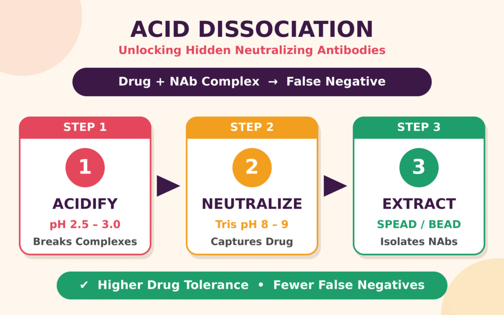
In clinical trial samples collected during chronic dosing regimens or near peak exposure (Cmax), circulating therapeutic antibodies may bind to NAbs and form stable drug-NAb complexes. These complexes can prevent NAbs from interacting with assay capture reagents because of steric hindrance, potentially producing false-negative results.
To address this interference, sample pretreatment protocols may incorporate acid dissociation procedures.
- Acidic Complex Disruption: Serum samples are incubated with low-pH organic acids, such as 300 mM acetic acid or glycine-HCl at pH 2.5–3.0, to disrupt the non-covalent ionic and hydrogen bonds within drug-NAb complexes.
- Neutralization and Competitive Capture: Acidified samples are mixed with an excess of biotinylated target or a magnetic capture phase. The sample pH is then rapidly restored to physiological conditions using a high-molarity Tris buffer at pH 8.0–9.0.
- SPEAD and BEAD Techniques: Solid-Phase Extraction with Acid Dissociation (SPEAD) and Biotin-Extraction Acid Dissociation (BEAD) incorporate biotinylated drug reagents during acidification to compete with unlabeled drug. Subsequent extraction using streptavidin-coated magnetic beads helps isolate purified NAbs before functional testing.
Soluble Target Interference Mitigation
Soluble target proteins, including circulating cytokines, shed cell-surface receptors, and soluble ligands, can complicate bioanalytical interpretation when present in patient serum. These circulating molecules may bind directly to biotherapeutic reagents in CLBAs or interact with cell-surface receptors in CBAs. Such interactions can suppress assay signals, mimic NAb activity, and lead to false-positive results.
Bioanalytical strategies for reducing target interference include the following:
- Target-Blocking Reagents: Non-competing monoclonal anti-target antibodies can be incorporated to selectively bind soluble target molecules without obstructing the primary therapeutic binding domain.
- Immunoaffinity Depletion: Matrix samples are incubated with magnetic beads coated with target-specific antibodies to remove soluble target proteins before assay analysis.
- Minimal Required Dilution (MRD) Tuning: Matrix sample dilution factors, such as 1:10 to 1:100, are optimized to reduce endogenous target concentrations below interfering thresholds while maintaining adequate NAb detection sensitivity.
When interpreting analytical results, it is also important to consider how structural heterogeneity and molecular impurities may affect the overall assessment of a biosimilar. See the approach to impurity profiling of biosimilars for further context.
Head-to-Head Comparative Interpretation of Neutralizing Antibody (NAb) Assays for Biosimilars
Head-to-head comparative interpretation of Neutralizing Antibody (NAb) Assays for Biosimilars involves evaluating clinical sample cohorts from biosimilar and reference product treatment arms using a unified bioanalytical platform to establish immunogenicity comparability. Regulatory frameworks require evidence that any observed differences in immunogenicity remain within predefined statistical boundaries and do not adversely affect clinical safety or efficacy.
Single-Assay Approach vs. Dual-Assay Strategies
Regulatory guidance from the FDA and EMA generally supports a single-assay testing strategy for comparative clinical trials. Under this approach, one validated NAb assay uses the biosimilar candidate as the capture or inhibition reagent to analyze blinded clinical samples from both the biosimilar and reference product treatment cohorts.
Implementing a single-assay format requires comprehensive pre-study comparability testing.
- Positive Control Parity: Purified surrogate positive controls, such as anti-idiotypic monoclonal or polyclonal antibodies, must demonstrate comparable signal inhibition curves against both the biosimilar candidate and the reference product.
- Sensitivity and Drug Tolerance Equivalency: The assay detection limits (LOD) and drug tolerance thresholds established using the biosimilar drug must fall within acceptable equivalence margins of ±20% relative to the reference product.
A well-designed comparative program should integrate immunogenicity findings with other structural and functional assessments. Review the framework for biosimilar comparability studies to understand how these evaluations contribute to the overall evidence package.
Comparative Clinical Data Interpretation and PK/PD Integration
Comparative immunogenicity datasets are evaluated using three integrated parameters.
- Incidence Rates and Risk Ratios: The overall incidence of NAbs is compared between the biosimilar and reference treatment arms. The upper boundary of the 95% confidence interval for the difference in incidence must remain within the predefined non-inferiority margins.
- Titer Progression and Onset Kinetics: The analysis determines whether NAb-positive subjects in the biosimilar group develop higher antibody titers, exhibit faster onset kinetics, or experience persistent rather than transient neutralizing activity compared with subjects in the reference treatment arm.
- Integration with PK/PD and Safety Outcomes: NAb positivity is evaluated alongside individual patient pharmacokinetic clearance rates and pharmacodynamic biomarker suppression. Changes in free drug exposure or biomarker attenuation among NAb-positive patients must demonstrate comparable clinical patterns across both treatment cohorts.
Under modernized regulatory frameworks that reduce the need for clinical efficacy trials, demonstrating that NAb development produces comparable PK/PD alterations for biosimilar and reference molecules provides important evidence supporting clinical safety equivalence.
For a broader view of how analytical evidence supports regulatory comparability, consult the guidance on ICH Q6B biological characterization.
Conclusion
In summary, Neutralizing Antibody (NAb) Assays for Biosimilars are a fundamental component of immunogenicity comparability packages. Their successful implementation requires robust bioanalytical validation, precise statistical cut point determination, and rigorous head-to-head comparative evaluation. Selecting appropriate cell-based or ligand-binding assay formats, applying scientifically sound statistical methods, and addressing matrix interferences help bioanalytical laboratories generate reliable comparability datasets that meet regulatory expectations.
Successful biosimilar development also depends on integrating immunogenicity findings with evidence from structural, physicochemical, and functional characterization. Learn more about comprehensive biosimilar analytical characterization services to support a scientifically robust comparability package.
To learn more about custom bioanalytical assay development, validated NAb testing platforms, and comparative immunogenicity support, visit the ResolveMass Laboratories Contact Page.
Frequently Asked Questions
Acid dissociation helps separate neutralizing antibodies from immune complexes formed with circulating therapeutic drugs in clinical serum samples. By temporarily lowering the sample pH, this technique disrupts non-covalent interactions and makes previously bound antibodies available for detection. Appropriate neutralization and subsequent sample processing help minimize drug interference and reduce the risk of false-negative results.
A floating cut point is calculated by applying a pre-established Normalization Factor (NF) to the negative control (NC) signal measured on the individual assay plate. Depending on the validated method, the factor is either added to or multiplied by the negative control value. This approach adjusts the positivity threshold to accommodate routine analytical variation between assay runs.
The Shapiro-Wilk test and skewness assessment help determine whether the assay signal data satisfy the assumptions required for parametric analysis. A dataset with a Shapiro-Wilk p-value of at least 0.05 and absolute skewness below 1.0 may meet the specified parametric criteria. If non-normality persists after appropriate data transformation, non-parametric percentile methods may be more suitable.
Soluble target proteins circulating in patient serum can interact with assay reagents or cellular receptors and interfere with the expected analytical response. This interference may suppress the measured signal in a way that resembles neutralization by anti-drug antibodies. Target-blocking reagents, immunoaffinity depletion, or optimized sample dilution can help minimize this interference and improve result specificity.
NAb assay sensitivity should be sufficient to detect low concentrations of a suitable positive control antibody in the relevant biological matrix. The required sensitivity depends on the therapeutic molecule, assay format, clinical context, and validated performance characteristics. A range of 100–500 ng/mL is sometimes used as a development target, but it should not be interpreted as a universal FDA or EMA requirement for every NAb assay.
Pharmacokinetic (PK) measurements and pharmacodynamic (PD) biomarkers can provide supporting evidence of the potential clinical effects of immunogenicity. Changes in drug exposure or biological activity may suggest neutralization, but they do not independently establish that NAbs are responsible for those effects. Whether a dedicated NAb assay is necessary depends on the product’s risk profile, mechanism of action, and applicable regulatory expectations.
Evolving FDA guidance and scientific approaches increasingly emphasize comprehensive analytical characterization, pharmacokinetic comparisons, and other relevant evidence when determining the need for comparative clinical efficacy studies. As the role of these studies changes, reliable immunogenicity assessments remain important for evaluating potential differences in safety and efficacy. Validated NAb assays can help identify clinically relevant neutralizing responses and support a scientifically justified biosimilar comparability assessment.
A single-assay design evaluates samples from both biosimilar and reference product treatment groups using the same validated analytical platform. This reduces the risk that differences in reagents, assay conditions, or analytical sensitivity will influence the comparison. When appropriately validated for both products, the approach supports a more consistent assessment of potential differences in immunogenicity.
Minimal Required Dilution (MRD) is the lowest sample dilution that provides acceptable control of non-specific matrix interference under the assay conditions. Selecting an appropriate MRD helps reduce background signals caused by serum components while retaining sufficient sensitivity to detect neutralizing antibodies. Excessive dilution may weaken low-titer antibody signals, whereas insufficient dilution can increase interference and compromise assay specificity.
Reference:
- Partridge, M. A., Kamen, L., Wu, B., Solberg, H., McNally, J., Stevenson, L., Gupta, S., Liu, S., Xu, W., Wu, Y., & White, J. (2025). Assessment of neutralizing antibody activity in clinical studies: Use of surrogate measurements instead of stand-alone assays. The AAPS Journal, 27(5), Article 132. https://doi.org/10.1208/s12248-025-01118-6
- U.S. Food and Drug Administration. (2019). Immunogenicity testing of therapeutic protein products—Developing and validating assays for anti-drug antibody detection: Guidance for industry. https://www.fda.gov/media/119788/download
- European Medicines Agency. (2017). Guideline on immunogenicity assessment of therapeutic proteins (Revision 1). https://www.ema.europa.eu/en/documents/scientific-guideline/guideline-immunogenicity-assessment-therapeutic-proteins-revision-1_en.pdf
- Ingenhoven, K., Kramer, D., Jensen, P. E., Hermanrud, C., Ryner, M., Deisenhammer, F., Pallardy, M., Menge, T., Hartung, H.-P., Kieseier, B. C., Bertotti, E., Creeke, P., Fogdell-Hahn, A., Warnke, C., & ABIRISK Consortium. (2017). Development and validation of an enzyme-linked immunosorbent assay for the detection of binding anti-drug antibodies against interferon beta. Frontiers in Neurology, 8, Article 305. https://doi.org/10.3389/fneur.2017.00305
- Cheng, C.-A., Jiang, A.-L., Liu, Y.-R., & Chang, L.-C. (2023). Investigation of immunogenicity assessment of biosimilar monoclonal antibodies in the United States. Clinical Pharmacology & Therapeutics, 114(6), 1274–1284. https://doi.org/10.1002/cpt.3033
Get In Touch With Us
About The Author