

Introduction
Covalently attaching functional peptide vectors to synthetic nucleic acids can substantially modify the pharmacokinetics and biodistribution of oligonucleotide therapeutics by influencing systemic clearance, tissue-specific extravasation, and interactions with cell-surface receptors. This hybrid molecular approach addresses several major biological transport barriers associated with unconjugated antisense oligonucleotides (ASOs) and small interfering RNAs (siRNAs), including rapid renal elimination, limited endothelial transport, and poor passive permeability across the plasma membrane. Through the covalent incorporation of cell-penetrating peptides (CPPs) or receptor-targeting homing ligands, peptide-oligonucleotide conjugates (POCs) can achieve more favorable hydrodynamic characteristics, controlled plasma protein interactions, and selective tissue tropism. A detailed understanding of the relationship among systemic metabolism, clearance mechanisms, organ retention, and intracellular endosomal escape kinetics is therefore essential for optimizing these targeted therapeutics during preclinical development and supporting their progression toward clinical translation.
Share via:
Quick Summary:
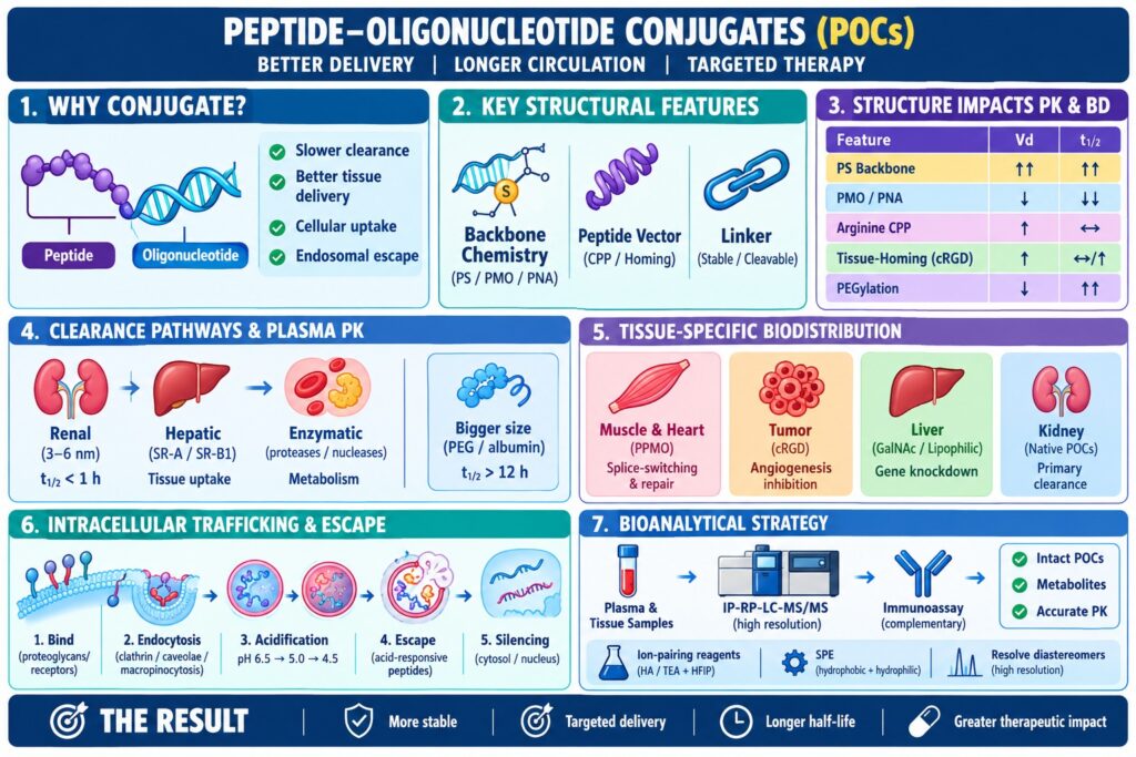
- Peptide–oligonucleotide conjugates (POCs) improve oligonucleotide delivery by reducing rapid renal clearance, enhancing tissue targeting, and promoting cellular uptake.
- Molecular design matters: oligonucleotide backbone chemistry, peptide vectors, and linker stability strongly influence pharmacokinetics, biodistribution, and therapeutic activity.
- Backbone modifications such as phosphorothioate (PS) can increase plasma protein binding and prolong circulation, while uncharged PMO/PNA molecules may require peptide conjugation to improve tissue exposure.
- Peptide vectors enable targeted delivery: CPPs such as arginine-rich peptides enhance cellular uptake, while homing peptides such as cRGD promote receptor-specific tissue accumulation.
- Clearance and biodistribution depend on renal filtration, hepatic uptake, metabolism, molecular size, and tissue-specific receptor interactions. PEGylation or albumin binding can significantly extend circulation time.
- Intracellular trafficking is critical: POCs undergo receptor binding, endocytosis, endosomal acidification, and escape before the oligonucleotide reaches the cytosol or nucleus for gene silencing or splice modulation.
- Advanced bioanalysis using IP-RP-LC-MS/MS, high-resolution MS, and hybridization-based assays helps quantify intact POCs, identify metabolites, and characterize pharmacokinetics and tissue distribution.
Molecular Architectures and Structural Determinants of Pharmacokinetics and Biodistribution
The chemical architecture of a peptide-oligonucleotide conjugate, including the nucleoside sugar and phosphate backbone chemistry, peptide vector sequence, and linker structure, represents a major determinant of its systemic pharmacokinetics and biodistribution. These structural characteristics influence whether the conjugate remains predominantly unbound in circulation and undergoes rapid renal filtration, associates reversibly with circulating plasma proteins to establish an intravascular reservoir, or preferentially interacts with cell-surface receptors expressed within specific tissues.
Learn more about the structural diversity and design strategies in Types of Peptide-Oligonucleotide Conjugates.
Oligonucleotide Backbone Modifications and Charge Dynamics
Modifications introduced into the nucleoside sugar and phosphate backbone strongly influence nuclease stability, overall electrostatic properties, and the extent of plasma protein binding. Phosphorothioate (PS) modifications substitute a non-bridging oxygen atom with sulfur, improving resistance to endo- and exonucleases while also promoting non-specific and reversible associations with plasma proteins, including serum albumin. This interaction with plasma proteins can increase the systemic area under the plasma concentration-time curve (AUC) by limiting the fraction of the conjugate available for glomerular filtration. Conversely, uncharged nucleic acid chemistries, including Phosphorodiamidate Morpholino Oligomers (PMOs) and Peptide Nucleic Acids (PNAs), generally exhibit minimal blood protein association and can therefore undergo rapid urinary elimination unless they are covalently attached to cationic peptide vectors capable of promoting tissue interactions.
Read our detailed comparison on Peptide vs. Antibody Oligonucleotide Conjugates.
Peptide Targeting Vectors and Homing Domains
Peptide ligands covalently linked to nucleic acid molecules play a central role in determining tissue distribution, endothelial transport, and cellular internalization mechanisms. Cationic cell-penetrating peptides (CPPs), especially arginine-rich sequences such as Tat, Antennapedia, and Pip6, can interact electrostatically with negatively charged cell-surface glycosaminoglycans (GAGs), promoting endocytic uptake across multiple tissue types, including striated muscle. In contrast, tissue-homing peptide sequences, such as cyclic RGD (cRGD) motifs, can selectively recognize α_vβ_3 and α_vβ_5 integrins that are overexpressed on tumor endothelial and parenchymal cells. This receptor-mediated interaction can redirect systemic exposure from non-specific reticuloendothelial clearance compartments toward disease-associated target tissues.
Discover the biological mechanisms of cellular transport in How Do Peptide-Oligonucleotide Conjugates Enter Cells?.
Linker Chemistry and Cleavage Kinetics
The chemical nature of the linker connecting the peptide and nucleic acid components determines the structural stability of the conjugate in circulation and influences when and where the active oligonucleotide moiety is released. Stable thioether and amide linkages preserve the covalent architecture of the conjugate during systemic circulation and can be advantageous when the presence of the intact peptide does not interfere sterically with hybridization to the target pre-mRNA or mRNA. Alternatively, cleavable linkers can be designed to respond to intracellular conditions. Disulfide bonds can undergo reduction in the presence of intracellular glutathione (1–10 mM), whereas acid-labile hydrazone bonds can respond to the acidic environment encountered during early and late endosomal maturation (pH 5.0–6.5). These mechanisms can promote more efficient intracellular liberation of the active payload.
Dive deeper into conjugation chemistry with Peptide-Oligonucleotide Conjugate Linker Chemistry.
| Structural Feature | Pharmacokinetic Effect | Impact on Distribution (Vd) | Impact on Half-Life (t1/2) |
|---|---|---|---|
| Phosphorothioate (PS) Backbone | High plasma protein binding; reduced glomerular filtration | Broad systemic distribution; deep tissue exposure | Prolonged terminal half-life |
| Uncharged PMO / PNA Backbone | Minimal serum protein binding; rapid renal filtration | Low distribution volume without peptide conjugate | Ultra-short circulation time (t1/2 < 1 h) |
| Arginine-Rich CPP Conjugation | Enhanced cell-surface glycosaminoglycan binding | Elevated uptake in liver, kidney, and skeletal muscle | Moderate; driven by tissue accumulation |
| Tissue-Homing Peptides (e.g., cRGD) | Receptor-mediated target cell recognition | Specific enrichment in target tumors or microvasculature | Variable; dependent on receptor clearance |
| High Molecular Weight PEGylation | Increases hydrodynamic radius beyond renal threshold | Confined initial distribution volume | Markedly extended circulation (t1/2 > 12 h) |
Systemic Clearance Pathways and Plasma Pharmacokinetics and Biodistribution
Systemic elimination of peptide-oligonucleotide conjugates occurs predominantly through renal glomerular filtration, hepatic reticuloendothelial uptake, and proteolytic or nuclease-mediated biotransformation in blood and tissue compartments. The resulting systemic clearance rate (CL) and volume of distribution (V_d) are determined by the balance between rapid urinary elimination of unbound conjugate and tissue retention resulting from peptide-mediated receptor interactions.
The functional glomerular pore size in human kidneys has a filtration cutoff of approximately 3 to 6 nanometers, which corresponds broadly to the size range of globular proteins around 30 to 50 kDa. Native oligonucleotides, generally approximately 5–10 kDa, together with small uncharged POCs, can remain below this effective filtration threshold and consequently exhibit rapid urinary clearance (t1/2 < 1 hour). Incorporation of high-molecular-weight polyethylene glycol (PEG 20–40 kDa), or peptide sequences capable of facilitating reversible albumin binding, increases the effective hydrodynamic volume of the conjugate. This can reduce renal filtration and substantially prolong terminal elimination half-lives, with reported values exceeding 12–16 hours.
Learn more about clearance, plasma stability, and exposure in Peptide-Oligonucleotide Conjugates Pharmacokinetics.
The systemic disposition of peptide-oligonucleotide conjugates can be described as a sequence of distinct biological phases:
- Intravascular Distribution Phase (t1/2α): The initial distribution phase occurs rapidly as the conjugate reaches equilibrium between the central blood compartment and highly vascularized parenchymal organs.
- Renal Glomerular Filtration: Unbound, low-molecular-weight POCs measuring below approximately 3–6 nm can pass through the renal glomerulus and enter the tubular lumen, resulting in rapid urinary elimination.
- Hepatic & Reticuloendothelial Clearance: Scavenger receptors (SR-A, SR-B1) expressed on liver sinusoidal endothelial cells and Kupffer cells can facilitate sequestration of phosphorothioate-containing and lipophilic conjugates.
- Proteolytic & Nuclease Biotransformation: Serum endopeptidases can cleave susceptible peptide sequences, while 3′- and 5′-exonucleases can degrade nucleic acid strands into shorter truncated species.
Tissue-Specific Biodistribution and Biological Barrier Permeation
The tissue-specific biodistribution of POC therapeutics is largely determined by the ability of the peptide component to facilitate trans-endothelial extravasation and interact selectively with cellular target receptors. Incorporation of cell-homing peptides can alter the distribution pattern of oligonucleotide therapeutics by reducing reliance on conventional renal and hepatic clearance pathways and directing therapeutic exposure toward disease-relevant tissues, including skeletal muscle, cardiac tissue, and solid tumors.
Discover targeted strategies in Peptide-Oligonucleotide Conjugates Drug Delivery.
Peptide-conjugated PMOs (PPMOs), including Pip6-PMO and PPMO-B, have demonstrated substantial bio-accumulation in skeletal muscle, diaphragm, and cardiac tissue following systemic administration. In mdx mouse models of Duchenne muscular dystrophy, PPMOs can traverse dense vascular endothelial barriers associated with striated muscle, resulting in sustained dystrophin restoration and functional correction. Similarly, integrin-targeted conjugates, including cRGD-PEG-oligonucleotides and cyclic RGD-albumin conjugates (RPAO), can preferentially accumulate within tumor microenvironments through interactions with α_vβ_3 integrins expressed by tumor endothelial and parenchymal cells. More advanced POCs incorporating lipophilic groups or specialized transcytosis peptides can further improve transport across tight junction-associated barriers, supporting broader tissue exposure within pulmonary, ocular, and central nervous system tissues.
Read about penetrating the blood-brain barrier in Peptide-Oligonucleotide Conjugates (POCs) in CNS Drug Delivery.
| Target Organ / Tissue | Conjugate Class | Dominant Uptake Mechanism | Pharmacodynamic Outcome |
|---|---|---|---|
| Skeletal & Cardiac Muscle | PPMO (e.g., Pip6-PMO, PPMO-B) | Cell-penetrating peptide-mediated translocation | Splice-switching & dystrophin restoration |
| Tumor Neovasculature | cRGD-Oligonucleotide / RPAO | α_vβ_3 Integrin receptor endocytosis | Angiogenesis inhibition & gene silencing |
| Liver (Hepatocytes) | GalNAc or Lipophilic-POCs | ASGPR-mediated uptake / LDL binding | High-potency gene knockdown |
| Kidney (Proximal Tubules) | Uncharged / Native POCs | Endocytic filtration & megalin capture | Primary clearance & accumulation site |
Intracellular Trafficking and Endosomal Escape Kinetics
Once internalized, peptide-oligonucleotide conjugates accumulate within cells primarily through endocytic pathways. However, their pharmacological activity is often limited by inefficient escape from endosomal compartments into the cytosol and nucleus. Endosomal sequestration can retain more than 98% of internalized conjugates within acidic vesicular compartments, thereby substantially restricting the amount of payload that becomes available for cytosolic or nuclear activity.
Following interaction with cell-surface proteoglycans or specific membrane receptors, POCs can enter cells through clathrin-mediated endocytosis, caveolae-dependent internalization, or macropinocytosis. During vesicular maturation, early endosomes with a pH of approximately 6.5 progressively transition into late endosomes with a pH of approximately 5.0–5.5 and subsequently into lysosomes with a pH below 4.5. During this progression, hydrolytic enzymes can degrade susceptible peptide linkers and nucleic acid backbones. To improve intracellular release, advanced peptide designs may incorporate amphiphilic, histidinylated, or membrane-disruptive sequences that respond to the progressively acidic endosomal environment. These peptide motifs can promote local membrane destabilization and facilitate release of the functional oligonucleotide into the cytosol, where it can subsequently interact with cytosolic RNAi complexes or reach the nucleus to engage pre-mRNA splicing machinery.
Explore key mechanics in Endosomal Escape in Peptide-Oligonucleotide Conjugate Therapeutics.
The intracellular trafficking process can be represented as a defined sequence of vesicular events:
- Cell Surface Engagement: The conjugate initially associates with cell-surface proteoglycans or integrins through electrostatic interactions or receptor-mediated recognition.
- Endocytic Internalization: Following surface engagement, the conjugate is internalized through clathrin-coated pits, caveolae, or macropinocytosis.
- Endosomal Acidification & Maturation: The vesicular environment becomes progressively more acidic, with pH decreasing from approximately 6.5 in early endosomes to approximately 5.0 in late endosomes.
- Endosomal Escape: Protonation of specific peptide motifs can promote membrane destabilization, allowing the active conjugate to escape from the endosomal compartment and enter the cytosol.
- Nuclear / Cytosolic Target Silencing: The released oligonucleotide interacts with complementary mRNA or pre-mRNA sequences to produce splice modulation or gene silencing.
Bioanalytical Strategies for Pharmacokinetics and Biodistribution Profiling
Reliable bioanalytical characterization of peptide-oligonucleotide conjugates in plasma and complex tissue homogenates requires sensitive analytical platforms capable of distinguishing intact conjugates from their biotransformation products. High-resolution Ion-Pairing Reversed-Phase Liquid Chromatography coupled with Tandem Mass Spectrometry (IP-RP-LC-MS/MS), together with hybridization-based immunoassays, provides complementary approaches for characterizing these complex bioconjugates. These methods support quantitative measurement of intact POCs while also enabling assessment of truncated metabolites across diverse biological matrices.
Because modified oligonucleotides contain a polyanionic backbone, IP-RP-LC-MS/MS typically employs volatile alkylamine ion-pairing reagents, such as 15 mM hexylamine (HA), triethylamine (TEA), or N,N-dimethylbutylamine, in combination with fluorinated counter-ions such as 50 mM hexafluoro-2-propanol (HFIP) in the aqueous mobile phase. This reagent combination helps neutralize the negative charges associated with the nucleic acid backbone, thereby promoting retention on C18 stationary phases while supporting efficient electrospray ionization (ESI) in negative ion mode. Sample preparation approaches, including solid-phase extraction (SPE), must simultaneously accommodate the hydrophobic peptide component and the hydrophilic, polyanionic nucleic acid chain. In addition, phosphorothioate backbone modifications introduce chiral centers and can generate hundreds of thousands of diastereomers. This stereochemical complexity can contribute to chromatographic peak broadening and creates a requirement for high-resolution LC-HRMS when accurate discrimination of n-1 and n-2 short-chain metabolites is necessary.
Understand modern bioanalytical techniques in Structural Characterization of Peptide-Oligonucleotide Conjugates.
Bioanalytical capabilities at ResolveMass Laboratories Inc. incorporate high-resolution IP-RP-LC-MS/MS platforms together with optimized solid-phase extraction strategies to support the differentiation of intact POCs from peptide-cleaved and nuclease-degraded metabolites during tissue pharmacokinetic investigations.
Prepare your asset for the clinic with Peptide-Oligonucleotide Conjugates in IND Submissions.
Conclusion
A comprehensive understanding and deliberate control of the pharmacokinetics and biodistribution of peptide-oligonucleotide conjugates are essential for realizing their therapeutic potential across neuromuscular, oncological, and extrahepatic disease indications. Strategic optimization of oligonucleotide chemistry, peptide targeting vector architecture, and linker stability can help minimize rapid renal filtration, prolong systemic plasma exposure, improve tissue delivery, and enhance endosomal escape. At the same time, advanced IP-RP-LC-MS/MS bioanalytical platforms provide the analytical sensitivity and resolution needed to characterize conjugate distribution, metabolism, and biotransformation during preclinical development and clinical investigations.
Read more about overcoming obstacles in Challenges in Peptide-Oligonucleotide Conjugates.
For expert bioanalytical method development, solid-phase sample preparation, and high-resolution LC-MS profiling of therapeutic conjugates, Contact ResolveMass Laboratories.
Frequently Asked Questions
Phosphorothioate (PS) modifications replace non-bridging oxygen atoms within the nucleic acid backbone with sulfur atoms, improving resistance to nuclease-mediated degradation. They also promote reversible interactions with serum proteins such as albumin. Increased protein association can reduce renal filtration and extend systemic exposure, thereby influencing tissue distribution and terminal half-life.
Although peptide-oligonucleotide conjugates can be efficiently internalized through endocytic pathways, most of the internalized material remains sequestered within endosomal compartments. More than 98% may remain trapped and eventually undergo lysosomal degradation. Consequently, limited release into the cytosol or nucleus represents a major barrier to achieving effective gene modulation.
Molecular size and hydrodynamic dimensions strongly influence the extent to which peptide-oligonucleotide conjugates undergo renal filtration. Molecules below the effective glomerular filtration threshold of approximately 3 to 6 nanometers can be cleared rapidly through the urine. Increasing hydrodynamic size through PEGylation or plasma protein binding can reduce filtration and prolong systemic circulation.
IP-RP-LC-MS/MS methods commonly use volatile alkylamines such as hexylamine (HA) or triethylamine (TEA) together with fluorinated alcohols such as hexafluoro-2-propanol (HFIP) or hexafluorotertbutanol (HFTP). These reagents facilitate interaction of the negatively charged oligonucleotide backbone with reverse-phase stationary phases. They also support suitable electrospray ionization conditions for sensitive LC-MS/MS analysis.
Peptide-conjugated Phosphorodiamidate Morpholino Oligomers (PPMOs) combine an uncharged PMO backbone with an arginine-rich cell-penetrating peptide. The peptide component improves cellular uptake, overcoming the limited membrane permeability of unconjugated PMOs. This enhanced delivery enables splice-switching oligomers to reach cardiac and skeletal muscle cells and promote dystrophin expression.
Hybridization-based assays, including dual-probe capture assays, provide highly sensitive measurement of oligonucleotide concentrations in biological matrices. However, these assays may have limited ability to distinguish intact POCs from structurally related degradation products. IP-RP-LC-MS/MS offers greater molecular selectivity by separating and identifying intact conjugates and truncated metabolites generated through peptidase or nuclease activity.
Phosphorothioate modifications introduce chiral centers at the internucleotide linkages, resulting in a very large number of possible diastereomeric forms within an oligonucleotide sequence. For a typical 20-mer, this can produce hundreds of thousands of stereochemical configurations. The resulting chromatographic complexity can cause peak broadening and requires optimized LC conditions and high-resolution mass spectrometry for reliable PK characterization.
Non-cleavable linkers preserve the peptide-oligonucleotide structure during systemic circulation and after cellular internalization, potentially influencing intracellular trafficking and target interactions. Cleavable linkers, such as disulfide or acid-labile bonds, are designed to respond to intracellular reducing or acidic conditions. Their cleavage can release the oligonucleotide payload and facilitate restoration of its intended target-binding and gene-modulating activity.
Unbound oligonucleotides with sufficiently small hydrodynamic dimensions can undergo rapid renal filtration, substantially limiting their systemic exposure. Reversible association with plasma proteins such as albumin increases the fraction retained within the circulation. This extended residence time can provide a longer window for tissue distribution, receptor interactions, and cellular uptake by target tissues.
Reference:
- Kang, H., Alam, M. R., Dixit, V., Fisher, M., & Juliano, R. L. (2008). Cellular delivery and biological activity of antisense oligonucleotides conjugated to a targeted protein carrier. Bioconjugate Chemistry, 19(11), 2182–2188. https://doi.org/10.1021/bc800270w
- Järver, P., Coursindel, T., El Andaloussi, S., Godfrey, C., Wood, M. J. A., & Gait, M. J. (2012). Peptide-mediated cell and in vivo delivery of antisense oligonucleotides and siRNA. Molecular Therapy—Nucleic Acids, 1(6), e27. https://doi.org/10.1038/mtna.2012.18
- Yu, B., Zhao, X., Lee, L. J., & Lee, R. J. (2009). Targeted delivery systems for oligonucleotide therapeutics. The AAPS Journal, 11(1), 195–203. https://doi.org/10.1208/s12248-009-9096-1
- Anand, P., Zhang, Y., Patil, S., & Kaur, K. (2025). Metabolic stability and targeted delivery of oligonucleotides: Advancing RNA therapeutics beyond the liver. Journal of Medicinal Chemistry, 68(7), 6870–6896. https://doi.org/10.1021/acs.jmedchem.4c02528
- Healy, J. M., Lewis, S. D., Kurz, M., Boomer, R. M., Thompson, K. M., Wilson, C., & McCauley, T. G. (2004). Pharmacokinetics and biodistribution of novel aptamer compositions. Pharmaceutical Research, 21(12), 2234–2246. https://doi.org/10.1007/s11095-004-7676-4
Get In Touch With Us
About The Author


