

Introduction
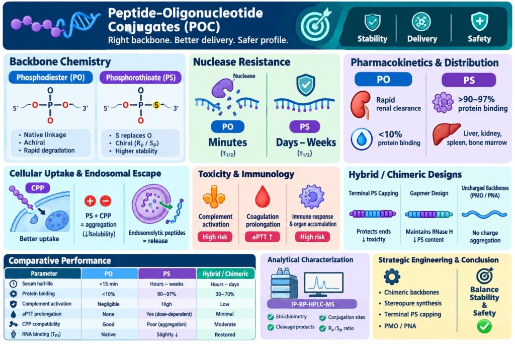
Selecting between a Phosphorothioate vs Phosphodiester Backbone for peptide-oligonucleotide conjugate (POC) design involves carefully balancing metabolic stability and bio-distribution with systemic toxicity and potential off-target immunotoxicity. The covalent attachment of synthetic cell-penetrating peptides (CPPs) or receptor-targeting peptides to therapeutic oligonucleotides can improve intracellular delivery across hydrophobic biological membranes. However, the underlying nucleic acid backbone chemistry remains a major determinant of the conjugate’s resistance to enzymatic degradation, its interaction with circulating plasma proteins, and its overall toxicological behavior. Natural phosphodiester (PO) linkages maintain native binding kinetics and generally exhibit minimal off-target protein interactions, but they are highly susceptible to rapid nucleolytic degradation in human plasma. In contrast, phosphorothioate (PS) modifications replace one non-bridging oxygen atom with a sulfur atom, substantially increasing exonuclease resistance while promoting plasma protein binding that reduces rapid renal elimination. This report evaluates the biophysical mechanisms, pharmacokinetic characteristics, endosomal trafficking behavior, and toxicological trade-offs that influence backbone selection during modern bioconjugate development.
Learn how advanced analytical and translational workflows accelerate bioconjugate candidates into early-phase clinical development by exploring Resolve Mass Preclinical Services.
Quick Summary:
- PO vs PS backbone: Phosphodiester (PO) offers native chemistry and low protein interaction, while phosphorothioate (PS) replaces oxygen with sulfur to improve stability.
- Nuclease resistance: PO backbones degrade rapidly in plasma, whereas PS modifications provide substantially greater resistance, extending persistence from minutes to days or weeks.
- Pharmacokinetics: PO shows rapid renal clearance and <10% protein binding; PS can exhibit >90–97% plasma protein binding, promoting longer circulation and broader tissue distribution.
- Cellular delivery: CPP–PS combinations may cause electrostatic aggregation and reduced solubility, while PMO/PNA neutral backbones can minimize charge-related interactions.
- Safety trade-offs: PS improves stability but may increase risks of complement activation, coagulation changes, immune stimulation, and organ accumulation, particularly at higher exposures.
- Hybrid strategies: Terminal PS capping, gapmer designs, stereopure PS chemistry, and PMO/PNA substitution can balance nuclease protection, delivery, and safety.
- Analytical characterization: IP-RP-HPLC-MS/HRMS can assess conjugate stoichiometry, cleavage products, peptide attachment sites, and Rₚ/Sₚ diastereomeric profiles.
Structural Mechanics of the Phosphorothioate vs Phosphodiester Backbone
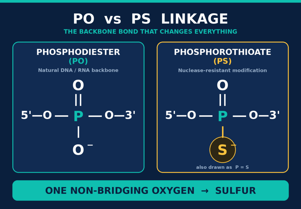
The fundamental difference between a Phosphorothioate vs Phosphodiester Backbone results from the replacement of one non-bridging oxygen atom in the internucleotide phosphate linkage with a sulfur atom. This seemingly small chemical modification changes the charge distribution, bond characteristics, and stereochemical properties of the oligonucleotide.
Within a conventional phosphodiester (PO) linkage, electron density is distributed across the two non-bridging oxygen atoms, producing a localized negative charge and a relatively compact ionic radius. When one of these non-bridging oxygen atoms is replaced by sulfur to generate a phosphorothioate (PS) linkage, the larger van der Waals radius of sulfur (~1.85 Å compared with ~1.40 Å for oxygen), together with its lower electronegativity, modifies the distribution of negative charge around the phosphorus-sulfur bond. This chemical alteration increases the overall lipophilic character of the nucleic acid strand and can strengthen hydrophobic interactions with cellular membranes and structural proteins.
The substitution of sulfur also converts the phosphorus atom into a chiral center, resulting in 2N possible diastereomers for an oligonucleotide containing N phosphorothioate linkages. These diastereomers occur as two distinct configurations at each linkage position:
- $R_p$ Diastereomer: The Rp configuration places the sulfur atom in a spatial arrangement that more closely resembles natural phosphodiesters, helping preserve substrate geometry favorable for target RNA cleavage through RNase H recruitment. Nevertheless, the Rp stereocenter is more susceptible to enzymatic hydrolysis by serum nucleases than the corresponding Sp configuration.
- $S_p$ Diastereomer: The Sp configuration introduces greater steric hindrance within nuclease active-site pockets, providing strong resistance against enzymatic cleavage. However, sequences containing continuous Sp linkages may modestly decrease the thermal melting temperature (Tm) of the target RNA duplex and may also reduce RNase H activation rates.
Recent advances in stereopure phosphorothioate synthesis use chiral auxiliaries to regulate Rp and Sp configurations during solid-phase synthesis. Precise control over backbone stereochemistry allows developers to optimize nuclease resistance while retaining the binding affinity and catalytic knockdown potency required for therapeutic activity.
Dive deeper into structural options, payload combinations, and chemical design paradigms by viewing Types of Peptide-Oligonucleotide Conjugates.
Nuclease Resistance and Plasma Stability Dynamics
Phosphorothioate modifications provide substantially greater enzymatic stability in blood and tissues, potentially extending stability from minutes to multiple days, whereas unmodified phosphodiester backbones can undergo rapid degradation by serum nucleases. This difference in metabolic persistence strongly influences whether a bioconjugate can remain intact during systemic circulation long enough to reach its intended target organ.
Endogenous nucleases, including 3′-exonucleases, 5′-exonucleases, and endonucleases, catalyze the hydrolysis of native phosphodiester bonds through nucleophilic attack. Divalent metal cations (Mg2+ or Zn2+) within catalytic centers participate in destabilizing the phosphorus-oxygen bond and supporting the hydrolytic reaction. Because the phosphodiester backbone can fit efficiently within these catalytic pockets, cleavage of terminal nucleotides can occur rapidly.
Replacing a non-bridging oxygen atom with sulfur interferes with this catalytic process. The altered electronic properties and larger atomic radius of sulfur can reduce efficient coordination of divalent cations within nuclease active sites. This combination of steric and electronic differences decreases the efficiency of nucleophilic attack by catalytic water molecules and increases the resistance of phosphorothioate linkages to enzymatic hydrolysis by several orders of magnitude. As a result, phosphodiester bioconjugates may exhibit systemic elimination half-lives (τ1/2) of less than 15 minutes, whereas phosphorothioate-containing bioconjugates can demonstrate tissue elimination half-lives extending from several days to weeks.
Discover how specialized degradation assays and bioanalytical testing safeguard bioconjugates against nuclease degradation at Peptide-Oligonucleotide Conjugate Stability Services.
Pharmacokinetics, Plasma Protein Binding, and Tissue Distribution of Phosphorothioate vs Phosphodiester Backbone Architectures
The incorporation of a Phosphorothioate vs Phosphodiester Backbone can substantially modify systemic pharmacokinetics by shifting the disposition of an oligonucleotide from rapid renal filtration toward extensive plasma protein binding and wider tissue distribution.
Unmodified phosphodiester oligonucleotides generally exhibit minimal plasma protein binding (<10%). Because these molecules are highly charged and hydrophilic, relatively small oligonucleotides can undergo rapid glomerular filtration and urinary excretion. This rapid renal clearance can substantially limit their ability to achieve sustained exposure in peripheral tissues.
Phosphorothioate modifications change this disposition by promoting non-specific, generally low-affinity interactions with circulating serum proteins, including human serum albumin (HSA), α1-acid glycoprotein, and immunoglobulins. More than 90% to 97% of circulating PS-modified oligonucleotides may exist in a protein-bound state. This protein-binding behavior produces several important pharmacological consequences:
- Prevention of Glomerular Filtration: Association with HSA, which has a molecular mass of approximately 66 kDa, produces a higher-molecular-weight complex that is less readily filtered across the glomerular barrier. This interaction therefore contributes to reduced rapid renal clearance.
- Broad Tissue Accumulation: Because the interaction with plasma proteins is reversible, with dissociation constants (Kd) in the micromolar range, the protein-bound bioconjugate can function as a circulating reservoir. Free bioconjugate can continuously dissociate from the protein complex and distribute into tissues, with substantial accumulation commonly observed in the liver (Kupffer cells and hepatocytes), kidneys (proximal tubule epithelial cells), spleen, and bone marrow.
- Gymnotic Uptake Facilitation: Strong surface interactions with plasma proteins can increase non-specific membrane association and facilitate unassisted cellular uptake, also known as gymnosis, through endocytic pathways in endothelial and parenchymal cells.
Understand how backbone modifications dictate half-life, systemic clearance, and organ deposition by reading Peptide-Oligonucleotide Conjugates Pharmacokinetics.
Cellular Uptake, CPP Interactions, and Endosomal Trapping Mechanisms
Covalent conjugation of cell-penetrating peptides to oligonucleotides can enhance intracellular delivery; however, the chemical nature of the underlying backbone substantially affects bioconjugate stability, aqueous solubility, cellular interactions, and subcellular distribution.
Examine how peptide targeting promotes receptor-mediated and adsorptive endocytosis by reading How Do Peptide-Oligonucleotide Conjugates Enter Cells?.
Cell-penetrating peptides are commonly enriched in basic amino acids, including arginine and lysine, which remain positively charged at physiological pH. When these peptides are combined with polyanionic phosphorothioate backbones, electrostatic attraction between oppositely charged groups may promote intramolecular collapse or intermolecular aggregation. Such interactions can decrease aqueous solubility and potentially conceal or alter the conformations of domains responsible for target recognition.
To minimize electrostatic cross-linking, contemporary bioconjugate designs may incorporate neutral backbone analogs, including Phosphorodiamidate Morpholino Oligomers (PMO) and Peptide Nucleic Acids (PNA). Substitution of charged phosphate linkages with uncharged morpholino or polyamide backbones removes charge-mediated aggregation with cationic peptides. This can help maintain bioconjugate solubility and support translocation across difficult biological barriers, including the blood-brain barrier.
Even when cellular entry is successfully achieved through receptor-mediated or adsorptive endocytosis, endosomal entrapment remains a significant limitation to therapeutic activity. Phosphorothioate backbones can exhibit substantial endosomal retention because of non-specific interactions with intravesicular membrane proteins. Addressing this sequestration may require incorporation of endosomolytic peptide domains, such as amphipathic HA2 or retro-inverso peptides, which can destabilize endosomal membranes under acidic conditions. This mechanism can promote release of the nucleic acid payload into the cytosol and nucleus.
Explore specialized domain designs and endosomolytic modifications aimed at overcoming vesicular sequestration at Endosomal Escape in POC Therapeutics.
Toxicological Profiles and Immunological Trade-offs of the Phosphorothioate vs Phosphodiester Backbone
The principal trade-off associated with a Phosphorothioate vs Phosphodiester Backbone is the balance between improved metabolic stability and the potential for systemic toxicological effects. Although phosphorothioate modification provides important nuclease resistance, extensive PS modification can also introduce several off-target effects.
Phosphodiester backbones are generally well tolerated and exhibit limited non-specific protein interactions in systemic circulation, although oligonucleotides containing particular unmethylated CpG motifs can stimulate Toll-like Receptor 9 (TLR9). Despite their favorable interaction profile, the rapid enzymatic degradation of phosphodiester backbones can substantially restrict their therapeutic persistence.
Phosphorothioate backbones are resistant to nuclease-mediated degradation but can produce specific toxicity concerns because of their non-specific interactions with plasma and cellular proteins:
- Alternative Complement Pathway Activation: High peak plasma concentrations (Cmax) of phosphorothioate oligonucleotides can promote transient activation of the alternative complement pathway. PS-ASOs can interact with Factor H, an important negative regulator of alternative complement activation. Interference with Factor H activity can facilitate cleavage of C3 into C3a and C3b and contribute to the generation of anaphylatoxins such as C5a, which may produce hemodynamic changes, vasodilation, and systemic hypotension.
- Inhibition of Coagulation Cascades: Phosphorothioate oligonucleotides can interact with proteins involved in the intrinsic coagulation pathway, particularly Factor XII and heparin-binding proteins. These interactions may result in a dose-dependent and transient prolongation of activated partial thromboplastin time (aPTT).
- Proinflammatory Immune Responses and Organ Accumulation: Administration of phosphorothioate oligonucleotides at high doses can promote non-sequence-specific immune stimulation. Reported effects can include splenomegaly, lymphoid hyperplasia, and enlargement of Kupffer cells within the liver. Histological evaluation may also demonstrate basophilic granule accumulation in renal proximal tubule cells and hepatic histiocytes, consistent with tissue deposition of the modified polymer.
Learn how leading platforms resolve immunogenicity, solubility, and off-target risks by reviewing Challenges in Peptide-Oligonucleotide Conjugates.
Strategic Chemical Engineering: Hybrid and Chimeric Phosphorothioate vs Phosphodiester Backbone Designs
To address the competing requirements of nuclease resistance and minimization of off-target toxicity, bioconjugate chemists can employ chimeric backbone architectures that incorporate both phosphorothioate and phosphodiester linkages within the same oligonucleotide.
Review synthetic chemistry routes and cleavable vs. non-cleavable linkers by visiting Peptide-Oligonucleotide Conjugate Linker Chemistry.
Limiting phosphorothioate modifications to selected regions reduces the total sulfur content while retaining sufficient protection against enzymatic degradation. This approach can help decrease protein-mediated toxicity while preserving the metabolic stability needed for therapeutic applications:
- Terminal End-Capping: Nucleases can degrade oligonucleotides from both the 3′ and 5′ termini. Introducing two to four phosphorothioate linkages at the 3′- and 5′-ends of an otherwise phosphodiester strand can provide protection against exonuclease-mediated degradation while reducing the overall extent of phosphorothioate modification. This strategy can thereby reduce complement activation and systemic immune stimulation relative to a fully phosphorothioate-modified structure.
- Gapmer Designs: In antisense gapmers, a central region composed of phosphodiester or phosphorothioate deoxyribonucleotides, which supports RNase H recruitment, is flanked by 2′-modified sugar regions such as 2′-O-methoxyethyl [2′-MOE] or Locked Nucleic Acid [LNA]. Selective incorporation of phosphorothioate linkages within these regions provides nuclease protection while limiting the overall phosphorothioate content. This design maintains target RNA cleavage while potentially reducing exposure to phosphorothioate-associated toxicological effects.
- Uncharged Linkage Substitutions: Substituting charged phosphorothioate linkages with neutral phosphorodiamidate morpholino (PMO) or peptide nucleic acid (PNA) backbones can reduce non-specific ionic interactions with plasma proteins. When these neutral backbones are conjugated with cell-penetrating peptides, they can minimize charge-mediated aggregation and provide favorable physicochemical and safety characteristics.
Evaluate the trade-offs between peptide- and antibody-guided oligonucleotide modalities at Peptide vs Antibody-Oligonucleotide Conjugates.
Comparative Performance Profile of Phosphorothioate vs Phosphodiester Backbone Designs
The operational, structural, pharmacokinetic, and toxicological characteristics of phosphodiester, fully phosphorothioate, and hybrid or chimeric backbone architectures can be summarized as follows:
| Property / Parameter | Phosphodiester (PO) Backbone | Phosphorothioate (PS) Backbone | Hybrid / Chimeric Architecture |
|---|---|---|---|
| Chemical Structure | Native diester linkage with non-bridging oxygens | Sulfur replaces one non-bridging oxygen | Terminal PS linkages surrounding an internal PO core |
| P-Chirality | Achiral phosphorus centers | Chiral centers generating Rp and Sp diastereomers | Controlled chiral centers at terminal PS sites |
| Serum Half-Life ($\tau_{1/2}$) | Very short (<15 minutes) | Extended (Hours in plasma; days to weeks in tissue) | Moderate-to-long (Hours in plasma; days in tissue) |
| Nuclease Resistance | Rapidly cleaved by exonucleases and endonucleases | High resistance to enzymatic cleavage | High resistance to exonucleases via terminal caps |
| Plasma Protein Binding | Minimal (<10%) | High (>90–97% bound to HSA and IgG) | Intermediate (30–70% dependent on PS content) |
| Renal Excretion Rate | Rapid clearance via glomerular filtration | Slow clearance due to protein binding retention | Balanced clearance profile |
| Alternative Complement Activation | Negligible risk | High risk (Cmax-dependent via Factor H inhibition) | Low risk due to lower sulfur density |
| aPTT Coagulation Prolongation | None | Concentration-dependent transient prolongation | Minimal impact at equivalent doses |
| CPP Conjugation Compatibility | Good solubility; minimal charge aggregation | High risk of electrostatic collapse with cationic CPPs | Moderate compatibility; requires linker optimization |
| RNA Hybridization Affinity (Tm) | Native target binding affinity | Slightly reduced Tm (~0.5°C lower per PS linkage) | Restored target affinity via sugar modifications |
Characterization of these bioconjugate architectures requires advanced analytical methods, including Ion-Pair Reversed-Phase Liquid Chromatography coupled with High-Resolution Orbitrap Mass Spectrometry (IP-RP-HPLC-MS). High-resolution LC-MS platforms allow analytical scientists to verify bioconjugate stoichiometry, monitor exonucleolytic cleavage products, identify and map peptide conjugation sites, and determine stereopure Rp/Sp diastereomeric ratios across different production batches.
Discover state-of-the-art mass spectrometry methods for structural validation by visiting Structural Characterization of Peptide-Oligonucleotide Conjugates.
Conclusion
Selecting the appropriate Phosphorothioate vs Phosphodiester Backbone configuration for peptide-oligonucleotide conjugate design requires a careful assessment of metabolic persistence and potential toxicological liabilities. Fully phosphorothioate-modified backbones provide substantial protection against nuclease degradation and increase systemic tissue retention through their interactions with plasma proteins. However, their polyanionic sulfur-containing structure can be associated with complement activation, prolonged coagulation times, and electrostatic aggregation when combined with cationic cell-penetrating peptides. In contrast, unmodified phosphodiester backbones generally avoid these protein-mediated interactions but are susceptible to rapid enzymatic degradation and renal clearance in vivo. Contemporary chemical engineering approaches address these competing properties through chimeric backbone configurations, stereopure synthesis, terminal PS capping, and the use of uncharged backbones such as PMO and PNA. Aligning backbone chemistry with the functional requirements of the peptide cargo enables bioconjugate developers to optimize therapeutic performance while supporting an appropriate safety profile.
For analytical consultation, characterization services, or custom bioanalytical assay development for therapeutic oligonucleotides and peptide bioconjugates, contact the analytical team at ResolveMass Laboratories through the Contact Us page.
Frequently Asked Questions
Natural phosphodiester oligonucleotides closely match the substrate characteristics recognized by endogenous nucleases, including 3′- and 5′-exonucleases and endonucleases. These enzymes use divalent metal ions such as Mg2+ and Zn2+ to facilitate hydrolysis of the phosphate ester linkage. Consequently, unmodified phosphodiester oligonucleotides can undergo rapid degradation in biological fluids.
Phosphorothioate modification increases oligonucleotide persistence by promoting reversible interactions with circulating plasma proteins, particularly human serum albumin. Protein association increases the effective molecular size of the complex and reduces rapid renal filtration. This altered disposition can substantially extend systemic exposure and tissue residence compared with unmodified phosphodiester oligonucleotides.
Phosphorothioate oligonucleotides can produce off-target effects because of their interactions with plasma and cellular proteins. At high peak plasma concentrations, they may contribute to alternative complement pathway activation through interactions involving Factor H and downstream complement components. They can also cause transient aPTT prolongation and dose-dependent inflammatory or tissue-related effects involving organs such as the liver and spleen.
Sulfur substitution at the phosphate linkage creates a stereogenic phosphorus center, producing Rp and Sp configurations. The Sp configuration can provide greater nuclease resistance because of steric effects within nuclease active sites, while the Rp configuration can more closely preserve substrate geometry relevant to RNase H-mediated target RNA cleavage. The distribution of these stereoisomers can therefore influence both stability and biological activity.
Phosphorothioate oligonucleotides retain a polyanionic backbone that can interact strongly with positively charged cell-penetrating peptides containing amino acids such as arginine and lysine. These electrostatic interactions may promote intramolecular compaction or intermolecular cross-linking. Such effects can decrease aqueous solubility and, under certain conditions, result in aggregation or precipitation of the bioconjugate.
PMO and PNA contain neutral backbone chemistries that substantially reduce the charge-driven interactions observed between cationic peptides and anionic oligonucleotides. Their neutral character can improve aqueous compatibility and reduce aggregation during peptide conjugation. These backbones also provide high resistance to nuclease-mediated degradation and can reduce certain non-specific interactions associated with charged phosphorothioate structures.
End-capped PS/PO architectures place a limited number of phosphorothioate linkages at the 3′- and 5′-termini while retaining phosphodiester or other selected chemistries within the internal sequence. The terminal PS segments provide protection against exonuclease-mediated degradation at vulnerable ends. Limiting PS incorporation can also reduce the overall extent of sulfur modification and thereby moderate protein-mediated and immunological effects.
Phosphorothioate substitution can alter the thermodynamic properties of an oligonucleotide-RNA duplex and may produce a modest reduction in thermal melting temperature (Tm). The magnitude of this effect depends on sequence composition, modification pattern, and the number and stereochemistry of PS linkages. Incorporating 2′-O-sugar modifications such as 2′-MOE or LNA can help maintain or enhance target-binding affinity.
Ion-Pair Reversed-Phase Liquid Chromatography coupled with High-Resolution Mass Spectrometry (IP-RP-HPLC-MS) is an important analytical approach for characterizing phosphorothioate-containing bioconjugates. It can support assessment of molecular composition, purity, peptide conjugation stoichiometry, and degradation products. High-resolution analysis can also assist in evaluating stereopure Rp/Sp diastereomer distributions across production batches.
Reference:
- Chang, Y.-S., Kim, Y.-K., Kwon, H.-S., Park, H.-W., Min, K.-U., Kim, Y.-Y., & Cho, S.-H. (2009). The effect of CpG-oligodeoxynucleotides with different backbone structures and 3′ hexameric deoxyriboguanosine run conjugation on the treatment of asthma in mice. Journal of Korean Medical Science, 24(5), 860–866. https://doi.org/10.3346/jkms.2009.24.5.860
- Crooke, S. T., Vickers, T. A., & Liang, X.-H. (2020). Phosphorothioate modified oligonucleotide-protein interactions. Nucleic Acids Research, 48(10), 5235–5253. https://doi.org/10.1093/nar/gkaa299
- Herkt, M., & Thum, T. (2021). Pharmacokinetics and proceedings in clinical application of nucleic acid therapeutics. Molecular Therapy, 29(2), 521–539. https://doi.org/10.1016/j.ymthe.2020.11.008
- Watanabe, T. A., Geary, R. S., & Levin, A. A. (2006). Plasma protein binding of an antisense oligonucleotide targeting human ICAM-1 (ISIS 2302). Oligonucleotides, 16(2), 169–180. https://doi.org/10.1089/oli.2006.16.169
- Eckstein, F. (2014). Phosphorothioates, essential components of therapeutic oligonucleotides. Nucleic Acid Therapeutics, 24(6), 374–387. https://doi.org/10.1089/nat.2014.0506
- Yoo, B. H., Bochkareva, E., Bochkarev, A., Mou, T.-C., & Gray, D. M. (2004). 2′-O-methyl-modified phosphorothioate antisense oligonucleotides have reduced non-specific effects in vitro. Nucleic Acids Research, 32(6), 2008–2016. https://doi.org/10.1093/nar/gkh516
Get In Touch With Us
About The Author


