

Introduction
Poly(lactic-co-glycolic acid)-PEG Block Copolymers influence the biological fate of nanoparticles by creating a hydrophilic, sterically protective outer layer that limits opsonization, decreases reticuloendothelial system-mediated clearance, and substantially prolongs systemic circulation time. In contrast, unmodified poly(lactic-co-glycolic acid) (PLGA) nanoparticles are rapidly identified as foreign entities by the innate immune system following intravenous administration. The hydrophobic polyester surface promotes the immediate adsorption of plasma proteins from the bloodstream, a phenomenon referred to as opsonization. This process labels the nanoparticles for recognition and removal by the Mononuclear Phagocyte System (MPS), particularly macrophages located in the liver (Kupffer cells) and spleen.
The covalent attachment of poly(ethylene glycol) (PEG) to PLGA produces amphiphilic diblock (PLGA-PEG) or triblock (PLGA-PEG-PLGA or PEG-PLGA-PEG) copolymer structures that spontaneously self-assemble into core-shell nanoparticles. Within these assemblies, the hydrophobic PLGA core serves as a reservoir for lipophilic therapeutic agents, while the hydrophilic PEG segments extend outward to form a protective surface corona.
This structural modification profoundly changes the physicochemical behavior of the nanocarrier by altering hydrodynamic size, concealing surface charge, and generating a steric barrier that discourages plasma protein adsorption. A thorough understanding of the physical chemistry underlying Poly(lactic-co-glycolic acid)-PEG Block Copolymers—including PEG chain density, molecular weight relationships, and spatial conformations—is essential for controlling drug release behavior, enhancing circulation persistence, and supporting passive or active tumor targeting through the Enhanced Permeation and Retention (EPR) effect.
Learn how base polymers differ in degradation and performance before PEGylation in our guide on PLA vs PLGA vs PCL Comparative Analysis.
Share via:
Article Summary:
- PLGA-PEG block copolymers improve nanoparticle performance by forming a hydrophilic PEG shell that reduces opsonization, minimizes immune clearance, and significantly extends circulation time compared with unmodified PLGA nanoparticles.
- PEGylation enhances surface properties by increasing hydrodynamic diameter, reducing zeta potential toward near-neutral values, lowering interfacial energy, and improving colloidal stability under physiological conditions.
- PEG chain density determines stealth efficiency, with low-density “mushroom” conformations providing partial protection, while high-density “brush” conformations create a continuous steric barrier that effectively blocks protein adsorption.
- The PEG corona suppresses protein corona formation and complement activation, reduces macrophage recognition, promotes dysopsonin adsorption, and enables better mucus penetration for improved mucosal drug delivery.
- PEGylated PLGA nanoparticles exhibit superior pharmacokinetics, including prolonged half-life, higher bioavailability (AUC), reduced systemic clearance, and enhanced passive tumor accumulation through the Enhanced Permeation and Retention (EPR) effect.
- Comprehensive characterization relies on analytical techniques such as ¹H-NMR, GPC/SEC, Dynamic Light Scattering (DLS), XPS, zeta potential analysis, iodine assay, and LC-MS/RDMS to evaluate structure, molecular weight, PEG surface density, and degradation behavior.
- Overall, PLGA-PEG block copolymers provide a versatile nanocarrier platform with improved stability, stealth properties, controlled drug delivery, and enhanced therapeutic targeting, making them highly valuable for advanced nanomedicine and targeted drug delivery applications.
Surface Chemistry Modulation of Poly(lactic-co-glycolic acid)-PEG Block Copolymers
Poly(lactic-co-glycolic acid)-PEG Block Copolymers modify nanoparticle surface characteristics by relocating the hydrodynamic shear plane outward, shielding charges associated with the hydrophobic core, and lowering the magnitude of the zeta potential. This transformation converts a hydrophobic, negatively charged surface into a hydrophilic, sterically stabilized interface that effectively resists non-specific biological adsorption.
Hydrodynamic Diameter vs. Core Dimensions in Poly(lactic-co-glycolic acid)-PEG Block Copolymers
When uncoated PLGA nanoparticles are characterized using transmission electron microscopy (TEM) or scanning electron microscopy (SEM), the measured dry particle size generally corresponds closely to the hydrodynamic diameter (Dh) obtained through Dynamic Light Scattering (DLS). However, Poly(lactic-co-glycolic acid)-PEG Block Copolymers display a significant difference between the solid core dimension and the measured hydrodynamic diameter.
In these systems, the hydrophobic PLGA segments associate to create a dense internal core, while the hydrophilic PEG chains project outward into the surrounding aqueous environment. These PEG chains interact strongly with water molecules through hydrogen bonding involving their ether oxygen atoms, resulting in the formation of a hydrated outer layer.
The presence of this hydration shell expands the apparent particle size, causing the hydrodynamic diameter to exceed the actual physical core radius by several nanometers. The magnitude of this increase is directly influenced by both the molecular weight (MW) of PEG and the density at which PEG chains are grafted onto the nanoparticle surface.
Discover how active ingredient characteristics impact core encapsulation in our overview of Encapsulating Hydrophilic vs Hydrophobic APIs in PLGA.
Zeta Potential Shielding and Slipping Plane Mechanics
The electrokinetic potential, commonly referred to as zeta potential (ζ), represents the effective surface charge measured at the hydrodynamic shear plane of a nanoparticle. Bare PLGA nanoparticles typically exhibit a strongly negative zeta potential, often ranging from -20 mV to -45 mV. This negative charge originates from exposed terminal carboxylic acid groups present along the polyester backbone.
Following PEG incorporation, the electrically neutral polyether chains effectively shield these charged functional groups from the surrounding environment. As PEG content increases, the location of the hydrodynamic slipping plane is displaced farther from the charged PLGA surface. Because electric potential decreases exponentially with distance from the source charge, the measured zeta potential becomes progressively less negative.
As a result, Poly(lactic-co-glycolic acid)-PEG Block Copolymers commonly exhibit near-neutral zeta potential values, generally falling between -5 mV and -15 mV. This reduction in surface charge diminishes electrostatic interactions with negatively charged plasma proteins and cellular membranes, thereby decreasing non-specific cellular uptake and reducing the likelihood of unwanted systemic interactions.
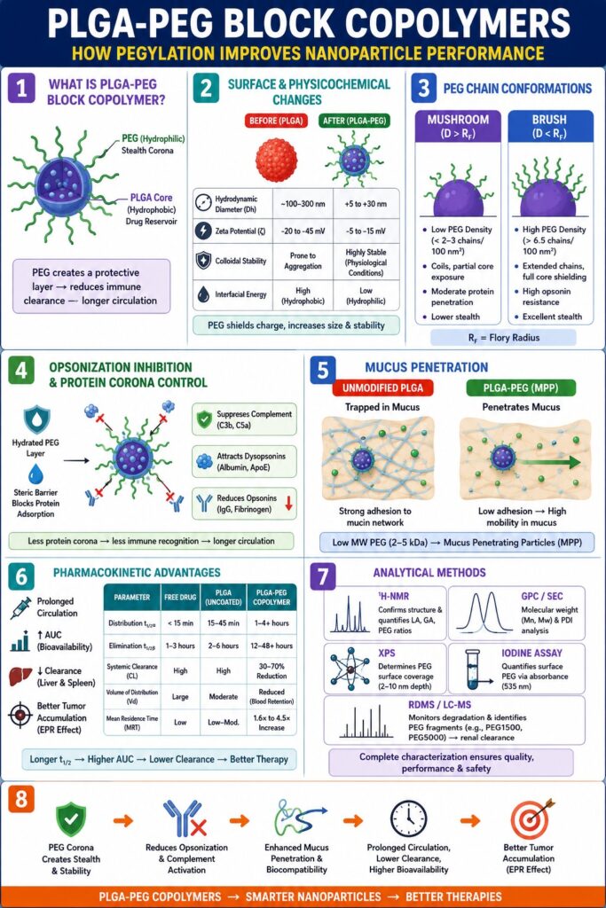
| Property / Parameter | Bare PLGA Nanoparticles | Poly(lactic-co-glycolic acid)-PEG Block Copolymer Nanoparticles | Biophysical Mechanism / Consequence |
|---|---|---|---|
| Hydrodynamic Diameter (Dh) | Approximately equivalent to dry core diameter (~100–300 nm) | Increased Dh relative to TEM core size (+5 to +30 nm) | Hydrated PEG chains absorb water and generate a flexible external shell. |
| Zeta Potential (ζ) | Strongly negative (-20 to -45 mV) | Near-neutral (-5 to -15 mV) | Outward displacement of the slipping plane masks terminal carboxylic acid groups. |
| Aqueous Colloidal Stability | Susceptible to aggregation in the presence of salts | Highly stable under physiological ionic conditions | Steric stabilization becomes the dominant mechanism rather than electrostatic repulsion. |
| Interfacial Energy | Elevated hydrophobic surface energy | Reduced interfacial energy due to the hydrophilic PEG layer | Hydrogen bonding between PEG ether oxygens and water lowers surface energy. |
Structural Regimes in Poly(lactic-co-glycolic acid)-PEG Block Copolymers: Mushroom vs. Brush Conformations
The surface organization of Poly(lactic-co-glycolic acid)-PEG Block Copolymers evolves from a mushroom configuration to a densely packed brush configuration as PEG grafting density surpasses the Flory overlap threshold. This transition plays a crucial role in determining whether circulating proteins can access the nanoparticle surface or are prevented from approaching it through steric exclusion.
Flory Radius and Polymer Chain Dynamics
The arrangement of PEG chains attached to the PLGA core is controlled by the Flory radius (RF) of the polymer in a favorable solvent such as water. The Flory radius describes the average spatial dimension occupied by an isolated polymer coil and can be expressed as:
RF = aN3/5
where a represents the monomer length (approximately 0.35 nm for ethylene glycol) and N denotes the degree of polymerization, corresponding to the number of repeating ethylene glycol units.
During the self-assembly of Poly(lactic-co-glycolic acid)-PEG Block Copolymers, the average spacing (D) between neighboring PEG attachment sites on the nanoparticle surface determines the extent of intermolecular interactions among polymer chains. The relationship between D and RF governs the conformational state adopted by the PEG corona.
The Mushroom Regime in Poly(lactic-co-glycolic acid)-PEG Block Copolymers
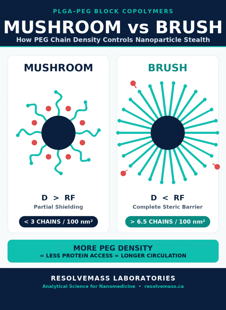
At relatively low PEG grafting densities, where the separation distance between adjacent PEG chains is greater than the Flory radius (D > RF), neighboring polymer chains remain largely independent and do not significantly overlap. Under these conditions, each PEG chain assumes a relaxed, coil-like conformation known as the mushroom regime.
Within the mushroom regime, PEG chains possess considerable conformational freedom and fluctuate independently in solution. However, because the chains are widely spaced, substantial portions of the hydrophobic PLGA surface remain exposed between individual polymer coils. These uncovered regions provide access points for opsonins and other plasma proteins, enabling them to penetrate the polymer layer and adsorb onto the nanoparticle surface.
As a consequence, nanoparticles exhibiting a mushroom-type PEG configuration possess only partial stealth characteristics. Although the PEG chains provide some degree of protection, the exposed hydrophobic domains remain vulnerable to protein adsorption, which can accelerate immune recognition and clearance from systemic circulation.
The Brush Regime and Steric Stabilization
As the PEG content within Poly(lactic-co-glycolic acid)-PEG Block Copolymers increases, the average spacing between adjacent grafting sites becomes smaller than the Flory radius (D < RF). Under these conditions, the available surface area is no longer sufficient to accommodate PEG chains in their relaxed coil conformation. Consequently, the polymer chains extend outward from the PLGA surface and transition into what is known as the brush regime.
In this densely packed brush configuration, steric crowding between neighboring PEG chains forces them to adopt an elongated orientation that projects away from the nanoparticle surface. The resulting arrangement forms a continuous hydrophilic barrier surrounding the particle. This hydrated barrier effectively prevents biomolecules from reaching the underlying PLGA core and is therefore essential for achieving robust stealth behavior.
Experimental investigations have shown that establishing a stable brush conformation on PLGA nanoparticles generally requires a PEG content of approximately 5 to 8 wt% using 5 kDa PEG. This level of PEGylation typically corresponds to surface densities greater than 6.5 PEG chains per 100 nm², which is sufficient to generate a continuous protective polymer corona.
Emulsifiers play a direct role in particle assembly. Read about Surfactants and Emulsifiers in PLGA Microsphere Fabrication.
| Structural Parameter | Mushroom Regime (D > RF) | Brush Regime (D < RF) |
|---|---|---|
| Surface Packing Density | Low (< 2–3 PEG chains / 100 nm²) | High (> 6.5 PEG chains / 100 nm²) |
| Chain Conformation | Relaxed, hemispherical random coils | Extended chains oriented perpendicular to the surface |
| Core Exposure | Partial exposure of hydrophobic PLGA regions | Complete shielding of the underlying PLGA core |
| Opsonin Resistance | Moderate; permits partial protein penetration | High; effectively repels plasma proteins through steric effects |
Mechanism of Opsonization Inhibition and Protein Corona Suppression by Poly(lactic-co-glycolic acid)-PEG Block Copolymers
Poly(lactic-co-glycolic acid)-PEG Block Copolymers minimize protein corona formation by generating a highly hydrated and dynamic steric barrier that inhibits the deposition of opsonins, including complement factor C5a and Immunoglobulin G. By reducing hydrophobic interactions that normally promote protein adsorption, these block copolymers shield the nanoparticle surface from detection by serum proteins and macrophage-associated scavenger receptors.
Thermodynamics of Steric Repulsion and Water Binding
When an opsonin protein approaches a nanoparticle composed of Poly(lactic-co-glycolic acid)-PEG Block Copolymers, penetration into the PEG corona requires compression of the tethered polymer chains. Such compression restricts the natural rotational and vibrational motion of the PEG backbones, leading to a reduction in conformational entropy (ΔS < 0).
At the same time, deformation of the hydrated PEG layer disrupts the associated water network and forces bound water molecules to leave the polymer matrix and re-enter the surrounding bulk solvent. This process requires additional energy and contributes to the overall resistance against protein adsorption.
The combined effects of entropy loss and dehydration create a positive free-energy barrier (ΔG > 0), which acts as a repulsive force that discourages proteins from approaching the nanoparticle surface. As a result, the PEG layer serves as an effective steric shield that limits biomolecular attachment and promotes prolonged circulation within the bloodstream.
Protein Corona Composition and Complement Cascade Evasion
In human plasma, unmodified PLGA nanoparticles rapidly acquire a hard protein corona composed of strongly binding opsonins such as Immunoglobulin G (IgG), complement proteins including C3b and C5a, fibrinogen, and fibronectin. The accumulation of these proteins promotes colloidal instability and activates both the classical and alternative complement pathways.
Poly(lactic-co-glycolic acid)-PEG Block Copolymers significantly influence both the amount and the composition of proteins that adsorb onto the nanoparticle surface. Rather than allowing extensive opsonin deposition, PEGylated nanoparticles selectively modify protein interactions in ways that favor prolonged circulation.
Complement Activation Suppression
Dense PEG coatings substantially reduce the conversion of complement component C3 into C3b and limit the production of the inflammatory mediator C5a. By suppressing these key complement activation events, PEGylated nanoparticles reduce downstream immune signaling and decrease recognition by immune cells responsible for nanoparticle clearance.
Dysopsonin Enrichment
PEG-modified surfaces preferentially attract dysopsonins, including serum albumin and selected apolipoproteins such as ApoE, while simultaneously reducing the adsorption of immune-targeting opsonins. These dysopsonins create a biologically compatible protein layer that further protects the nanoparticle surface without promoting rapid uptake by macrophages.
Mucus Penetration via Charge Passivation
In addition to improving systemic circulation, Poly(lactic-co-glycolic acid)-PEG Block Copolymers significantly influence the transport of nanoparticles across mucosal barriers. Unmodified PLGA nanoparticles frequently become immobilized within biological mucus layers, including cervicovaginal, gastrointestinal, and pulmonary mucus. This entrapment occurs because hydrophobic interactions and electrostatic attractions promote adhesion between the nanoparticle surface and the densely interconnected mucin network.
Applying a dense coating of low-molecular-weight PEG, typically ranging from 2 to 5 kDa, produces formulations known as Mucus-Penetrating Particles (MPP). The PEG layer passivates surface charge and minimizes hydrophobic adhesive interactions that would otherwise cause nanoparticle retention within mucus.
As a result, MPP formulations are capable of diffusing through dense mucus structures at rates approaching those observed in pure aqueous environments. This enhanced mobility improves nanoparticle transport across mucosal barriers and can significantly increase the effectiveness of localized and systemic drug delivery strategies that rely on mucosal administration routes.
Learn how surface modification enhances localized delivery in PLGA in CNS Drug Delivery & Blood-Brain Barrier.
Pharmacokinetic Enhancements and Biofate of Poly(lactic-co-glycolic acid)-PEG Block Copolymers
The incorporation of Poly(lactic-co-glycolic acid)-PEG Block Copolymers substantially improves systemic pharmacokinetic performance by prolonging circulation half-life (t₁/₂), increasing the area under the concentration-time curve (AUC), and reducing clearance through the liver and spleen. These modifications transform conventional burst-release nanoparticle systems into long-circulating delivery platforms capable of sustaining therapeutic drug levels for extended periods.
Circulation Kinetics: Elimination Half-Life and Bioavailability
Following intravenous administration, free drug molecules and unmodified PLGA nanoparticles are generally removed from circulation rapidly, often exhibiting distribution half-lives measured in minutes. A major contributor to the rapid elimination of conventional PLGA nanoparticles is uptake by hepatic Kupffer cells, which can account for approximately 80–90% of total nanoparticle sequestration within the body.
By minimizing immediate recognition by macrophages, Poly(lactic-co-glycolic acid)-PEG Block Copolymers significantly modify key pharmacokinetic parameters:
Terminal Elimination Half-Life (t₁/₂β)
The terminal elimination half-life is substantially prolonged, frequently demonstrating a two- to five-fold increase—or even greater—when compared with corresponding non-PEGylated nanoparticle formulations.
Area Under the Concentration-Time Curve (AUC)
Prolonged retention within the bloodstream results in a significant increase in AUC, reflecting improved systemic exposure and enhanced overall bioavailability of the encapsulated therapeutic agent.
Systemic Clearance (CL)
Systemic clearance is markedly reduced, indicating that the nanocarrier remains within the vascular compartment for longer periods instead of being rapidly captured and accumulated in non-target organs.
| Pharmacokinetic Parameter | Free Drug Solution | Uncoated PLGA Nanoparticles | Poly(lactic-co-glycolic acid)-PEG Block Copolymer Nanoparticles | Primary Kinetic / Physiological Driver |
|---|---|---|---|---|
| Distribution Half-Life (t₁/₂α) | Extremely rapid (< 15 min) | Rapid (15–45 min) | Prolonged (1–4+ hours) | Reduced interaction with vascular walls and decreased endothelial filtration. |
| Elimination Half-Life (t₁/₂β) | Short (1–3 hours) | Short to moderate (2–6 hours) | Extended (12–48+ hours) | Reduced MPS recognition and lower hepatic uptake prolong circulation. |
| Systemic Clearance (CL) | High renal and metabolic clearance | High hepatic phagocytic clearance | Markedly reduced (30–70% reduction) | Lower opsonization decreases recognition by hepatic scavenger receptors. |
| Volume of Distribution (Vd) | Large due to non-specific tissue distribution | Moderate because of liver and spleen accumulation | Reduced, remaining primarily within blood plasma | Hydrophilic PEG layers limit passage across healthy non-fenestrated endothelium. |
| Mean Residence Time (MRT) | Low | Low to moderate | Multi-fold increase (1.6× to 4.5×) | Extended circulation supports sustained therapeutic exposure. |
See how sustained release kinetics translate to commercial products in PLGA Long-Acting Injectable Formulation.
Tumor Accumulation via Passive Targeting (EPR Effect)
Prolonged circulation is a critical prerequisite for maximizing the benefits of the Enhanced Permeation and Retention (EPR) effect in solid tumors. Tumor-associated blood vessels are characterized by abnormal architecture, including enlarged inter-endothelial gaps that typically range from 100 to 700 nm, along with inefficient lymphatic drainage.
Unmodified PLGA nanoparticles are often eliminated from circulation before they can effectively exploit these vascular abnormalities. In contrast, Poly(lactic-co-glycolic acid)-PEG Block Copolymer nanoparticles remain in the bloodstream for significantly longer periods, enabling repeated passage through tumor vasculature.
This prolonged vascular residence increases the probability of nanoparticle extravasation through tumor fenestrations and promotes gradual accumulation within the tumor interstitium. Consequently, local drug concentrations can increase within diseased tissues while systemic exposure to healthy organs is reduced, thereby improving therapeutic selectivity and minimizing off-target toxicity.
Analytical Methods for Evaluating Poly(lactic-co-glycolic acid)-PEG Block Copolymers
Comprehensive characterization of Poly(lactic-co-glycolic acid)-PEG Block Copolymers requires the application of multiple complementary analytical techniques. Methods such as dynamic light scattering, nuclear magnetic resonance spectroscopy, mass spectrometry, and zeta potential analysis provide critical information regarding copolymer composition, molecular architecture, surface properties, and nanoparticle morphology.
Chemical Structure, Stoichiometry, and Molecular Weight Profiling
The successful synthesis of Poly(lactic-co-glycolic acid)-PEG Block Copolymers depends on precise control of both the poly(lactic acid) (LA) to poly(glycolic acid) (GA) ratio and the overall balance between PLGA and PEG segments. Analytical characterization is therefore essential for confirming structural integrity and composition.
Proton Nuclear Magnetic Resonance (¹H-NMR)
Proton Nuclear Magnetic Resonance (¹H-NMR) is widely employed to verify molecular structure and determine block composition. Characteristic resonance signals include δ ≈ 5.2 ppm for methine (-CH-) protons associated with LA units, δ ≈ 4.8 ppm for methylene (-CH₂-) protons of GA units, and δ ≈ 3.65 ppm for the repeating methylene (-CH₂CH₂O-) protons of PEG chains.
Quantitative integration of these signals enables accurate determination of the molar proportions of LA, GA, and PEG within the synthesized block copolymer, providing direct confirmation of copolymer stoichiometry.
Gel Permeation Chromatography / Size Exclusion Chromatography (GPC/SEC)
Gel Permeation Chromatography (GPC), also referred to as Size Exclusion Chromatography (SEC), is used to determine molecular weight distributions, including number-average molecular weight (Mn), weight-average molecular weight (Mw), and polydispersity index (PDI).
This technique confirms successful diblock or triblock copolymer formation and verifies the absence of residual homopolymer impurities that could negatively affect nanoparticle performance and reproducibility.
Learn more about controlling particle size distribution in PLGA PDI in Pharmaceutical Applications.
Quantifying Surface Density and Polymer Degradation Kinetics
Accurate assessment of biological performance requires the quantification of PEG surface density (Γ) as well as monitoring of polymer degradation pathways under physiologically relevant conditions.
X-ray Photoelectron Spectroscopy (XPS)
X-ray Photoelectron Spectroscopy (XPS) examines the elemental composition of the outermost 2–10 nm layer of the nanoparticle surface. The technique quantifies ether-oxygen (C-O-C) signals associated with PEG relative to polyester-derived carbon signals such as C=O and C-O.
This information provides direct evidence of PEG surface coverage and confirms successful presentation of PEG chains at the nanoparticle interface.
Colorimetric Iodine Assay
The colorimetric iodine assay is used to quantify surface-accessible PEG chains. In this method, triiodide (I₃⁻) ions interact with polyether segments to form a colored complex that exhibits measurable absorbance at 535 nm.
The intensity of the resulting signal provides an estimate of the amount of PEG exposed on the nanoparticle surface and helps evaluate PEG grafting efficiency.
Reduced-Dimensional Mass Spectrometry (RDMS)
Reduced-Dimensional Mass Spectrometry (RDMS) and advanced LC-MS methodologies enable the characterization of polydisperse block copolymers within complex biological matrices.
These analytical approaches demonstrate that PLGA-PEG copolymers gradually undergo ester bond hydrolysis following administration. Degradation generates stable PEG1500 or PEG5000 fragments, which are subsequently eliminated primarily through renal filtration pathways. Such studies provide valuable insights into polymer stability, metabolic fate, and clearance mechanisms.
Explore comprehensive analytical testing workflows under PLGA Characterization for RLD Equivalence.
Conclusion
Poly(lactic-co-glycolic acid)-PEG Block Copolymers represent a major advancement in the ability to control nanoparticle surface properties, thermodynamic behavior, and systemic pharmacokinetic performance. The replacement of hydrophobic PLGA surfaces with highly hydrated PEG coronas shifts the electrokinetic slipping plane, reduces apparent surface charge, and creates an entropy-driven steric barrier that limits interactions with serum opsonins.
The transition from a sparsely populated mushroom configuration to a densely packed brush conformation further enhances stealth characteristics by minimizing protein corona formation, suppressing complement activation, and reducing recognition by Kupffer cells and splenic macrophages. These effects collectively contribute to prolonged circulation and improved biological performance.
The resulting pharmacokinetic benefits include extended circulation half-life, lower systemic clearance, and enhanced passive accumulation within diseased tissues through the Enhanced Permeation and Retention (EPR) effect. As nanomedicine continues to evolve toward targeted therapeutics, controlled-release systems, and advanced mucosal delivery platforms, precise optimization of block copolymer composition, PEG grafting density, and molecular architecture remains increasingly important.
To learn more about proving equivalency for generic drug submissions, explore Q1/Q2 Polymer Equivalence Assessment.
Comprehensive analytical characterization is essential for ensuring reproducible performance, predictable biofate, and regulatory confidence in these advanced nanocarrier systems. To learn more about advanced polymer characterization, molecular weight profiling, and nano-formulation analytical services, visit the ResolveMass Laboratories Contact Page and connect with a specialized research scientist.
Frequently Asked Questions
PEGylation reduces the measured zeta potential by masking the charged functional groups present on the PLGA surface. The hydrated PEG corona shifts the electrokinetic slipping plane farther away from the nanoparticle core, where the electrical potential is naturally lower. As a result, the apparent surface charge becomes closer to neutral, reducing electrostatic interactions with plasma proteins, blood components, and cell membranes.
These block copolymers prevent protein corona formation by creating a highly hydrated and flexible steric barrier around the nanoparticle. When proteins approach the surface, they must compress the PEG layer and disrupt its associated water molecules, an energetically unfavorable process. This steric and thermodynamic resistance discourages protein adsorption, helping the nanoparticle avoid immune recognition and prolonging its circulation time in the bloodstream.
Effective mucus penetration and prolonged circulation generally require PEG surface coverage sufficient to establish a stable brush conformation. In many PLGA-PEG systems, this is achieved using approximately 5–8 wt% of 5 kDa PEG, corresponding to surface densities above 6.5 PEG chains per 100 nm². At this density, the PEG chains form a continuous protective layer that minimizes adhesive interactions with mucus components and serum proteins.
PEGylated block copolymers significantly reduce systemic clearance by limiting recognition and uptake by macrophages in the liver and spleen. This allows nanoparticles to remain in circulation for longer periods and decreases their rapid accumulation in non-target tissues. As a result, the area under the concentration-time curve (AUC) increases, elimination half-life is prolonged, and the apparent volume of distribution remains more closely associated with the vascular compartment.
The Accelerated Blood Clearance (ABC) phenomenon is associated with the immune system’s response to repeated administration of PEGylated nanoparticles. Following initial exposure, some individuals may generate anti-PEG antibodies, particularly IgM antibodies. During subsequent administrations, these antibodies can bind to PEG chains, activate complement pathways, and promote rapid removal of the nanoparticles by Kupffer cells and other phagocytic cells, leading to reduced circulation time.
Optimization of block ratios involves balancing degradation behavior, drug release characteristics, and stealth performance. Increasing the lactic acid (LA) content generally enhances hydrophobicity and slows polymer degradation, resulting in longer drug release durations. Higher glycolic acid (GA) content accelerates hydrolysis and polymer breakdown. The PLGA-to-PEG ratio must also be carefully adjusted to ensure adequate drug-loading capacity while maintaining sufficient PEG coverage for prolonged circulation and biological stability.
Yes, the distal end of PEG chains can be functionalized with reactive groups that enable attachment of targeting molecules. Functional groups such as Maleimide (MAL), NHS-esters, Alkynes, and Amines can be used to conjugate antibodies, peptides, Fab fragments, folic acid, or other targeting ligands. This strategy combines the stealth properties of PEGylation with active targeting capabilities, improving nanoparticle localization at specific biological sites.
Accurate measurement of PEG surface density typically requires the use of complementary analytical techniques. Proton Nuclear Magnetic Resonance (¹H-NMR) provides information on overall copolymer composition and block ratios, while X-ray Photoelectron Spectroscopy (XPS) evaluates PEG coverage at the nanoparticle surface. Additional methods, including colorimetric iodine assays, hydrodynamic size analysis, and advanced surface characterization techniques, can further quantify PEG density and confirm the formation of an effective steric barrier.
Reference:
- Zhou, H., Fan, Z., Li, P. Y., Deng, J., Arhontoulis, D. C., Li, C. Y., Bowne, W. B., & Cheng, H. (2018). Dense and dynamic polyethylene glycol shells cloak nanoparticles from uptake by liver endothelial cells for long blood circulation. ACS Nano, 12(10), 10130–10141. https://doi.org/10.1021/acsnano.8b04947
- Rabanel, J.-M., Hildgen, P., & Banquy, X. (2014). Assessment of PEG on polymeric particles surface, a key step in drug carrier translation. Journal of Controlled Release, 185, 71–87. https://doi.org/10.1016/j.jconrel.2014.04.017
- Xu, Q., Ensign, L. M., Boylan, N. J., Schön, A., Gong, X., Yang, J.-C., Lamb, N. W., Cai, S., Yu, T., Freire, E., & Hanes, J. (2015). Impact of surface polyethylene glycol (PEG) density on biodegradable nanoparticle transport in mucus ex vivo and distribution in vivo. ACS Nano, 9(9), 9217–9227. https://doi.org/10.1021/acsnano.5b03876
Get In Touch With Us
About The Author


