Introduction
The successful clinical development and regulatory approval of hybrid biotherapeutics depend on advanced analytical strategies for the Structural Characterization of Peptide Oligonucleotide Conjugates. These innovative molecules are created by covalently linking sequence-specific gene-modulating oligonucleotides, including antisense oligonucleotides (ASOs), small interfering RNA (siRNA), or steric-blocking morpholino oligomers, with functional targeting peptides or cell-penetrating peptides (CPPs). This molecular design improves tissue-specific delivery while overcoming one of the greatest limitations of nucleic acid therapeutics, namely poor intracellular uptake.
Learn more about the fundamentals of these therapeutic hybrids as you begin your research.
Despite these therapeutic advantages, peptide-oligonucleotide conjugates present substantial analytical challenges. Their hybrid architecture combines highly acidic, negatively charged oligonucleotides with highly basic, positively charged peptide sequences, resulting in complex physicochemical behavior. Conventional single-technique characterization methods are unable to adequately resolve the extensive molecular heterogeneity introduced during synthesis and chemical conjugation. Consequently, mass spectrometry has become the preferred analytical platform for bridging these distinct chemical properties. Modern mass spectrometry workflows enable scientists to distinguish truncated products, identify regioisomeric conjugate variants, determine the precise conjugation site, and establish product purity and safety with sub-parts-per-million mass accuracy.
Explore the common obstacles encountered during analysis to better prepare your lab protocols.
Share via:
Article Summary:
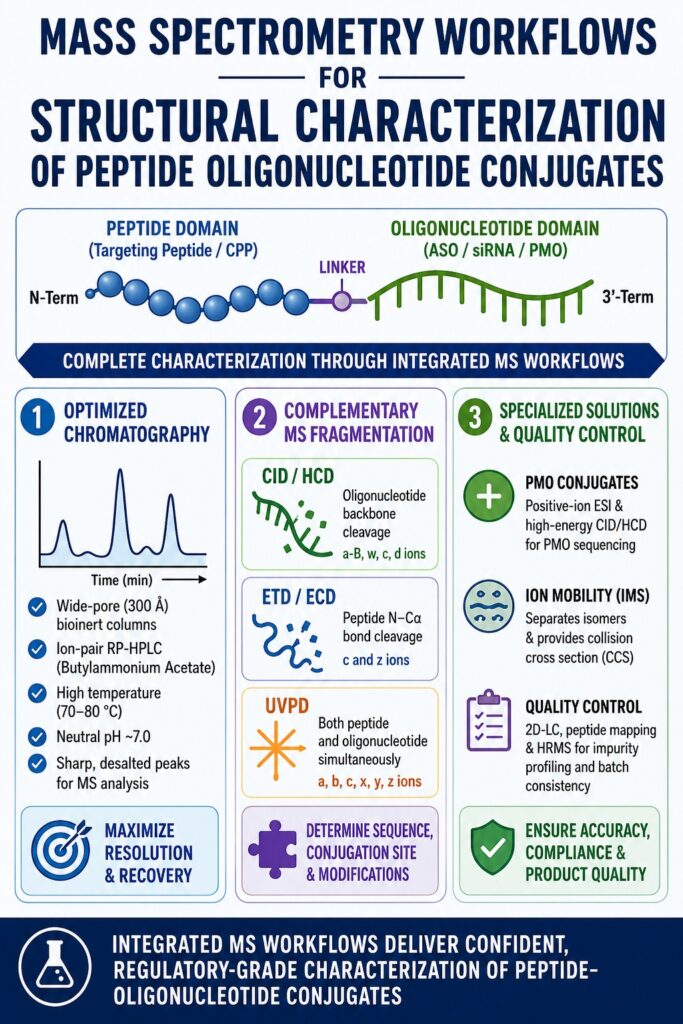
- Peptide-oligonucleotide conjugates (POCs) combine therapeutic peptides with oligonucleotides to improve targeted drug delivery, but their hybrid structure makes analytical characterization highly complex.
- Optimized ion-pair reversed-phase chromatography (IP-RP-HPLC) using wide-pore bioinert columns, elevated temperatures (70–80°C), and volatile ion-pairing reagents enables efficient separation and improved recovery of these hybrid biomolecules.
- Careful control of chromatographic conditions, particularly neutral mobile phase pH (~7.0) and bioinert stationary phases, minimizes sample adsorption, reduces aggregation, and enhances peak quality for accurate mass spectrometry analysis.
- Comprehensive structural characterization requires multiple MS/MS fragmentation techniques. CID/HCD primarily sequence the oligonucleotide region, ETD/ECD provide detailed peptide sequencing and conjugation site identification, while UVPD simultaneously fragments both domains for complete molecular analysis.
- Neutral morpholino (PMO) conjugates require specialized positive-ion electrospray ionization, higher collision energies, and ion mobility spectrometry (IMS) to accurately distinguish isomers, sequence molecules, and characterize complex structural variants.
- Regulatory-quality analysis relies on advanced multidimensional workflows, including two-dimensional liquid chromatography (2D-LC), peptide mapping, and high-resolution mass spectrometry (HRMS) to detect impurities, verify conjugation sites, and ensure batch consistency.
- Integrated mass spectrometry workflows are essential for product quality and regulatory compliance, supporting impurity profiling, structural verification, and the safe development of next-generation peptide-oligonucleotide therapeutics under GLP, GMP, and ICH validation standards.
Optimizing Chromatography for the Structural Characterization of Peptide Oligonucleotide Conjugates
Efficient chromatographic separation of peptide-oligonucleotide conjugates requires carefully optimized conditions that include wide-pore bioinert reversed-phase columns, elevated operating temperatures, and volatile alkylamine ion-pairing reagents. This specialized chromatographic configuration minimizes non-specific adsorption, disrupts secondary structural interactions, and produces sharp, desalted peaks that are ideally suited for electrospray ionization mass spectrometry.
Peptide-oligonucleotide conjugates (POCs) possess two biomolecular domains with fundamentally different physicochemical characteristics. The oligonucleotide region contains a highly hydrophilic, polyanionic phosphodiester or phosphorothioate backbone, whereas the peptide region frequently incorporates highly basic amino acids, such as arginine-rich CPPs, or hydrophobic targeting sequences. Conventional reversed-phase chromatography provides insufficient ionic interaction to effectively retain hydrophilic oligonucleotides, while standard anion-exchange chromatography cannot adequately distinguish differences arising from peptide hydrophobicity. Consequently, neither separation mode independently provides the resolution necessary for comprehensive characterization.
To address these limitations, Ion-Pair Reversed-Phase High-Performance Liquid Chromatography (IP-RP-HPLC) serves as an adaptable mixed-mode separation strategy. Volatile alkylamine ion-pairing reagents are introduced into the aqueous mobile phase, where they dynamically adsorb onto the hydrophobic stationary phase and create a positively charged surface. This positively charged layer electrostatically interacts with the negatively charged phosphate groups of the oligonucleotide domain, thereby enhancing retention. Separation selectivity can then be precisely optimized by adjusting alkylamine hydrophobicity and concentration, gradient composition, mobile phase pH, and column temperature.
Discover how synthesis methods influence chromatographic profiles in your daily operations.
The Impact of Mobile Phase pH and Temperature on Separation
Both peptide and oligonucleotide components readily undergo intermolecular and intramolecular self-association, forming secondary structures, hairpins, and electrostatic aggregates. Peptide-oligonucleotide conjugates containing arginine-rich CPPs are especially susceptible to aggregation because of strong electrostatic interactions between guanidinium side chains and negatively charged phosphate groups.
To minimize these non-covalent interactions, chromatographic separations are typically performed at elevated column temperatures ranging from 70°C to 80°C. Higher temperatures disrupt secondary structures, enhance mass transfer, improve chromatographic efficiency, and facilitate clear separation of the desired full-length conjugate from truncated intermediates and residual starting materials.
Mobile phase pH also plays a critical role in chromatographic performance. Maintaining neutral conditions around pH 7.0 provides the most effective balance between separation efficiency and column stability. Operating below pH 6.0 significantly reduces the resolution of closely related conjugate impurities, whereas pH values exceeding 8.0 accelerate silica degradation, shorten column lifetime, and increase sample carry-over throughout the chromatographic system.
| Chromatographic Parameter | Standard Condition | Optimized Condition | Technical and Mass Spectrometric Rationale | References |
|---|---|---|---|---|
| Pore Size | 80 Å to 120 Å | 300 Å (30 nm) | Eliminates mass transfer limitations for large hybrid biopolymers while reducing steric hindrance and peak tailing. | [cite: 1, 2, 3] |
| Column Hardware | Stainless steel | Bioinert organic-silica or PEEK-lined hybrid | Prevents electrostatic adsorption of acidic and basic biomolecules onto exposed metal surfaces. | [cite: 2, 3, 4] |
| Column Temperature | 30°C to 40°C | 70°C to 80°C | Disrupts secondary structures, Watson-Crick base pairing, and molecular aggregation to maximize chromatographic recovery. | [cite: 1, 2, 3, 21] |
| Ion-Pairing Reagent | 100 mM TEAA or 15 mM TEA / 400 mM HFIP | 100 mM Butylammonium Acetate (BAA) | Provides superior electrostatic shielding and improved separation of cationic peptide-linked oligonucleotides. | [cite: 2, 3, 4] |
| Mobile Phase B | 100% Methanol | 100% Acetonitrile | Improves elution efficiency of hydrophobic peptide domains using shallow chromatographic gradients. | [cite: 2, 4, 21] |
| System pH | pH 6.0 | pH 7.0 | Minimizes conjugate degradation, protects silica stationary phases, and reduces carry-over. | [cite: 2, 3, 4] |
Understand how to optimize your linker chemistry to improve molecular stability.
Stationary Phase Selection in the Structural Characterization of Peptide Oligonucleotide Conjugates
Selecting an appropriate stationary phase is essential for achieving accurate structural characterization of peptide-oligonucleotide conjugates. Wide-pore bioinert stationary phases minimize steric exclusion while preventing metal-induced sample adsorption during chromatographic analysis. A 300 Å C18 or C4 bioinert stationary phase constructed with an organic-silica hybrid coating consistently delivers excellent recovery and sharp peak profiles for highly basic chimeric biomolecules.
Traditional reversed-phase columns typically utilize silica particles with pore diameters ranging from 80 Å to 120 Å, making them suitable for small molecules and conventional peptides. However, these pore sizes are insufficient for bulky peptide-oligonucleotide conjugates, preventing efficient diffusion into the stationary phase and leading to restricted mass transfer, severe peak tailing, and progressive column fouling. Transitioning to wide-pore stationary phases with approximately 300 Å pore diameters accommodates the significantly larger hydrodynamic volume of hybrid conjugates, allowing efficient interaction with the stationary phase and substantially improving chromatographic resolution.
Conventional stainless-steel columns also introduce analytical complications because exposed metallic surfaces act as coordination sites that interact strongly with negatively charged phosphate groups. These interactions frequently cause irreversible adsorption, sample loss, distorted peak shapes, and persistent ghost peaks during subsequent blank injections.
For these reasons, analytical laboratories routinely employ specialized bioinert hardware, including PEEK-lined columns or advanced organic-silica hybrid column technologies such as YMC Accura. Such bioinert configurations ensure maximum analyte recovery, particularly when analyzing trace-level impurities or conducting microscale pharmacokinetic investigations.
METAL-LINED COLUMN (Adsorption & Peak Tailing) BIOINERT COATED COLUMN (High Recovery & Sharp Peaks)
       Active Metal Sites (Fe/Ti)                           Organic-Silica Bioinert Layer
     [M---Fe+]  [M---Fe+]  [M---Fe+]                      [M] --------> [M] --------> [M]
   ===================================                 ===================================
   |  O     O     O     O     O      |                 |  O     O     O     O     O      |
   | / \   / \   / \   / \   / \     |                 | / \   / \   / \   / \   / \     |
   | | |   | |   | |   | |   | |     |                 | | |   | |   | |   | |   | |     |
   ===================================                 ===================================
See why bioinert hardware is critical for your data integrity and reproducibility.
Gas-Phase Dissociation Mechanisms in the Structural Characterization of Peptide Oligonucleotide Conjugates
Comprehensive structural characterization of peptide-oligonucleotide conjugates requires the integration of multiple tandem mass spectrometric fragmentation techniques. By combining collision-induced, electron-mediated, and photon-mediated dissociation mechanisms, researchers can independently sequence the peptide component, characterize the oligonucleotide region, and accurately localize the conjugation site.
Learn about the biological mechanism of action to better guide your structural analysis.
Tandem mass spectrometry (MS/MS) operates by activating isolated precursor ions in the gas phase, causing covalent bond cleavage along the molecular backbone. Because peptide amide bonds and oligonucleotide phosphodiester linkages possess markedly different chemical stabilities, no single fragmentation method provides complete structural information. Conditions optimized for peptide fragmentation often fail to efficiently cleave nucleic acid backbones, whereas methods optimized for oligonucleotide sequencing frequently leave peptide structures largely intact. Therefore, comprehensive characterization requires multiple complementary fragmentation approaches.
             PEPTIDE DOMAIN                           OLIGONUCLEOTIDE DOMAIN
          (Targeting Peptide / CPP)                     (ASO / siRNA)

N-Term -- [aa1] -- [aa2] -- [aa3] ---- Linker ---- [Nuc1] -- [Nuc2] -- [Nuc3] -- 3'-Term

ETD/ECD ---------> c/z ions (Peptide)

CID/HCD ----------------------------------------> a-B, w, c, d ions (Oligonucleotide)

UVPD -----------------------------> Simultaneous fragmentation of both domains
Collision-Induced Dissociation (CID) and Higher-Energy Collisional Dissociation (HCD)
CID and HCD are thermal activation techniques that induce fragmentation through repeated collisions between precursor ions and inert gas molecules such as helium, nitrogen, or argon. These collisions gradually convert kinetic energy into internal vibrational energy, causing preferential cleavage at the molecule’s most labile bonds.
Within peptide-oligonucleotide conjugates, phosphodiester bonds are significantly more susceptible to fragmentation than peptide amide bonds. Consequently, CID and HCD predominantly generate oligonucleotide-specific fragments, including neutral base losses and backbone cleavages that produce a-B, w, c, and d ion series.
Although these fragmentation patterns provide excellent sequence confirmation for the oligonucleotide domain, peptide sequencing remains limited because the peptide region typically survives as a large intact modification attached to fragmented nucleotides. As a result, CID and HCD alone cannot comprehensively define peptide sequences or precisely localize conjugation sites.
Electron Transfer Dissociation (ETD) and Electron Capture Dissociation (ECD)
ETD and ECD represent non-ergodic fragmentation techniques that operate by transferring low-energy electrons from donor radical anions, such as fluoranthene, to multiply protonated precursor ions. Electron transfer initiates radical-mediated cleavage of peptide N–Cα bonds.
Because fragmentation occurs rapidly without extensive vibrational energy redistribution, ETD and ECD preferentially cleave peptide backbones while preserving chemically labile structures. Within peptide-oligonucleotide conjugates, these methods generate complementary c- and z-type peptide ions while maintaining the integrity of the oligonucleotide backbone, chemical linker, and sensitive post-translational modifications, including phosphorylation, glycosylation, and lipidation. This enables precise identification of the amino acid residue involved in conjugation.
Ultraviolet Photodissociation (UVPD)
Ultraviolet Photodissociation (UVPD) is an advanced fragmentation technique that employs high-energy ultraviolet laser irradiation, typically at wavelengths of 193 nm or 213 nm, to excite trapped precursor ions directly into electronically excited states.
Unlike low-energy thermal activation, UVPD simultaneously fragments both peptide and oligonucleotide backbones, producing exceptionally rich tandem mass spectra.
The resulting fragmentation includes:
- Peptide Backbone: Cleavage of amide, N–Cα, and Cα–C bonds generates comprehensive a/x, b/y, and c/z ion series.
- Oligonucleotide Backbone: Produces extensive sequence-defining fragments while preserving chemically fragile modifications.
- Conjugation Site Analysis: Rapid fragmentation preserves critical linker information, allowing accurate mass determination on both sides of the conjugation junction.
| Fragmentation Technique | Activation Source | Primary Cleavage Sites | Key Fragment Ions | Targeted Application | Limitations | References |
| CID / HCD | Low-energy collisions with inert gases | Phosphodiester and glycosidic bonds | a-B, w, c, d ions | Oligonucleotide sequencing and impurity profiling | Limited peptide fragmentation; loss of labile modifications | [cite: 8, 9, 10, 24] |
| ETD / ECD | Electron transfer from donor radical anions | Peptide N–Cα bonds | c and z ions | Peptide sequencing and conjugation site identification | Requires highly charged precursor ions; lower efficiency for acidic conjugates | [cite: 8, 9, 10, 24] |
| UVPD | High-energy UV laser irradiation | Both peptide and oligonucleotide backbones | a, b, c, x, y, z ions; a, w, d oligonucleotide ions | Comprehensive single-analysis sequencing | Complex spectra requiring specialized instrumentation | [cite: 9, 10, 26] |
Explore specialized delivery platforms for PMOs to understand the therapeutic context of these molecules.
Analytical Challenges and Solutions for Neutral Morpholino (PMO) Heteroconjugates
Neutral morpholino conjugates require positive-ion electrospray ionization because their phosphorodiamidate backbone lacks the negative charge necessary for efficient ionization in conventional negative-ion mode. Consequently, analytical workflows rely on positive-mode electrospray, optimized high-energy fragmentation, and ion mobility spectrometry to achieve complete structural characterization.
Phosphorodiamidate Morpholino Oligomers (PMOs) are an increasingly important class of steric-blocking antisense therapeutics. Unlike native DNA and RNA, PMOs replace the conventional ribose or deoxyribose sugar with a neutral six-membered morpholine ring, while the negatively charged phosphodiester linkage is substituted with an uncharged phosphorodiamidate bond.
NATIVE PHOSPHODIESTER LINKAGE             PHOSPHORODIAMIDATE LINKAGE

        O−                                      N(CH3)2
        |                                          |
    O=P–O                                     O=P–O
        |                                          |
        O                                          O
      Pentose Sugar                          Morpholine Ring
This chemical architecture significantly enhances resistance to enzymatic degradation and prevents sequencing by traditional exonuclease-based methods. However, the absence of intrinsic negative charge also makes conventional negative-ion mass spectrometry ineffective.
Positive-Ion Electrospray and Collision Energy Optimization
Analysis of PMOs and peptide-conjugated PMOs (PPMOs) requires operation in positive electrospray ionization mode using multiply protonated precursor ions. Volatile acids such as formic acid or dilute trifluoroacetic acid promote protonation of morpholine nitrogen atoms and basic peptide residues.
Because phosphorodiamidate bonds exhibit greater chemical stability than phosphodiester linkages, substantially higher collision energies are necessary during CID and HCD fragmentation. Under optimized conditions, controlled cleavage of phosphorodiamidate bonds generates sequence-defining fragment ions that enable accurate PMO sequencing and identification of process-related impurities, including n−1 deletion products.
The Role of Ion Mobility Spectrometry (IMS)
One of the greatest analytical challenges associated with PMO conjugates is distinguishing positional and structural isomers that possess identical molecular masses and frequently co-elute during liquid chromatography.
Ion Mobility Spectrometry (IMS) overcomes this limitation by separating ions according to their size, three-dimensional shape, and charge distribution while they travel through a buffer gas under an applied electric field. Measurement of the Collision Cross Section (CCS) enables separation of species that are indistinguishable by mass alone.
IMS facilitates the identification of:
- Full-length conjugates and closely related truncated impurities.
- Positional isomers containing peptide attachments at different morpholino linkage sites.
- Distinct conformational states arising from alternative gas-phase folding of peptide-oligonucleotide hybrids.
Quality Control, Regulatory Compliance, and Impurity Profiling Standards
Regulatory approval of peptide-oligonucleotide conjugates requires high-resolution mass spectrometry platforms capable of achieving mass accuracy below 5 ppm while detecting impurities present at concentrations as low as 0.05% of the total UV area. Analytical procedures must comply with ICH Q2 (R1/R2) validation guidelines and incorporate multidimensional LC-MS workflows together with peptide mapping to verify structural integrity and ensure batch-to-batch consistency.
As peptide-oligonucleotide conjugates progress from early development into commercial manufacturing, comprehensive quality control and stability-indicating analytical methods become increasingly important. Because both peptide and oligonucleotide components contribute independently to impurity formation and metabolic behavior, analytical strategies must evaluate each component simultaneously.
According to International Council for Harmonisation (ICH) Q2 guidelines, validated analytical methods must reliably separate, identify, and quantify all relevant impurities, including sequence-related deletions, residual starting materials, oxidation products, deamidation products, and other degradation species.
Review best practices for ensuring conjugate stability as part of your ICH Q2 validation strategy.
Two-Dimensional Liquid Chromatography (2D-LC) Workflows
Because peptide-oligonucleotide conjugates often generate highly heterogeneous product mixtures, single-dimensional chromatographic methods generally lack sufficient resolving power.
Advanced quality control laboratories therefore employ orthogonal two-dimensional liquid chromatography coupled with high-resolution mass spectrometry.
- First Dimension (D1): Strong Anion Exchange Chromatography (SAX-HPLC) separates compounds according to charge differences, effectively resolving truncated sequences and n−1 deletion products.
- Second Dimension (D2): Ion-Pair Reversed-Phase HPLC (IP-RP-HPLC) or Hydrophilic Interaction Liquid Chromatography (HILIC) separates analytes according to hydrophobicity and polarity.
- Online Desalting: The second-dimension column simultaneously removes non-volatile salts introduced during SAX-HPLC before sample introduction into the electrospray ionization source.
Evaluate how these conjugates perform in preclinical models to validate your analytical findings.
Peptide Mapping and High-Resolution Mass Spectrometry (HRMS)
Verification of peptide primary structure and confirmation of conjugation sites commonly rely on bottom-up peptide mapping workflows. Under carefully controlled digestion conditions, enzymes such as trypsin, chymotrypsin, or lysyl endopeptidase cleave the peptide domain into smaller fragments suitable for LC-MS/MS analysis.
The resulting peptide fragments are analyzed using high-resolution Orbitrap or Q-TOF mass spectrometers. Exact mass measurements and tandem MS fragmentation patterns are compared with theoretical databases to reconstruct peptide sequences, identify low-level degradation products such as oxidation and deamidation, verify conjugation sites, and confirm manufacturing consistency across production batches.
Conclusion
Advanced mass spectrometry workflows play a central role in the comprehensive Structural Characterization of Peptide Oligonucleotide Conjugates. Successfully analyzing these complex hybrid biomolecules requires an integrated analytical strategy that combines wide-pore bioinert reversed-phase chromatography with high-resolution mass spectrometry. The complementary application of CID, HCD, ETD, ECD, and UVPD provides complete structural information by independently sequencing peptide and oligonucleotide domains, accurately identifying conjugation sites, and comprehensively profiling process-related impurities.
If you need to scale up your production, our specialized services can assist in maintaining high-quality outputs.
Establishing validated analytical workflows within strict GLP and GMP environments ensures compliance with global regulatory requirements while supporting the safe and efficient development of next-generation hybrid biotherapeutics. Robust analytical characterization ultimately accelerates therapeutic development, improves product quality, and strengthens confidence in regulatory submissions.
For specialized analytical validation, method development, and high-resolution mass spectrometry services supporting peptide-oligonucleotide conjugate development, visit the ResolveMass Contact Us page.
Frequently Asked Questions (FAQs)
Wide-pore stationary phases, typically around 300 Å (30 nm), provide sufficient space for large peptide-oligonucleotide conjugates to enter the pores and interact effectively with the stationary phase. Conventional columns with pore sizes between 80 Å and 120 Å restrict the movement of these larger biomolecules, resulting in poor chromatographic performance. This limitation often leads to peak broadening, tailing, and reduced recovery. Using wide-pore columns significantly improves separation efficiency, impurity resolution, and overall analytical accuracy.
Peptide-oligonucleotide conjugates containing highly basic cell-penetrating peptides frequently form secondary structures and electrostatic aggregates that interfere with chromatographic separation. Operating the column at elevated temperatures, generally between 70°C and 80°C, disrupts these intermolecular interactions and denatures stable conformations. This results in narrower chromatographic peaks, improved mass transfer, and better separation between the desired conjugate and closely related impurities or reaction by-products.
Although TEAA and TEA/HFIP are widely used for conventional oligonucleotide analysis, they may not provide optimal chromatographic performance for peptide-linked conjugates containing highly basic amino acids. Butylammonium Acetate (BAA), particularly at pH 7.0, offers enhanced electrostatic shielding and stronger interaction with C18 stationary phases. This optimized ion-pairing system produces sharper peaks, improved baseline stability, higher resolution, and reduced sample carry-over during LC-MS analysis.
Standard stainless-steel chromatographic hardware contains exposed metallic surfaces that can strongly interact with the negatively charged phosphate groups of oligonucleotides. These unwanted interactions often cause irreversible sample adsorption, distorted peak shapes, reduced recovery, and increased carry-over between analyses. Bioinert column technologies, including PEEK-lined hardware and specialized organic-silica coatings, eliminate direct metal contact and provide more reliable, reproducible, and accurate analytical results.
During CID analysis, the phosphodiester backbone of the oligonucleotide is considerably more susceptible to fragmentation than the peptide amide backbone. As a result, fragmentation predominantly occurs within the nucleic acid portion, producing characteristic a-B, w, c, and d ion series useful for oligonucleotide sequencing. The peptide component generally remains largely intact, making comprehensive peptide sequencing difficult without incorporating complementary fragmentation techniques.
Electron Transfer Dissociation (ETD) selectively fragments peptide N–Cα bonds through a radical-driven mechanism rather than thermal activation. This approach preserves sensitive structures such as the oligonucleotide backbone, chemical linker, and post-translational modifications while generating informative c- and z-type peptide ions. Consequently, ETD allows accurate peptide sequencing and enables researchers to determine the exact amino acid residue involved in conjugation with high confidence.
UVPD employs high-energy ultraviolet laser pulses, commonly at 193 nm or 213 nm, to rapidly excite precursor ions and induce fragmentation across multiple bond types. Unlike conventional collision-based methods, UVPD simultaneously cleaves both peptide and oligonucleotide backbones, generating diverse fragment ion series within a single experiment. This comprehensive fragmentation enables complete sequence verification while accurately defining the conjugation junction between both molecular domains.
Reference:
- Marchetti, N., & Gilar, M. (2025). High-resolution HPLC for separating peptide–oligonucleotide conjugates. Molecules, 30(11), 2506. https://doi.org/10.3390/molecules30112506
- Kashyap, S., & Mohapatra, P. R. (2013). Pulmonary alveolar microlithiasis. Lung India, 30(2), 143–147. https://doi.org/10.4103/0970-2113.110424
- Jensen, O. N., Kulkarni, S., Aldrich, J. V., & Barofsky, D. F. (1996). Characterization of peptide-oligonucleotide heteroconjugates by mass spectrometry. Nucleic Acids Research, 24(19), 3866–3872. https://doi.org/10.1093/nar/24.19.3866
- Klabenkova, K., Fokina, A., & Stetsenko, D. (2021). Chemistry of peptide-oligonucleotide conjugates: A review. Molecules, 26(17), Article 5420. https://doi.org/10.3390/molecules26175420
- Lundin, K. E., Gissberg, O., & Smith, C. I. E. (2003). Oligonucleotide therapies: The past and the present. Human Gene Therapy, 14(11), 1065–1076. https://doi.org/10.1089/104303403322167682

