


Introduction
A specialized Subvisible and Visible Particle Testing Service provides essential physical characterization and quality control data needed to establish the safety, stability, and comparability of biopharmaceutical products. High-resolution particulate characterization at ResolveMass Laboratories Inc. evaluates therapeutic proteins across the entire particle size spectrum, from sub-micron subvisible aggregates to macroscopic particulates. Particulates in biopharmaceutical injectables can pose serious clinical risks, including microvascular occlusion, phlebitis, and the activation of anti-drug antibody (ADA) immune responses in patients. For biosimilar developers, demonstrating analytical similarity to a reference medicinal product (RMP) requires more than meeting baseline compendial requirements. It involves establishing comprehensive particulate profile comparability under accelerated stress conditions, during real-time stability studies, and throughout storage in the final container-closure system.
Explore our biosimilar characterization services for comprehensive analytical support in biopharmaceutical development.
Share via:
Quick Summary:
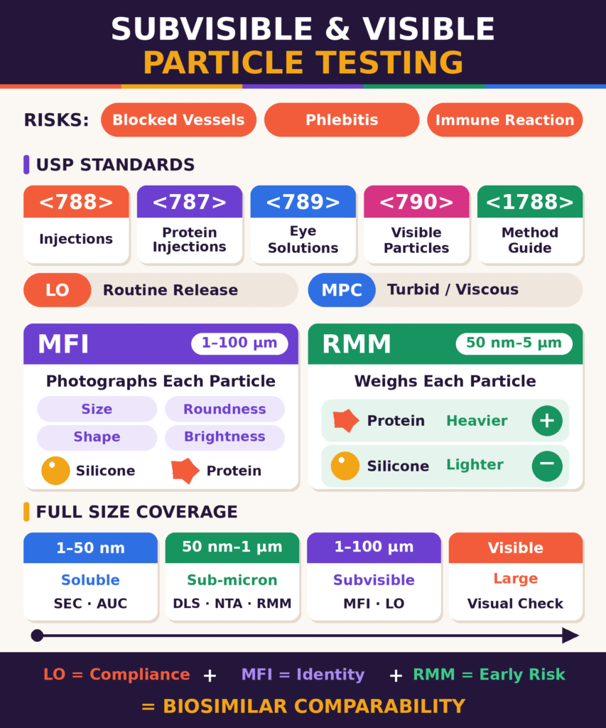
- Why it matters: Particles in injectable biologics can cause vascular occlusion, phlebitis and immune (anti-drug antibody) responses. For biosimilars, you have to show the particle profile is comparable to the reference product, not just that it meets compendial limits.
- Compendial framework: USP <788>, harmonized with EP 2.9.19 and JP 6.07, sets subvisible particle limits for injections. Related chapters cover protein injections (<787>), ophthalmic solutions (<789>), visible particles (<790>) and method guidance (<1788>).
- LO vs. MPC: Light Obscuration is the preferred routine method. Microscopic Particle Count is the alternative when turbidity, viscosity or other formulation properties interfere with LO.
- Micro-Flow Imaging (MFI): MFI images particles in roughly the 1–100 µm range. It catches translucent protein aggregates that LO may undercount, and it uses size, circularity, aspect ratio and intensity to help tell protein aggregates from silicone oil droplets.
- Resonant Mass Measurement (RMM): RMM measures the buoyant mass of sub-micron particles (about 50 nm–5 µm). Denser protein particles give a positive signal and lighter silicone oil droplets give a negative one, so the two can be separated where optical methods struggle.
- Orthogonal strategy: Combining methods covers the full size range. SEC and AUC handle soluble aggregates, DLS, NTA and RMM handle sub-micron particles, MFI and LO handle subvisible particles, and visual inspection handles visible particles. Results should be read together, not treated as interchangeable counts.
- Regulatory value: LO covers compliance, MFI covers particle identity, and RMM covers early aggregation risk. Together they give the evidence needed to demonstrate biosimilar particulate comparability to the FDA, EMA and Health Canada.
Compendial Subvisible and Visible Particle Testing Service Requirements: Harmonized USP, EP, and JP Standards
Compendial particle testing establishes acceptance criteria for subvisible particulates in parenteral drug formulations to help prevent clinical complications, including vascular occlusion and potential immunogenic reactions. Compliance requires standardized light obscuration or microscopic particle counting methods aligned with applicable pharmacopoeial standards.
Pharmacopoeial harmonization through the Pharmacopoeial Discussion Group (PDG) has aligned key requirements across the United States Pharmacopeia (USP), European Pharmacopoeia (EP 2.9.19), and Japanese Pharmacopoeia (JP General Test 6.07). USP <788>, titled Subvisible Particulate Matter in Injections, includes two primary analytical procedures: Method 1 (Light Obscuration, LO) and Method 2 (Microscopic Particle Count, MPC).
Method 1 is the preferred compendial method for routine testing when the sample is suitable for light obscuration. It operates by drawing liquid samples through an optical sensor, where particles obstruct a light beam and generate signals used to estimate particle size and count. Method 2 involves filtering the drug product through a membrane filter and counting the retained particles under microscopic illumination. This method can be appropriate when optical properties, such as turbidity, high viscosity, or other formulation characteristics, interfere with reliable light obscuration measurements.
| Pharmacopoeial Chapter | Product Scope | Primary Analytical Method | Standard Acceptance Criteria and Size Limits |
|---|---|---|---|
| USP <788> | Parenteral injections, including particles ≥10 µm and ≥25 µm | Light Obscuration (LO) or Microscopic Particle Count (MPC) | Small-volume injections (100 mL or less): LO ≤6,000 particles/container at ≥10 µm and ≤600 particles/container at ≥25 µm. Large-volume injections (more than 100 mL): ≤25 particles/mL at ≥10 µm and ≤3 particles/mL at ≥25 µm. |
| USP <787> | Subvisible particulate matter in therapeutic protein injections | Light Obscuration (LO) and Microscopic Particle Count (MPC), as applicable | Provides requirements and testing considerations specific to therapeutic protein injections. Applicability and acceptance criteria should be checked against the current official chapter and product-specific requirements. |
| USP <789> | Particulate matter in ophthalmic solutions | Light Obscuration (LO) and microscopic methods, as applicable | Compendial limits include ≤50 particles/mL at ≥10 µm, ≤5 particles/mL at ≥25 µm, and ≤2 particles/mL at ≥50 µm, subject to the applicable chapter requirements. |
| USP <790> | Visible particulate matter in injections | Direct visual inspection under controlled lighting | Injections must meet the applicable compendial visual-inspection requirements. Inspection conditions and procedures should follow the current chapter and approved product-specific protocol. |
| USP <1788> | Methods for determining particulate matter in injections and ophthalmic solutions | Light obscuration, microscopy, and related characterization approaches | Provides informational and methodological guidance. It should not be treated as an independent source of universal batch-release limits. |
Compendial requirements and testing approaches should be selected according to the dosage form, formulation properties, applicable pharmacopoeia, and approved product specifications. For low-fill or high-value monoclonal antibody products, sample availability may constrain the amount of material available for analysis. Any reduced-volume or single-unit sampling approach must be scientifically justified and consistent with the applicable compendial requirements and an appropriate sampling plan.
Learn how critical quality attributes in biosimilars guide analytical assessment and product quality evaluation.
USP <790> addresses visible particulate inspection, while USP <788> addresses subvisible particulate matter in injections. Visual inspection is used to identify visible particles under controlled conditions, but it does not establish particle identity or provide detailed structural and morphological information. Where necessary, complementary analytical techniques can help characterize the source and nature of detected particulates.
Micro-Flow Imaging (MFI) in Subvisible and Visible Particle Testing Service
Micro-Flow Imaging (MFI) uses dynamic flow imaging analysis to capture digital images of subvisible particles, commonly within the approximately 1–100 µm range, depending on the instrument and analytical configuration. By assessing morphological characteristics, including circularity, intensity, and aspect ratio, MFI can help differentiate proteinaceous aggregates from silicone oil droplets and foreign particulate matter.
Within a Subvisible and Visible Particle Testing Service, MFI addresses an important limitation of light obscuration when analyzing therapeutic proteins. Light obscuration determines particle size by measuring the reduction in transmitted light as a particle passes through a sensing zone. However, proteinaceous aggregates may have refractive indices relatively close to those of aqueous formulation buffers. This limited refractive-index contrast can make some translucent protein aggregates difficult to detect accurately using light obscuration alone, potentially resulting in undercounting or inaccurate sizing. MFI complements light obscuration by capturing brightfield images of particles flowing through a microfluidic flow cell, enabling their detection and morphological characterization.
Advanced image-analysis software extracts morphological parameters for each detected particle:
- Equivalent Circular Diameter (ECD): The diameter of a circle with the same projected area as the particle image.
- Circularity: A measure of geometric roundness calculated as 4π × Area / Perimeter2, where a value approaching 1 indicates a more circular projected shape.
- Aspect Ratio: The ratio of particle width to length, used to quantify particle elongation and help identify elongated or fibrillar structures.
- Mean Intensity and Edge Gradient: Image-based optical measurements that describe particle brightness and edge characteristics, supporting differentiation among particle populations.
These parameters can support image classification to distinguish proteinaceous aggregates from intrinsic silicone oil droplets. Silicone oil microdroplets originating from lubricated prefilled syringes or autoinjectors may appear as highly spherical structures with characteristic edge and intensity patterns. Protein aggregates may exhibit irregular geometries, lower circularity, or diffuse edges. However, these features can overlap, and morphology alone does not definitively establish particle composition. Classification rules should therefore be validated for the formulation and sample matrix.
MFI can provide additional information about particle populations in heavily siliconized drug delivery systems. When particle identity is important, combining MFI with complementary techniques can improve confidence in distinguishing protein aggregates, silicone oil droplets, and extrinsic particulates.
Explore native mass spectrometry for biosimilars to understand complementary approaches for investigating intact protein structures and molecular assemblies.Discover how aggregation analysis in biosimilars supports the investigation of protein aggregates and product stability.
Resonant Mass Measurement (RMM) for Sub-Micron Particle Discrimination
Resonant Mass Measurement (RMM) quantifies and differentiates sub-micron particles, commonly within an approximate range of 50 nm to 5 µm depending on the instrument configuration, according to their individual buoyant mass as they pass through a Suspended Microchannel Resonator. This physical measurement can distinguish silicone oil microdroplets from denser therapeutic protein particles in a size range where conventional optical imaging may have limited sensitivity.
RMM operates using a Suspended Microchannel Resonator (SMR), which consists of a fluidic microchannel embedded within a vibrating mechanical resonator. As a liquid sample passes through the microchannel, individual particles alter the effective mass of the resonator, producing a shift in its resonant frequency. The magnitude and direction of the frequency shift are related to the particle’s buoyant mass, represented by the following equation:
mb = Vp(ρp − ρm)
Where:
- mb = particle buoyant mass
- Vp = particle volume
- ρp = particle density
- ρm = density of the surrounding fluid matrix
RMM differentiates particle populations according to their density relative to the surrounding formulation matrix:
- Protein Aggregates: Proteinaceous particles are generally denser than aqueous buffer matrices. When their density exceeds the fluid density, they have positive buoyant mass. Under the conventional resonator sign convention, this produces a frequency shift associated with an increase in effective resonator mass.
- Silicone Oil Droplets: Silicone oil droplets generally have a lower density than aqueous buffer matrices. Their buoyant mass is negative, producing a frequency response in the opposite direction to that of particles denser than the surrounding fluid.

The exact frequency-shift sign depends on the instrument’s measurement convention and configuration. In practice, particle density, fluid density, viscosity, sample composition, and instrument calibration must be considered when interpreting RMM measurements.
This physical distinction enables RMM to count and size sub-micron particles with different buoyant-mass characteristics within the same formulation sample. RMM can provide valuable characterization data for prefilled syringe biosimilar development, where silicone oil droplets and protein aggregation must be evaluated as distinct potential contributors to the particulate profile.
Explore native mass spectrometry for biosimilars to understand complementary approaches for investigating intact protein structures and molecular assemblies.
Orthogonal Characterization Frameworks for Biosimilar Comparability
Establishing biopharmaceutical similarity between a biosimilar candidate and a reference medicinal product requires an orthogonal characterization strategy that covers relevant particle populations across multiple size ranges. Combining compendial methods with complementary analytical techniques helps minimize the blind spots associated with relying on a single measurement technology.
A comprehensive characterization framework can coordinate multiple analytical platforms across distinct particle-size regimes:
- Soluble Aggregate Regime (approximately 1–50 nm): Size-Exclusion Chromatography (SEC-HPLC) and Sedimentation Velocity Analytical Ultracentrifugation (SV-AUC) can quantify or characterize soluble dimers, trimers, and higher-order oligomers, subject to method capability.
- Sub-Micron Particle Regime (approximately 50 nm–1 µm): Dynamic Light Scattering (DLS), Nanoparticle Tracking Analysis (NTA), and Resonant Mass Measurement (RMM) can help characterize particle size distributions, concentration, and potential differences between silicone droplets and proteinaceous particles.
- Subvisible Particle Regime (approximately 1–100 µm): Micro-Flow Imaging (MFI) provides morphological classification, while Light Obscuration (LO) generates compendial particle-count data when the method is suitable for the formulation.
- Visible Particle Regime (macroscopic particles): Visual inspection under the applicable USP requirements evaluates visible particulate contamination and product appearance. The lower detectable size depends on the inspection procedure and particle characteristics.
| Analytical Method | Typical or Approximate Size Range | Primary Target and Application | Compendial Status | Major Analytical Advantage | Primary Technical Limitation |
|---|---|---|---|---|---|
| Light Obscuration (LO) | Approximately 2–150 µm, depending on the instrument | Routine lot-release and stability testing | Official compendial method under USP <788>, where applicable | High throughput, automated operation, and broad regulatory recognition | Detection and sizing may be affected by refractive-index contrast, particle shape, formulation properties, and other matrix effects. |
| Microscopic Particle Count (MPC) | Typically ≥10 µm for compendial counting applications; method-dependent | Samples for which light obscuration is unsuitable, including certain opaque or viscous formulations | Official compendial alternative under USP <788>, where applicable | Direct optical examination of particles retained on a membrane | Lower throughput, labor-intensive procedures, and potential analyst-to-analyst variability |
| Micro-Flow Imaging (MFI) | Approximately 1–100 µm, depending on configuration | Morphological classification and subvisible particle characterization | Supplementary characterization technique; consult applicable USP informational chapters | Provides particle images and morphological information, including for some translucent particles | Potential flow-cell clogging at high particle densities and lower throughput than LO |
| Resonant Mass Measurement (RMM) | Approximately 50 nm–5 µm, depending on configuration | Differentiation of sub-micron particles with different buoyant masses | Complementary characterization technique | Buoyant-mass measurement can distinguish particles denser or less dense than the surrounding fluid | Microchannel clogging, sample-matrix effects, and the need for appropriate calibration and method suitability assessment |
| Nanoparticle Tracking Analysis (NTA) | Approximately 30 nm–1 µm, depending on the instrument and sample | Sub-micron sizing and concentration measurement | Complementary characterization technique | Can characterize polydisperse particle populations and estimate particle concentration | Results depend on light-scattering properties, viscosity, dilution, and detection settings. |
| Visual Inspection | Visible particles; detection depends on size, contrast, and inspection conditions | Release inspection for macroscopic visible particles | Compendial visual-inspection requirements under USP <790>, as applicable | Supports assessment of visible particulate contamination across inspected units | Qualitative or semi-quantitative assessment; manual inspection may be affected by analyst fatigue and particle visibility. |
Combining MFI, RMM, and LO can provide a more comprehensive particulate profile for biosimilar characterization and regulatory submissions:
- Compendial Release: LO verifies whether particle counts meet the applicable USP <788> acceptance criteria for particles ≥10 µm and ≥25 µm, where the method is suitable for the product.
- Morphological Characterization: MFI helps investigate whether elevated particle counts in prefilled syringes may be associated with silicone oil droplets, proteinaceous aggregates, or other particle populations.
- Sub-Micron Risk Assessment: RMM provides buoyant-mass-based particle characterization across its validated operating range, supporting the evaluation of sub-micron particulate populations and potential early aggregation-related changes.
The integrated strategy should be designed around the product’s formulation, container-closure system, manufacturing process, and stability profile. Method selection, sample preparation, particle-size coverage, and acceptance criteria should be justified scientifically. Results from complementary techniques should be interpreted together rather than assuming that their reported particle counts are directly interchangeable.
Learn about biosimilar comparability studies and how analytical evidence supports comparisons between a biosimilar candidate and its reference product.
This approach can support regulatory submissions to agencies such as the FDA, EMA, and Health Canada by providing evidence that the biosimilar candidate has a particulate profile comparable to that of the reference medicinal product. The relevance of any observed differences should be evaluated in the context of analytical uncertainty, product quality, stability, and patient safety.
Review the ICH Q6B guidelines for biological characterisation to understand key considerations for biological product specifications and characterization.
Conclusion
A scientifically validated Subvisible and Visible Particle Testing Service is essential for characterizing particulate matter, assessing product stability, and supporting biosimilar comparability. Compliance with applicable USP requirements establishes the necessary compendial testing framework, while advanced characterization techniques, including Micro-Flow Imaging (MFI) and Resonant Mass Measurement (RMM), provide additional scientific insight into complex particulate stability concerns.
Implementing orthogonal workflows that combine USP compendial testing, MFI morphological imaging, and RMM buoyant-mass profiling enables a broader assessment of particulate populations across different size ranges. By helping distinguish silicone oil microdroplets from therapeutic protein aggregates, these methods can support investigations into formulation stability, container-closure interactions, and potential product-quality risks.
ResolveMass Laboratories Inc. provides specialized analytical expertise and characterization support for biopharmaceutical development programs. Appropriate method selection, scientifically justified testing protocols, and comprehensive analytical documentation can help developers evaluate particulate comparability throughout product development and stability assessment.
Explore our end-to-end biosimilar analytical package for regulatory submissions to support comprehensive analytical characterization and regulatory documentation.
To discuss customized testing protocols or request analytical support, visit the ResolveMass Laboratories Contact Page.
Frequently Asked Questions
Light Obscuration (LO) determines particle size by measuring the interruption of light as particles pass through an optical sensor. Translucent therapeutic protein aggregates may have refractive indices similar to those of formulation buffers, making them difficult to detect accurately. Consequently, LO may underestimate their number or size and cannot reliably distinguish protein aggregates from silicone oil droplets or other contaminants.
Micro-Flow Imaging (MFI) captures detailed brightfield images of individual particles and evaluates their morphological characteristics using specialized software. Silicone oil droplets generally exhibit high circularity (greater than 0.90), clearly defined dark edges, and bright central regions, whereas protein aggregates often have irregular shapes, lower circularity (less than 0.85), and variable internal intensity. These differences help classify and quantify the two particle populations separately.
The harmonized USP revision aligned particulate testing requirements with European Pharmacopoeia (EP 2.9.19) and Japanese Pharmacopoeia (JP 6.07). Important changes include the explicit inclusion of intramuscular and subcutaneous injectables, smaller sample volumes of 1–5 mL, and provisions for single-unit container testing. Scientifically justified alternative diluents are also permitted under the revised framework.
Resonant Mass Measurement (RMM) detects and characterizes particles within an approximate size range of 50 nm to 5 μm. This capability complements techniques such as SEC-HPLC, which evaluates soluble aggregates, and Micro-Flow Imaging (MFI), which generally examines particles measuring 1–100 μm. RMM is particularly useful for differentiating sub-micron protein aggregates from silicone oil droplets based on their buoyant mass.
For small-volume parenterals containing less than 100 mL, Method 1 (Light Obscuration) specifies limits of no more than 6,000 particles per container measuring ≥ 10 μm and 600 particles per container measuring ≥ 25 μm. Method 2 (Microscopic Particle Count) has corresponding limits of 3,000 and 300 particles per container, respectively. The applicable acceptance criteria should be confirmed against the current official pharmacopoeial chapter and the relevant product requirements.
Subvisible particles measuring approximately 1–100 μm are important critical quality attributes (CQAs) because they can affect both product safety and immunogenicity. After administration, particulates may contribute to complications such as microvascular occlusion and phlebitis. Protein aggregates may also promote immune responses by presenting repetitive epitope structures, potentially triggering anti-drug antibody (ADA) production, reduced therapeutic efficacy, or hypersensitivity reactions.
USP establishes compendial requirements for visible particulate matter, including the expectation that injectable products remain essentially free of visible particles when examined under specified conditions. Inspection typically involves black and white backgrounds with controlled illumination of approximately 2,000–3,750 lux. USP provides supplementary guidance on visual inspection approaches, including manual, semi-automated, and automated procedures, as well as defect classification and lifecycle risk management.
Method 2 is appropriate when the formulation’s characteristics interfere with reliable particle detection by Light Obscuration (LO). Examples include highly viscous or turbid solutions, colored preparations, emulsions, liposomal formulations, and samples containing microbubbles that generate optical artifacts. Microscopic examination allows particles retained on a membrane filter to be evaluated directly when LO results may be unreliable.
Informational chapters provide scientific guidance and methodological recommendations rather than independently enforceable compendial acceptance criteria. Nevertheless, their recommendations can support regulatory expectations for instrument qualification, particle size measurement, and orthogonal characterization during biosimilar development. Approaches such as Micro-Flow Imaging (MFI) and Dynamic Image Analysis (DIA) can strengthen analytical similarity assessments and help investigate out-of-specification (OOS) results.
Reference:
- Zhang, B., Flores, K., Horvath, N., Gonzalez Lopez, N., Sawant, N., Falcone, E., Kwok, S. C., Minocha, S., & Radhakrishnan, V. (2026). Control strategy for subvisible particulates of therapeutic protein injections in pre-filled syringes using flow imaging. Journal of Pharmaceutical Sciences, 115(3), 104168. https://doi.org/10.1016/j.xphs.2026.104168
- United States Pharmacopeia. (2026). 〈788〉 Particulate matter in injections (Stage 4 Harmonization; official August 1, 2026). In United States Pharmacopeia–National Formulary (USP–NF). https://www.usp.org/sites/default/files/usp/document/harmonization/20250725HSm99586.pdf
- United States Pharmacopeia. (n.d.). 〈788〉 Particulate matter in injections. In United States Pharmacopeia–National Formulary (USP–NF). https://www.uspnf.com/sites/default/files/usp_pdf/EN/USPNF/revisionGeneralChapter788.pdf
- Gross-Rother, J., Blech, M., Preis, E., Bakowsky, U., & Garidel, P. (2020). Particle detection and characterization for biopharmaceutical applications: Current principles of established and alternative techniques. Pharmaceutics, 12(11), 1112. https://doi.org/10.3390/pharmaceutics12111112
- Sharma, D. K., Oma, P., & Krishnan, S. (2009). Silicone microdroplets in protein formulations—Detection and enumeration. Pharmaceutical Technology, 33(4), 74–79. https://www.pharmtech.com/view/silicone-microdroplets-protein-formulations-detection-and-enumeration
- Frahm, G. E., Pochopsky, A. W. T., Clarke, T. M., & Johnston, M. J. W. (2016). Evaluation of microflow digital imaging particle analysis for sub-visible particles formulated with an opaque vaccine adjuvant. PLOS ONE, 11(2), e0150229. https://doi.org/10.1371/journal.pone.0150229
Get In Touch With Us
About The Author