

Introduction
Characterizing Peptides in PLGA Microsphere and Depot Formulations requires sophisticated orthogonal analytical methodologies to assess complex drug release kinetics, quantify covalent peptide-polymer acylation adducts, and establish comprehensive long-term stability profiles. Poly(lactic-co-glycolic acid) (PLGA) matrices are regarded as a leading polymer platform for long-acting injectable (LAI) depot drug delivery systems because of their established biocompatibility, predictable biodegradability, and extensive regulatory history. The encapsulation of therapeutic peptides—such as octreotide, leuprolide, goserelin, exenatide, salmon calcitonin (sCT), and human parathyroid hormone (hPTH)—within PLGA microspheres or solid implants enables sustained systemic exposure over prolonged periods ranging from several weeks to multiple months.
Explore ResolveMass Laboratories’ peptide physicochemical characterization services for comprehensive analytical evaluation of therapeutic peptides.
However, the microenvironment that develops within a degrading PLGA matrix creates significant chemical stability challenges. As ester bonds within the PLGA backbone undergo hydrolytic cleavage, accumulated lactic and glycolic acid monomers, together with short-chain oligomers, produce a highly acidic local microclimate (μpH < 3.0). This localized acidic environment, combined with reactive ester intermediates, facilitates nucleophilic substitution reactions between primary amine groups present on the peptide and the degrading polymer matrix. This degradation pathway, referred to as peptide acylation, generates heterogeneous adducts that can decrease native peptide potency, modify pharmacokinetic profiles, and potentially contribute to immunogenic responses.
To address these formulation challenges, robust bioanalytical techniques are necessary to monitor peptide degradation pathways, resolve complex acylation-related mass shifts, characterize microenvironmental changes, and establish biorelevant in vitro release testing (IVRT) protocols. Advanced characterization methodologies—including high-resolution LC-MS/MS, MALDI-TOF MS, capillary electrophoresis, and USP Apparatus 4 flow-through dissolution—are critical for optimizing formulation stability, maintaining batch-to-batch reproducibility, and supporting stringent chemistry, manufacturing, and controls (CMC) regulatory submissions.
Learn more about advanced peptide characterization CRO deliverables for structured analytical characterization programs.
Share via:
Quick Summary:
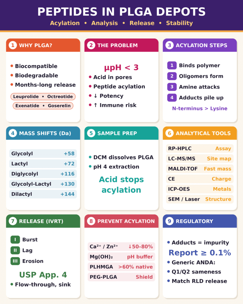
- Why PLGA depots matter: PLGA microspheres and implants are a leading platform for long-acting injectables. They let peptides like leuprolide, octreotide, goserelin and exenatide release slowly over weeks to months.
- The core problem is acylation: As PLGA degrades, lactic and glycolic acids build up inside the pores and drop the local pH below 3. Reactive oligomers then attack the peptide’s amine groups, mainly the N-terminus and lysine. This forms covalent adducts that can reduce potency and raise immunogenicity risk.
- Adducts have predictable mass shifts: They show up as +58 Da (glycolyl), +72 Da (lactyl), and dimer additions of +116, +130 and +144 Da. This makes them traceable by mass spectrometry.
- Characterization takes orthogonal methods:
- Extraction with DCM and a pH 4 buffer stops acylation during sample prep.
- RP-HPLC, LC-MS/MS, MALDI-TOF and capillary electrophoresis quantify and map the adducts.
- SEM, laser diffraction and ICP-OES profile particle structure and excipient content.
- Release follows three phases: an initial burst, then a diffusion-controlled lag, then erosion-driven release. USP Apparatus 4 (flow-through cell) gives the most reproducible long-term in vitro release data because it maintains sink conditions and prevents particle aggregation.
- Acylation can be reduced through formulation:
- Divalent cations such as Ca²⁺ and Zn²⁺ cut adducts by 50–80%.
- Basic buffers like Mg(OH)₂ neutralize the acidic microclimate.
- Alternative polymers help: PLHMGA gives over 60% native peptide release, and PEG-PLGA adds a steric shield.
- Regulators treat adducts as impurities: Adducts at or above 0.1% must be identified and quantified. Generic depot ANDAs must also show Q1/Q2 sameness and matching release and impurity profiles against the reference listed drug.
Mechanisms of Peptide Acylation inside PLGA Depots
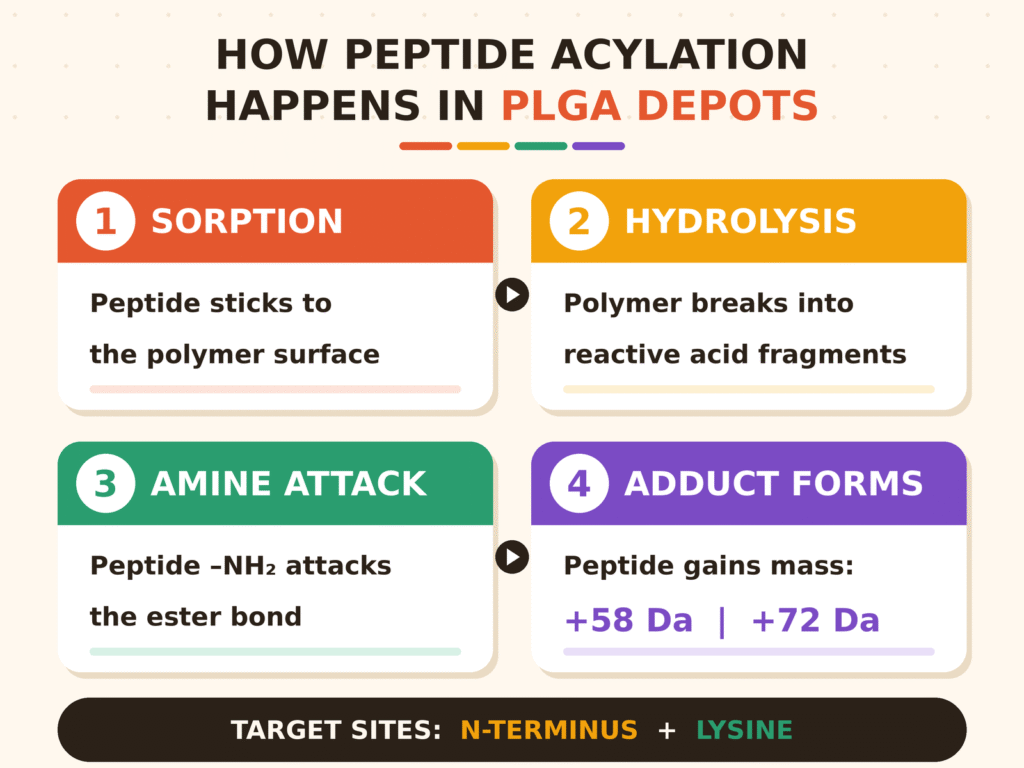
Peptide acylation within PLGA depots primarily occurs through nucleophilic aminolysis, in which unprotonated primary amines on the peptide attack reactive ester carbonyl carbons present on the polymer backbone or on soluble oligomeric degradation products. This chemical conjugation process generates covalent lactyl, glycolyl, or oligomeric adducts that compromise the structural integrity and biological activity of the therapeutic peptide.
The secondary structure and primary amino acid sequence of an individual peptide determine its susceptibility to acylation. The principal reactive sites include the unprotonated N-terminal α-amine group and the ε-amine groups of lysine residues. Since the pKa of an N-terminal α-amine (~7.5–8.0) is lower than that of a lysine ε-amine (~10.5), the N-terminus demonstrates greater reactivity under moderately acidic to neutral local pH conditions, where a larger proportion of the amine remains in its unprotonated (–NH₂) state.
Discover ResolveMass expertise in peptide sequencing and structural characterization to support detailed evaluation of peptide identity and sequence-related modifications.
The physical mechanism of acylation occurs through a defined sequence of events:
- Sorption and Local Pre-Concentration: Peptides become adsorbed onto hydrophobic PLGA polymer surfaces through a combination of electrostatic and hydrophobic interactions. This process produces an elevated local concentration of peptide at the polymer interface, increasing opportunities for chemical interaction.
- Hydrolytic Generation of Reactive Oligomers: Bulk polymer degradation generates soluble, low-molecular-weight glycolic acid and lactic acid oligomers. Compared with intact polymer chains, these short-chain species exhibit lower steric hindrance and greater mobility, facilitating nucleophilic interaction with peptide molecules adsorbed onto the polymer.
- Nucleophilic Attack and Conjugation: The unprotonated amine performs a nucleophilic attack on the ester carbonyl carbon of the oligomer or polymer chain. An intermediate complex is formed, followed by polymer chain cleavage and covalent transfer of the acyl moiety to the peptide.
- Mass-Shift Cascade: Continued polymer degradation generates multi-acylated adducts with predictable molecular weight increases associated with individual glycolic acid (+58.01 Da) or lactic acid (+72.02 Da) additions.
| Adduct Type | Mass Shift (Δm) | Parent Polymer Unit | Target Amino Acid Group |
|---|---|---|---|
| Glycolyl Adduct | +58.01 Da | Glycolic acid residue (C₂H₂O₂) | N-terminal α-NH₂, Lysine ε-NH₂ |
| Lactyl Adduct | +72.02 Da | Lactic acid residue (C₃H₄O₂) | N-terminal α-NH₂, Lysine ε-NH₂ |
| Diglycolyl Adduct | +116.01 Da | Glycolic acid dimer (C₄H₄O₄) | Primary amines (N-term, Lysine) |
| Glycolyl-Lactyl Adduct | +130.03 Da | Glycolic-Lactic co-dimer (C₅H₆O₄) | Primary amines (N-term, Lysine) |
| Dilactyl Adduct | +144.04 Da | Lactic acid dimer (C₆H₈O₄) | Primary amines (N-term, Lysine) |
The microenvironmental pH (μpH) within degrading microspheres has a complex influence on acylation kinetics. Hydrolysis of ester bonds generates terminal carboxylic acid groups that accumulate within internal pores, reducing the internal μpH to levels as low as 2.0–3.0. Highly acidic conditions promote additional autocatalytic polymer cleavage, resulting in the generation of abundant oligomeric acylation substrates. Nevertheless, extreme acidity also causes extensive protonation of peptide primary amines (–NH₃⁺), reducing their nucleophilicity and temporarily suppressing the acylation process. As degradation continues and buffer salts diffuse into the polymer matrix, localized micro-domains undergo intermediate increases in pH toward neutral conditions. Within these transitional pH regions, the simultaneous availability of unprotonated peptide amines and accumulated reactive oligomers can produce rapid acylation.
Advanced Methodologies for Characterizing Peptides in PLGA Microsphere and Depot Formulations
Characterizing Peptides in PLGA Microsphere and Depot Formulations requires a comprehensive combination of high-resolution liquid chromatography, tandem mass spectrometry, capillary electrophoresis, and elemental analysis techniques to distinguish parent molecules from degradation-derived adducts. These orthogonal methodologies enable pharmaceutical scientists to isolate, identify, and quantitatively evaluate minor chemical modifications occurring within complex polymeric matrices.
See how multi-attribute monitoring (MAM) for peptide characterization can support broader monitoring of critical peptide quality attributes.
Extraction Protocols and Sample Preparation
Quantitative extraction of peptides from hydrophobic PLGA matrices depends on biphasic organic-aqueous solvent systems that can completely dissolve the polymer while minimizing the possibility of artifactual acylation. The use of methylene chloride or dichloromethane for dissolving the PLGA matrix, together with an acidic aqueous extraction buffer such as 0.1 M acetate buffer at pH 4.0, facilitates efficient recovery of both parent peptides and acylated adducts for subsequent chromatographic analysis. Maintaining an acidic aqueous phase throughout liquid-liquid extraction protonates primary amine groups, thereby effectively stopping the acylation reaction during sample preparation and minimizing the formation of artificial adducts.
Spectroscopic and Chromatographic Techniques for Characterizing Peptides in PLGA Microsphere and Depot Formulations
Reversed-phase high-performance liquid chromatography coupled with tandem mass spectrometry (RP-HPLC-MS/MS) and MALDI-TOF MS enables accurate molecular weight determination and site-specific fragment mapping of acylated peptide adducts. Capillary electrophoresis provides an orthogonal charge-based separation approach that can distinguish acylated variants according to the reduction in positive amine charges associated with chemical modification.
RP-HPLC employing C18 columns and acidic mobile phases (0.1% trifluoroacetic acid in water with acetonitrile gradients) separates parent peptides from modified derivatives according to relatively subtle differences in hydrophobicity. Electrospray ionization mass spectrometry (ESI-MS/MS), combined with collision-induced dissociation (CID), fragments the peptide backbone and enables the precise localization of acylation sites on specific amino acid residues. Capillary zone electrophoresis (CZE) complements LC-MS through separation of peptide variants according to their net charge-to-mass ratios, allowing charge-neutralized acylated species to be distinguished from native positively charged peptides.
Explore 2D NMR for peptide characterization for complementary structural information alongside chromatographic and mass spectrometric techniques.
Physical Matrix and Microstructural Profiling
Physical characterization of PLGA depot systems includes assessment of particle size distribution, surface porosity, internal pore networks, and elemental distribution of excipients. Scanning electron microscopy (SEM) provides visualization of structural erosion, while inductively coupled plasma-optical emission spectroscopy (ICP-OES) enables quantitative measurement of stabilizing inorganic cations.
Laser diffraction analysis determines particle size distribution, which directly affects the initial surface area available for exposure and influences burst release characteristics. Nitrogen adsorption porous gas analysis and SEM cross-sectional imaging provide information regarding surface pore diameters and the development of internal channels over time. ICP-OES can be used to monitor the retention and leaching rates of co-encapsulated divalent metal salts, helping confirm that protective excipients remain within the formulation during the critical early stages of depot degradation.
| Analytical Methodology | Target Parameter / Measurement | Key Technical Advantage | Method Limitations |
|---|---|---|---|
| RP-HPLC (UV/Vis) | Drug assay, loading efficiency, purity | High precision, standard quantitative assay | Limited resolution for complex multi-acylated structural isomers |
| LC-MS/MS (ESI) | Structural adduct mapping, sequence identity | High mass accuracy, detailed peptide fragment mapping | Requires volatile mobile phase additives (e.g., TFA, formic acid) |
| MALDI-TOF MS | High-throughput mass profiling | Rapid identification of large oligomeric-peptide adducts | Lower quantitative accuracy compared to triple-quadrupole LC-MS |
| Capillary Electrophoresis (CE) | Charge-variant resolution | Separates species based on net charge and hydrodynamic radius | Lower volumetric sensitivity for highly dilute release samples |
| ICP-OES | Divalent cation content (Ca²⁺, Zn²⁺, Mn²⁺) | Precise quantification of stabilizing inorganic additives | Requires complete matrix digestion or inorganic acid extraction |
| USP Apparatus 4 | In vitro drug release kinetics (IVRT) | Continuous flow, maintains sink conditions, minimizes aggregation | Method optimization requires precise cell packing geometry |
In Vitro Release Testing (IVRT) and Dissolution Dynamics
In vitro release testing (IVRT) for PLGA peptide depots evaluates the extended tri-phasic dissolution profile to characterize drug release kinetics and demonstrate batch-to-batch consistency. The use of standardized release apparatuses provides biorelevant hydrodynamic conditions while minimizing artifactual particle aggregation during extended testing periods.
Learn more about peptide stability analytical methods used to investigate degradation and stability behavior in complex peptide products.
Peptide release from PLGA microspheres generally follows a characteristic tri-phasic pattern. Phase I corresponds to the initial burst release, which results from the rapid dissolution of peptide molecules located on the microsphere surface or within macropores connected to the surface. Phase II represents a diffusion-controlled lag phase in which water penetrates the relatively dense polymer matrix and initiates ester hydrolysis while the overall structural framework remains substantially intact. Phase III represents the erosion-accelerated release phase, which occurs after polymer chain cleavage reduces the molecular weight below a critical threshold. This process promotes matrix swelling, pore collapse, rapid mass loss, and accelerated liberation of the encapsulated peptide cargo.
For reproducible IVRT profiles during multi-week testing periods, USP Apparatus 4 (Flow-Through Cell System) provides important technical advantages compared with conventional shake-flask or rotating paddle approaches. In traditional closed-container systems, hydrophobic microspheres may float, aggregate, or adhere to vessel surfaces, producing non-uniform diffusion layers and localized microclimates that can influence release rates. USP Apparatus 4 minimizes these artifacts by positioning the microspheres within a bed of uniform glass beads inside a vertically oriented flow cell.
A pulsating piston pump delivers temperature-controlled dissolution buffer, such as phosphate-buffered saline containing 0.02% Tween 80 at pH 7.4 and 37°C, upward through the particle bed at controlled pulse rates, for example, 120 ± 10 pulses/min. This continuous laminar flow maintains dynamic sink conditions, minimizes particle agglomeration, more closely represents subcutaneous hydrodynamic transport, and supports automated media switching for accelerated or biorelevant dissolution models.
Formulation Mitigation Strategies to Prevent Peptide Acylation
Preventing peptide acylation within PLGA depots requires targeted formulation strategies that may include the incorporation of inorganic divalent cation salts, microclimate pH-buffering agents, and hydrophilic polymer modifications. These approaches can interfere with peptide-polymer sorption, neutralize localized acidity, or eliminate reactive ester linkages that contribute to acylation.
Divalent cationic salts—including calcium chloride (CaCl₂), zinc chloride (ZnCl₂), and manganese chloride (MnCl₂)—can function as potent acylation inhibitors. These inorganic cations competitively interact with carboxylate end-groups on PLGA chains, neutralizing negative surface charges and interfering with the initial electrostatic sorption of positively charged peptides. Additionally, transition metal ions such as zinc (Zn²⁺) can form reversible coordination complexes with peptide amine groups, generating steric shielding that directly reduces the accessibility of primary amines to nucleophilic attack by polymer ester groups. To minimize premature salt leaching during formulation, divalent cations should be incorporated into both the primary inner aqueous phase (W₁) and the outer continuous water phase (W₂) during double-emulsion processing. This approach helps maintain high entrapment efficiency while reducing microsphere surface porosity.
Microclimate pH control provides another effective stabilization strategy. Co-encapsulation of poorly soluble basic excipients, including calcium hydroxide (Ca(OH)₂) or magnesium hydroxide (Mg(OH)₂), can neutralize the glycolic and lactic acid monomers that accumulate within degrading pores. Maintaining the internal pore pH closer to neutral conditions can slow autocatalytic polymer hydrolysis, reduce the accumulation of reactive oligomeric intermediates, and help preserve peptide structural integrity throughout prolonged release cycles.
Polymer backbone engineering provides a structural strategy for reducing acylation. Replacing conventional PLGA with poly(D,L-lactide-co-hydroxymethyl glycolide) (PLHMGA) incorporates hydrophilic hydroxyl groups along the polymer chain. PLHMGA hydrolyzes into non-reactive, water-soluble products and can provide more than 60% native unmodified peptide release with substantially lower acylation rates compared with conventional PLGA matrices. Likewise, diblock copolymers such as PEG-PLGA incorporate hydrophilic poly(ethylene glycol) segments that create hydrated steric barriers, thereby reducing hydrophobic peptide sorption and subsequent acylation.
| Stabilization Strategy | Primary Chemical Mechanism | Impact on Peptide Acylation | Key Process & Formulation Considerations |
|---|---|---|---|
| Divalent Cations (CaCl₂, ZnCl₂) | Disrupts sorption, forms metal-peptide complexes | Reduces acylation adducts by >50–80% | Requires dual-phase (W₁/W₂) salt addition to prevent leaching |
| Basic Buffers (Ca(OH)₂, Mg(OH)₂) | Neutralizes internal acidic microclimate (μpH) | Suppresses acid-catalyzed ester degradation | Excessive buffering may accelerate bulk matrix erosion rates |
| Polymer Substitution (PLHMGA) | Replaces reactive glycolide ester bonds with hydroxyls | Prevents acylation; yields >60% native release | Requires novel polymer synthesis and regulatory evaluation |
| PEGylation (PEG-PLGA) | Steric shielding of reactive primary amines | Decreases covalent polymer-peptide adduct formation | Potential reduction in biological activity if PEG coupling is covalent |
Regulatory Standards and Regulatory Guidance for PLGA Formulations
Regulatory agencies consider peptide acylation products to be drug-related impurities that require appropriate identification, risk assessment, and quantitative specification limits within pharmaceutical regulatory submissions. Comprehensive characterization data are therefore necessary to demonstrate product safety, minimize the potential for immunogenic responses, and support ANDA or NDA approval.
Review the regulatory requirements for GLP-1 peptide characterization to understand key analytical considerations for peptide characterization programs.
Because acylated variants constitute chemically modified entities, regulatory guidance requires developers to identify and quantify degradation adducts that exceed established reporting thresholds, typically ≥0.1%. Changes to the primary structure of a peptide can influence receptor binding affinity, alter biological half-life, or potentially stimulate immunogenic anti-drug antibody (ADA) responses in patient populations. For generic long-acting injectables, including ANDA applications involving leuprolide, octreotide, or goserelin depots, applicants must establish qualitative and quantitative sameness (Q₁/Q₂ equivalence) compared with the Reference Listed Drug (RLD). This assessment requires evidence of comparable polymer degradation rates, equivalent release kinetics, matching microclimate conditions, and similar impurity profiles across the complete intended release period.
For impurity-focused analytical support, explore GLP-1 peptide impurity characterization services from ResolveMass Laboratories.
Conclusion
Characterizing Peptides in PLGA Microsphere and Depot Formulations is essential for understanding complex degradation pathways, managing acylation impurities, and maintaining predictable therapeutic performance. By integrating orthogonal mass spectrometry techniques, flow-through dissolution testing, and targeted stabilization chemistry, pharmaceutical developers can address matrix-related instability and advance long-acting peptide depot products toward successful commercialization. Implementing robust analytical protocols during the early stages of depot development supports structural preservation, maximizes native drug release, and helps satisfy global regulatory expectations for complex parenteral formulations.
Connect with ResolveMass Laboratories for GLP-1 peptide characterization CRO services and comprehensive analytical support for complex peptide development programs.
For technical consultations, custom depot formulation characterization, and bioanalytical support for long-acting injectables, visit the ResolveMass Contact Us Page.
Frequently Asked Questions
Acylation adducts can be evaluated using reversed-phase HPLC combined with high-resolution LC-MS/MS or MALDI-TOF mass spectrometry after extracting the peptide from the polymer matrix. LC-MS/MS can identify characteristic mass increases, including +58.01 Da for glycolyl and +72.02 Da for lactyl modifications. Tandem MS analysis can further help determine the specific amino acid residues undergoing modification.
The microenvironmental pH (μpH) inside degrading PLGA microspheres can decrease substantially as acidic monomers and oligomers accumulate within internal pores. Values below 3.0 can promote autocatalytic polymer degradation and increase the formation of mobile oligomeric species. These reactive degradation products can subsequently contribute to peptide acylation.
Divalent cationic salts, including CaCl₂, ZnCl₂, and MnCl₂, can interfere with peptide-polymer interactions by binding with PLGA carboxylate groups and modifying surface interactions. Zinc ions may additionally coordinate with peptide functional groups and reduce the accessibility of primary amines. This can limit nucleophilic interaction between peptide amines and reactive PLGA ester groups.
USP Apparatus 4 (Flow-Through Cell System) provides controlled continuous flow and dynamic sink conditions during PLGA microsphere release testing. The system reduces particle aggregation and minimizes physical disturbances that can affect release measurements. It also supports controlled media exchange and reproducible hydrodynamic conditions during extended IVRT studies.
Peptides containing accessible primary amines are generally more vulnerable to acylation within PLGA matrices. Important reactive sites include unprotonated N-terminal α-amines and lysine ε-amine side chains. Examples of peptides that may undergo this modification include octreotide, leuprolide, salmon calcitonin (sCT), human parathyroid hormone (hPTH), and exenatide.
PLGA molecular weight and the LA ratio influence the rate and extent of polymer degradation, which can affect peptide acylation. Lower molecular weight polymers and formulations containing higher glycolide (GA) proportions generally degrade more rapidly. Faster degradation can increase local concentrations of acidic degradation products and reactive oligomers, potentially accelerating acylation.
Real-time IVRT evaluates peptide release under physiologically relevant conditions, typically using pH 7.4 and 37°C over extended periods. Accelerated IVRT applies conditions such as higher temperatures or modified pH to obtain release information within a shorter timeframe. Accelerated methods must be appropriately correlated and validated against real-time release behavior.
PEGylation may reduce peptide acylation by introducing hydrophilic PEG segments that create steric and hydration barriers around reactive peptide groups. This can decrease direct interaction between primary amines and reactive ester groups within the PLGA matrix. However, the formulation must be carefully designed because PEG modification can potentially influence biological activity or receptor binding affinity.
Generic drug applications, including ANDA submissions, require comprehensive characterization and assessment of acylated peptide impurities. Developers should establish the identity, quantity, stability behavior, and batch-to-batch consistency of these degradation products relative to the Reference Listed Drug (RLD). Validated stability-indicating LC-MS methods can provide supporting evidence for structural characterization and impurity control.
Reference:
- Na, D. H., Youn, Y. S., Lee, S. D., Son, M. W., Kim, W. B., DeLuca, P. P., & Lee, K. C. (2003). Monitoring of peptide acylation inside degrading PLGA microspheres by capillary electrophoresis and MALDI-TOF mass spectrometry. Journal of Controlled Release, 92(3), 291–299. https://doi.org/10.1016/S0168-3659(03)00366-3
- Lefol, L. A., Sodano, A., Bawuah, P., Zeitler, J. A., Verin, J., Danede, F., Willart, J. F., Siepmann, J., & Siepmann, F. (2025). Release mechanisms of PLGA microparticles prepared using a microfluidics device or a beaker. International Journal of Pharmaceutics: X, 10, 100366. https://doi.org/10.1016/j.ijpx.2025.100366
- Hennink, W. E. (2012). Controlled release of octreotide and assessment of peptide acylation from poly(D,L-lactide-co-hydroxymethyl glycolide) compared to PLGA microspheres. Pharmaceutical Research, 29(1), 110–120. https://pubmed.ncbi.nlm.nih.gov/21744173/
- Lucke, A., Kiermaier, J., & Göpferich, A. (2002). Peptide acylation by poly(alpha-hydroxy esters). Pharmaceutical Research, 19(2), 175–181. https://pubmed.ncbi.nlm.nih.gov/11883645/
- Rawat, A., & Burgess, D. J. (2011). USP apparatus 4 method for in vitro release testing of protein loaded microspheres. International Journal of Pharmaceutics, 409(1–2), 178–184. https://pubmed.ncbi.nlm.nih.gov/21376792/
- Zhang, Y., Sophocleous, A. M., & Schwendeman, S. P. (2009). Inhibition of peptide acylation in PLGA microspheres with water-soluble divalent cationic salts. Pharmaceutical Research, 26(8), 1986–1994. https://pubmed.ncbi.nlm.nih.gov/19533307/
Get In Touch With Us
About The Author


