

Introduction
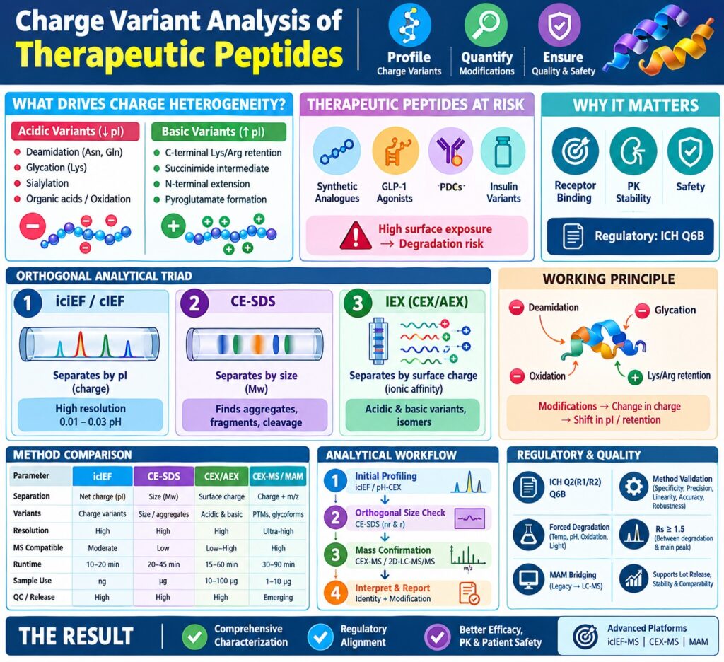
Charge Variant Analysis of Therapeutic Peptides is a critical analytical framework for profiling, quantifying, and monitoring charged proteoforms and post-translational modifications (PTMs) throughout biopharmaceutical development and quality control. This analytical approach assesses chemical and enzymatic modifications—including deamidation, oxidation, C-terminal cleavage, and glycation—that can modify peptide charge distribution and consequently influence bioactivity, pharmacokinetic stability, and safety characteristics. Therapeutic peptides, including synthetic analogues, glucagon-like peptide-1 (GLP-1) receptor agonists, peptide-drug conjugates (PDCs), and recombinant insulin variants, contain highly exposed primary sequences that can be particularly vulnerable to structural degradation during synthesis, downstream purification, and long-term storage. Since even minor changes in molecular surface charge can influence receptor binding kinetics and immunogenic potential, comprehensive charge characterization is an important component of regulatory assessment under International Council for Harmonisation (ICH) Q6B guidelines. A thorough analytical characterization strategy therefore depends on an orthogonal analytical triad comprising Imaged Capillary Isoelectric Focusing (icIEF), Capillary Electrophoresis-Sodium Dodecyl Sulfate (CE-SDS), and Ion-Exchange Chromatography (IEX).
Learn more about GLP-1 Analog Peptide Sequencing Workflows to support your therapeutic peptide development.
Share via:
Quick Summary:
- Charge variant analysis is essential for profiling therapeutic peptide heterogeneity, PTMs, degradation, and product quality throughout development and QC.
- Acidic variants mainly arise from deamidation, glycation, sialylation, oxidation, and organic-acid adducts, while basic variants can result from Lys/Arg retention, succinimide formation, N-terminal extensions, and related processing changes.
- icIEF/cIEF separates peptide variants by isoelectric point (pI) and provides high-resolution detection of small charge differences, making it valuable for charge profiling.
- CE-SDS separates peptides by molecular size, helping distinguish aggregates, fragments, backbone cleavage, and structural variants from true charge modifications.
- IEX (CEX/AEX) separates peptides based on surface charge and electrostatic interactions; salt- and pH-gradient methods support robust charge-variant analysis and preparative isolation.
- An orthogonal workflow combining icIEF, CE-SDS, IEX, and advanced CEX-MS/MAM provides comprehensive characterization of charge variants, size variants, PTMs, and molecular changes.
- Regulatory and quality control require validated, stability-indicating methods aligned with ICH Q2(R1/R2) and Q6B, supported by forced degradation, method bridging, lot release, stability, and comparability testing.
Molecular Drivers of Charge Heterogeneity in Therapeutic Peptides
Charge heterogeneity in therapeutic peptides is primarily driven by spontaneous chemical degradation pathways and enzymatic post-translational modifications that modify net molecular charge (Δq) or the distribution of charge across the molecular surface. These structural changes can shift the analyte’s isoelectric point (pI) and alter its retention behavior on charged stationary phases, resulting in distinguishable acidic or basic charge variants.
Acidic variants are proteoforms that display a lower pI during isoelectric focusing or elute before the principal native peak during cation-exchange chromatography (CEX). The major chemical pathways responsible for the formation of acidic species include:
- Asparagine and Glutamine Deamidation: Non-enzymatic hydrolysis converts uncharged asparagine (Asn) or glutamine (Gln) residues into negatively charged aspartic acid (Asp), iso-aspartic acid (isoAsp), or glutamic acid (Glu). This transformation introduces a negative charge (Δq = -1).
- Glycation: Reducing hexose sugars can become covalently attached to unprotonated lysine (Lys) side chains or N-terminal primary amines. This modification masks positively charged groups and results in a net charge change (Δq = -1).
- Sialylation: Incorporation of terminal N-acetylneuraminic acid residues into glycosylated peptides introduces additional negatively charged carboxylate groups.
- Organic Acid Adducts and Oxidation: Covalent association with residual processing reagents or photo-oxidative modification of sulfur-containing residues can alter the local electrostatic environment of the peptide.
Request our complete Full Characterization Data Package for a GLP-1 Receptor Agonis ANDA to streamline regulatory submissions.
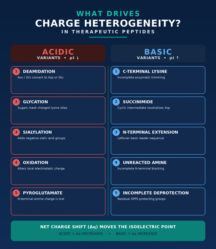
Basic variants are proteoforms characterized by a higher pI or later elution than the main peak during CEX analysis. The principal mechanisms responsible for the formation of basic variants include:
- C-Terminal Basic Residue Retention: Incomplete enzymatic carboxypeptidase processing during recombinant expression can result in the retention of basic lysine or arginine (Arg) residues, producing a positive charge change (Δq = +1).
- Succinimide Intermediate Formation: Dehydration involving aspartic acid or asparagine can generate a cyclic succinimide intermediate. This process neutralizes a negative carboxyl group and can therefore behave as a basic variant relative to Asp.
- N-Terminal Extension and Incomplete Deprotection: Basic leader sequences that remain after processing or incomplete removal of protecting groups during solid-phase peptide synthesis (SPPS) can generate additional basic species.
- N-Terminal Pyroglutamate Formation: Cyclization of N-terminal glutamine or glutamic acid residues removes the primary amine charge and modifies localized dipole interactions.
Ensure full structural accuracy using our specialized Sequence Confirmation Services for Synthetic Therapeutic Peptides.
| Post-Translational Modification / Degradation | Chemical Mechanism | Net Charge Change (Δq) | pI Shift Direction | Variant Classification |
|---|---|---|---|---|
| Asparagine Deamidation | Hydrolysis of Asn to Asp / isoAsp through a succinimide intermediate | -1 | Decrease | Acidic Variant |
| Glutamine Deamidation | Hydrolysis of Gln to Glu | -1 | Decrease | Acidic Variant |
| Glycation | Covalent attachment of reducing sugars to Lys side chains | -1 (loss of protonated amine) | Decrease | Acidic Variant |
| Sialylation | Addition of terminal N-acetylneuraminic acid | -1 per sialic acid residue | Decrease | Acidic Variant |
| C-Terminal Lysine Retention | Incomplete carboxypeptidase processing | +1 per basic residue | Increase | Basic Variant |
| Succinimide Intermediate | Cyclization of Asn/Asp within the peptide backbone | +1 (relative to Asp) | Increase | Basic Variant |
| N-Terminal Pyroglutamate | Cyclization of N-terminal Gln or Glu | -1 (loss of primary amine) | Decrease | Acidic Variant |
| N-Terminal Unreacted Amine | Incomplete N-terminal blocking or processing | +1 | Increase | Basic Variant |
Capillary Isoelectric Focusing (cIEF and icIEF) for Charge Variant Analysis of Therapeutic Peptides
Capillary Isoelectric Focusing (cIEF and icIEF) separates therapeutic peptide charge variants within a continuous pH gradient according to their characteristic isoelectric points (pI), where their net electrical charge reaches zero. The method offers high charge resolution and can distinguish proteoforms with pI differences of approximately 0.01 to 0.03 pH units without depending on chromatographic retention mechanisms.
In conventional cIEF, analytes and carrier ampholytes are introduced into a capillary and exposed to a high-voltage electric field until each peptide migrates to the location corresponding to its characteristic pI (net charge z = 0). After the focusing process, a secondary chemical or pressure-based mobilization step transports the focused zones past a single-point UV detector. However, this mobilization process may contribute to local zone broadening, hydrodynamic distortion, and longer overall analysis times.
Imaged Capillary Isoelectric Focusing (icIEF) avoids the need for mobilization by using a fluorocarbon-coated fused-silica capillary in combination with a charge-coupled device (CCD) camera. The CCD system records whole-column UV absorbance, commonly at 280 nm or 220 nm, in real time. Because the focused peptide zones remain stationary, the approach supports reliable baseline integration and improved reproducibility of peak areas.
The resolving capability (ΔpI) achieved by capillary isoelectric focusing is controlled by the balance between electrophoretic migration and axial thermal diffusion:
ΔpI ∝ √[D · (dpH/dx) / E · (-dz/dpH)]
where D represents the solute diffusion coefficient, dpH/dx describes the pH-gradient slope along the capillary axis, E denotes the applied electric field strength, and dz/dpH represents the charge-regulation capacity, or the slope of the charge-pH relationship, of the peptide near its pI.
Optimization of therapeutic peptide icIEF methods requires appropriate selection of carrier ampholytes, such as broad-range pH 3–10 ampholytes supplemented with narrow-range ampholytes, together with polymeric anti-loss additives such as methylcellulose to suppress electroosmotic flow (EOF) and minimize adsorption to the capillary wall. Calibrated pI peptide markers are also required for reliable pI assignment. More recent use of volatile ampholyte systems and electro-fluidic splitter interfaces has facilitated direct online icIEF-mass spectrometry (icIEF-MS), providing the capability to structurally identify focused charge peaks in real time.
Review our end-to-end Peptide Physicochemical Characterization Services for advanced charge and identity profiling.
Capillary Electrophoresis-Sodium Dodecyl Sulfate (CE-SDS) in Peptide Characterization
Capillary Electrophoresis-Sodium Dodecyl Sulfate (CE-SDS) separates denatured therapeutic peptides according to hydrodynamic size and apparent molecular weight by masking their intrinsic charge through a relatively uniform negative SDS charge density. It provides an important size-based orthogonal method within charge variant characterization, helping differentiate size variants, backbone cleavage products, and aggregates from modifications that genuinely alter molecular charge.
For CE-SDS analysis, peptide samples are thermally denatured in the presence of excess sodium dodecyl sulfate at elevated temperatures ranging from 70 °C to 90 °C. Analysis can be performed under non-reducing (nrCE-SDS) or reducing (rCE-SDS) conditions, with reducing conditions typically using dithiothreitol or 2-mercaptoethanol. SDS molecules associate non-covalently with hydrophobic regions of the peptide at an approximately constant mass ratio, producing denatured complexes with a relatively uniform mass-to-charge relationship. These complexes migrate through a replaceable hydrophilic gel polymer matrix, such as polyacrylamide, dextran, or agarose-based networks, contained within a narrow-bore capillary under a constant electric field. Electrophoretic mobility (μep) through the sieving matrix is inversely associated with the logarithm of molecular weight (Mw):
μep ∝ 1 / log(Mw)
Non-reducing CE-SDS is used to assess the structural integrity of intact peptide therapeutics and can resolve non-covalently associated fragments, disulfide-linked homodimers, and higher-order multimeric aggregates. Reducing CE-SDS disrupts inter-chain disulfide bridges, allowing individual subunits to be quantified and facilitating more precise assessment of backbone cleavage sites. Coupling CE-SDS with Laser-Induced Fluorescence (LIF) detection through pre-column fluorophore derivatization, for example using 5-carboxytetramethylrhodamine succinimidyl ester, can achieve limits of detection (LOD) in the low nanogram-per-milliliter range. This sensitivity makes the technique particularly useful for formulations containing low concentrations of therapeutic peptides.
Prevent stability loss by leveraging our dedicated Peptide Aggregation Analysis workflows.
Ion-Exchange Chromatography (CEX and AEX) Elution Strategies
Ion-Exchange Chromatography (IEX), particularly Cation-Exchange Chromatography (CEX), separates therapeutic peptide charge variants according to surface charge density and their electrostatic interactions with charged stationary-phase ligands. The technique is widely used for preparative isolation of variants, lot release testing, and direct coupling with native mass spectrometry.
Stationary phases used for peptide charge separations generally contain rigid, non-porous or small-pore polymeric particles in the 3–10 μm range. These particles may be functionalized with strong acidic groups, such as sulfonate/sulfopropyl groups used in strong cation exchange (SCX), or weak acidic groups, such as carboxylates used in weak cation exchange (WCX). Non-porous stationary phases reduce limitations associated with intra-particle diffusion, thereby producing narrow chromatographic peaks and high peak capacities for samples containing complex charge distributions.
Two principal elution approaches are used in ion-exchange separations:
- Salt-Gradient Elution: The mobile phase is maintained at a relatively constant pH while ionic strength is progressively increased. For example, a gradient from 0 to 500 mM NaCl or KCl can be applied in 20 mM sodium phosphate or MES buffer. The increasing concentration of counterions competes for charged sites on the stationary phase and sequentially displaces peptides according to their net surface charge density. Salt-gradient methods generally provide strong analytical robustness, although fractions often require offline desalting before mass spectrometry characterization.
- pH-Gradient Elution: A linear pH gradient is generated using multiple buffer components across the pI range of the target peptide. As the pH of the mobile phase approaches or exceeds the peptide’s pI, its net positive charge progressively approaches zero, resulting in rapid elution from the cation-exchange stationary phase. pH-gradient methods can provide enhanced resolution when analyzing structural proteoforms with only minor differences in surface charge.
The development of volatile buffer systems based on ammonium acetate, ammonium formate, or triethylammonium carbonate has made direct online coupling between CEX and native mass spectrometry possible. CEX-MS enables real-time structural characterization of acidic and basic variants while avoiding multiple fraction-collection steps and manual peptide-mapping workflows.
Learn about high-resolution mass profiling with Native Mass Spectrometry for Therapeutic Peptide Characterization.
Method Comparison and Orthogonal Strategies for Charge Variant Analysis of Therapeutic Peptides
A comprehensive orthogonal strategy incorporating icIEF, CE-SDS, and IEX is necessary for complete characterization of therapeutic peptides because no individual analytical technique can independently resolve every charge variant and size-related modification. Combining these analytical modes provides broader coverage of charge heterogeneity, aggregation, fragmentation, and site-specific post-translational modifications.
| Parameter | Imaged Capillary Isoelectric Focusing (icIEF) | Capillary Electrophoresis-Sodium Dodecyl Sulfate (CE-SDS) | Ion-Exchange Chromatography (CEX/AEX) | On-Line Native CEX-MS / Multi-Attribute Monitoring (MAM) |
|---|---|---|---|---|
| Separation Principle | Net charge in a dynamic pH gradient (pI) | Hydrodynamic size / apparent mass (Mw) | Surface charge density and electrostatic affinity | Chromatographic surface charge combined with gas-phase m/z |
| Primary Variant Resolved | Charge variants with pI shifts down to 0.01 | Size variants, aggregates, fragments, and backbone clips | Acidic and basic charge variants and isomers | Site-specific PTMs, glycoforms, and point mutations |
| Resolving Power | High (pI-based separation) | High (mass-based sieving resolution) | High (surface dipole and charge distribution) | Ultra-high (orthogonal chromatographic and MS resolution) |
| MS Compatibility | Moderate (requires volatile ampholytes/interfaces) | Low (incompatible with SDS surfactant without removal) | Low with salt gradients to high with volatile pH gradients | Direct high-resolution online coupling |
| Analysis Runtime | 10–20 minutes per sample | 20–45 minutes per sample | 15–60 minutes per sample | 30–90 minutes per sample |
| Sample Consumption | Very low (approximately nanograms) | Low (approximately micrograms) | Moderate (approximately 10–100 μg) | Low to moderate (approximately 1–10 μg) |
| QC & Release Suitability | High (widely adopted QA/QC platform) | High (standard lot release method for purity) | High (gold-standard profile release assay) | Emerging (rapidly growing for commercial CQA control) |
Implementation of a comprehensive testing workflow can be organized into several targeted analytical stages:
- Initial Profiling: Samples are first assessed using icIEF or pH-gradient CEX to establish the baseline charge variant distribution and determine the relative percentage of individual peaks.
- Orthogonal Size Assessment: When new or progressively increasing pre-peaks or post-peaks appear during stability studies, CE-SDS is performed under both non-reducing and reducing conditions. This helps establish whether the observed variant results from a charge modification, backbone cleavage, or aggregation.
- Mass Confirmation and Characterization: When peak identities must be established, volatile CEX-MS or heart-cutting 2D-LC-MS/MS, involving SCX coupled with Reversed-Phase LC-MS, can be used to determine intact molecular masses and localize post-translational modifications to specific amino acid residues.
Discover specialized techniques for complex architectures via Cyclic Peptide Characterization and explore CD Spectroscopy for Peptide Secondary Structure Characterization.
Regulatory Compliance, Method Validation, and Quality Control Integration
Regulatory compliance for charge variant analysis of therapeutic peptides requires appropriate method validation in accordance with International Council for Harmonisation (ICH) Q2(R1/R2) and Q6B guidelines. Validation establishes that analytical procedures demonstrate suitable specificity, precision, linearity, accuracy, and robustness. These characteristics are essential for reliable monitoring of critical quality attributes (CQAs) during lot release, stability studies, and process comparability assessments.
The stability-indicating capability of an analytical method is typically established through forced degradation studies. During these studies, peptide samples are exposed to controlled stress conditions, including elevated temperature, acidic and basic pH conditions for hydrolysis, oxidative stress using H2O2, and photo-exposure. Chromatographic and electrophoretic methods should provide sufficient separation between degradation products and the intact active therapeutic peak, with a minimum resolution factor of Rs ≥ 1.5.
As products progress toward late-stage commercialization, laboratories may transition from legacy profile-based approaches, such as offline CEX peak-area integration, to automated Multi-Attribute Monitoring (MAM) based on high-resolution LC-MS. Analytical method bridging studies are used to establish equivalency between the legacy and newer platforms. These studies demonstrate that MAM can monitor relevant CQAs with sensitivity and precision comparable to or greater than established gel-based and chromatographic release assays.
Modernize your quality control with Multi-Attribute Monitoring (MAM) for Peptide Characterization.
Conclusion
Charge Variant Analysis of Therapeutic Peptides represents an essential element of biopharmaceutical development because it supports the control of therapeutic efficacy, pharmacokinetic consistency, and patient safety. An orthogonal analytical strategy incorporating icIEF, CE-SDS, and volatile CEX-MS enables researchers and quality control specialists to characterize complex proteoforms comprehensively while maintaining alignment with stringent regulatory expectations.
As biopharmaceutical pipelines increasingly incorporate sophisticated engineered peptide architectures, conventional single-dimension profiling approaches are being complemented by advanced hyphenated platforms, including online icIEF-MS, volatile CEX-MS, and Multi-Attribute Monitoring. These multidimensional analytical strategies can simplify method development, reduce lot release timelines, and provide enhanced molecular-level insight throughout the drug development lifecycle.
🔗 Complete Quality Checklist: Download our essential Peptide Characterization CRO Deliverables Checklist to guide your next study.
For specialized analytical characterization, regulatory-compliant method development, and advanced charge variant profiling services, contact our technical team at ResolveMass Laboratories Contact Page.
Frequently Asked Questions
CE-SDS provides an orthogonal size-based assessment because charge-based methods may not always distinguish fragmentation or aggregation from genuine charge modifications. SDS denatures the peptide and masks its intrinsic charge, allowing separation according to hydrodynamic size. This helps determine whether an altered analytical peak represents a true charge variant, backbone cleavage product, or aggregate.
Deamidation converts an uncharged asparagine or glutamine residue into aspartic acid or glutamic acid, introducing a negative charge (Δq = -1). The resulting decrease in positive surface charge weakens the interaction between the peptide and the CEX stationary phase. Consequently, deamidated species generally elute before the principal peptide peak and appear as acidic variants.
Direct icIEF-MS coupling is complicated by carrier ampholytes, non-volatile salts, and polymeric additives such as methylcellulose used during icIEF analysis. These components can suppress electrospray ionization (ESI) and contribute to contamination of the MS ion source. Volatile ampholytes, specialized microfluidic interfaces, or online heart-cutting two-dimensional liquid chromatography approaches can help overcome these limitations.
Acidic charge variants have a lower net positive charge or greater negative character than the main peptide species, generally resulting in lower pI values and earlier CEX elution. Basic variants exhibit relatively greater positive charge and therefore tend to have higher pI values. In CEX analysis, these basic species commonly demonstrate longer retention than the principal peptide peak.
Native CEX-MS enables charge variants to be analyzed directly as they emerge from the chromatographic column using MS-compatible volatile buffers such as ammonium acetate. This approach reduces the need for fraction collection, concentration, desalting, and subsequent sample preparation. As a result, it can minimize handling-related artifacts while accelerating mass confirmation and structural characterization.
Carrier ampholytes establish the continuous pH gradient required for peptide focusing when an electric field is applied during cIEF. For peptides with very low or high pI values, broad-range mixtures such as pH 3–10 may provide insufficient local resolution. Narrow-range ampholytes or suitable synthetic chemical modifiers can therefore be incorporated to refine the gradient and improve separation around the target pI.
Non-reducing CE-SDS preserves covalent disulfide linkages while disrupting non-covalent interactions through denaturation. Non-covalent aggregates can dissociate into monomeric species, whereas intermolecular disulfide-linked dimers and higher-order covalent aggregates remain associated. Because these species have greater apparent molecular size, they migrate more slowly than the monomeric peptide.
Multi-Attribute Monitoring (MAM) using LC-MS/MS offers site-specific measurement of post-translational modifications and provides molecular information that profile-based IEX cannot directly deliver. However, complete replacement of IEX requires extensive validation, analytical bridging, and demonstrated suitability for the relevant CQAs. Complementary techniques may still be necessary for charge variants, aggregates, and conformationally distinct species.
Trifluoroacetic acid (TFA) is frequently used during synthetic peptide purification and can remain associated with positively charged residues such as lysine, arginine, and histidine. Residual TFA may partially shield these positive charges and consequently alter CEX retention behavior. Counterion exchange, such as replacing TFA with acetate or chloride, can improve profile consistency before charge variant characterization.
Reference:
- Shah, A., Cui, W., Harrahy, J., & Ivanov, A. R. (2024). Characterization of charge variants, including post-translational modifications and proteoforms, of bispecific antigen-binding protein by cation-exchange chromatography coupled to native mass spectrometry. Talanta, 266(Pt. 1), 125062. https://doi.org/10.1016/j.talanta.2023.125062
- Cao, M., De Mel, N., Shannon, A., Prophet, M., Wang, C., Xu, W., Niu, B., Kim, J., Albarghouthi, M., Liu, D., Meinke, E., Lin, S., Wang, X., & Wang, J. (2019). Charge variants characterization and release assay development for co-formulated antibodies as a combination therapy. mAbs, 11(3), 489–499. https://doi.org/10.1080/19420862.2019.1578137
- Schairer, J., Höchsmann, A., Bauer, B., Höfer, V., Römer, J., & Neusüß, C. (2025). Ion-exchange chromatography, capillary isoelectric focusing, and capillary zone electrophoresis coupled to mass spectrometry for charge variant analysis of monoclonal antibodies. mAbs, 17(1), 2537116. https://doi.org/10.1080/19420862.2025.2537116
- Shi, R. L., Xiao, G., Dillon, T. M., Ricci, M. S., & Bondarenko, P. V. (2020). Characterization of therapeutic proteins by cation exchange chromatography-mass spectrometry and top-down analysis. mAbs, 12(1), 1739825. https://doi.org/10.1080/19420862.2020.1739825
- Schlecht, J., Moritz, B., Kiessig, S., & Neusüß, C. (2023). Characterization of therapeutic mAb charge heterogeneity by iCIEF coupled to mass spectrometry (iCIEF–MS). Electrophoresis, 44(5–6), 540–548. https://doi.org/10.1002/elps.202200170
- Wagner, E., Colas, O., Chenu, S., Goyon, A., Murisier, A., Cianferani, S., François, Y., Fekete, S., Guillarme, D., D’Atri, V., & Beck, A. (2020). Determination of size variants by CE-SDS for approved therapeutic antibodies: Key implications of subclasses and light chain specificities. Journal of Pharmaceutical and Biomedical Analysis, 184, 113166. DOI
Get In Touch With Us
About The Author


