

Introduction
De Novo Peptide Sequencing by Tandem Mass Spectrometry is a sophisticated analytical proteomics technique used to determine the precise primary amino acid sequence of a peptide directly from tandem mass spectrometry data, without depending on existing genomic or protein sequence databases. In contrast to database-driven search approaches that compare experimental spectra with theoretical spectra generated from known protein libraries, de novo sequencing interprets the exact mass differences between consecutive fragment ions produced during peptide backbone fragmentation. As a result, De Novo Peptide Sequencing by Tandem Mass Spectrometry serves as a core analytical methodology for identifying previously uncharacterized proteins, characterizing highly variable biotherapeutic antibody regions, studying non-model organisms, and mapping novel post-translational modifications (PTMs).
The transition from conventional Edman degradation to liquid chromatography coupled with tandem mass spectrometry (LC-MS/MS) has transformed the analysis of primary protein structures. Edman degradation requires highly purified peptides with accessible N-termini and is inherently limited in throughput. In contrast, contemporary high-resolution tandem mass spectrometers achieve sub-parts-per-million (ppm) mass accuracy and can acquire thousands of fragmentation spectra every hour from highly complex biological samples.
The ability to determine primary protein structures directly from raw mass spectral data is essential in biopharmaceutical development, structural proteomics, and biomarker research. In situations where genomic reference databases are inadequate because of somatic hypermutation, incomplete genome annotation, non-ribosomal peptide biosynthesis, or extensive post-translational processing, de novo sequencing provides a direct, data-driven, and highly reliable route for sequence determination.
Need high-precision structural verification for GLP-1 therapeutics? Explore our detailed guide on Peptide Sequencing of GLP-1 Drugs for advanced LC-MS/MS sequence validation.
Article Summary:
- De Novo Peptide Sequencing determines the amino acid sequence of peptides directly from LC-MS/MS data without relying on protein or genomic databases, making it ideal for unknown or novel proteins.
- It is essential for analyzing non-model organisms, monoclonal antibodies, endogenous peptides, venoms, non-ribosomal peptides, and complex post-translational modifications (PTMs) where database searching is insufficient.
- The technique identifies peptide sequences by interpreting fragment ion series (b, y, a, c, z, d, and w ions) generated during tandem mass spectrometry and measuring mass differences between adjacent fragments.
- Modern workflows combine high-resolution LC-MS/MS, multiple enzyme digestion, complementary fragmentation methods (HCD, CID, ETD, ECD, EThcD), and advanced computational analysis to maximize sequence accuracy and coverage.
- Transformer-based AI models such as Casanovo and InstaNovo have significantly improved de novo sequencing by learning complex fragmentation patterns, providing higher accuracy than traditional graph-based algorithms.
- Key analytical challenges include missing fragment ions, leucine/isoleucine differentiation, near-isobaric amino acids, and chimeric spectra, which require advanced fragmentation strategies and computational deconvolution.
- De novo peptide sequencing plays a critical role in biopharmaceutical development, antibody characterization, structural proteomics, biomarker discovery, and regulatory sequence verification by enabling reliable identification of previously unknown peptide and protein sequences.
Primary Technical Triggers: When Is De Novo Peptide Sequencing by Tandem Mass Spectrometry Required?
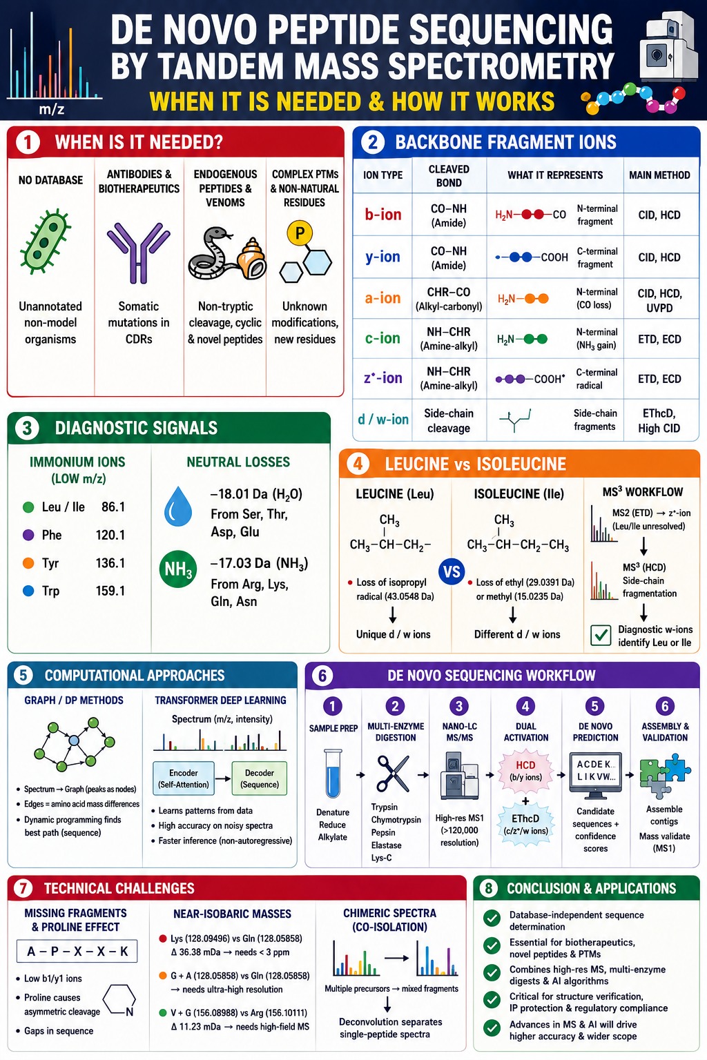
De Novo Peptide Sequencing by Tandem Mass Spectrometry becomes necessary whenever reference proteome databases are unavailable, incomplete, or inherently incapable of predicting the target peptide sequence from genomic information. It functions as a critical analytical solution when conventional bioinformatic search strategies cannot provide a reliable sequence assignment.
| Analytical Scenario | Primary Bottleneck in Database Searching | Technical Rationale for De Novo Sequencing |
|---|---|---|
| Unannotated & Non-Model Organisms | Complete absence of sequenced genomes or proteomes in public repositories (e.g., NCBI, UniProt). | Direct determination of amino acid sequences from spectral mass intervals without relying on database-derived assumptions. |
| Monoclonal Antibodies & Biotherapeutics | Random somatic hypermutations within Complementarity-Determining Regions (CDRs). | Accurate mapping of hypervariable antigen-binding loops, engineered variants, and non-germline mutations. |
| Endogenous Peptides & Venom Profiling | Non-tryptic proteolytic processing, unannotated pro-peptides, and cyclic modifications. | Characterization of unconstrained cleavage patterns, cyclic peptides, and previously unknown toxin structures. |
| Complex PTM Mapping & Non-Natural Residues | Exponential expansion of search space resulting in elevated false-discovery rates. | Direct localization and interpretation of delta-mass shifts on fragment ions without predefined modification settings. |
Unsequenced Non-Model Organisms and Metaproteomics
Proteomes derived from unsequenced non-model organisms lack comprehensive protein databases, making traditional search engines such as SEQUEST, Mascot, and Andromeda ineffective. During the analysis of newly identified environmental microorganisms, agricultural species, or other non-model organisms, database-based matching fails because the corresponding protein sequences are absent from available repositories. De novo sequencing overcomes this limitation by deriving amino acid sequences directly from the observed mass differences between fragment ions in the tandem mass spectrum.
Monoclonal Antibody and Fab Region Characterization
Monoclonal antibodies are generated through somatic recombination (V(D)J gene rearrangement) and undergo random somatic hypermutation within B cells, creating highly diverse sequences within their Complementarity-Determining Regions (CDRs). Since these hypervariable regions frequently differ from germline DNA templates, conventional database searches cannot accurately reconstruct the authentic amino acid sequence of isolated antibodies or Fab fragments. De novo sequencing provides a robust approach for characterizing recombinant antibody therapeutics, patient-derived immunoglobulins, and engineered antibody variants with high precision.
Endogenous Peptides, Venom Peptidomics, and Non-Ribosomal Peptides
Endogenous peptides, neuropeptides, animal venoms originating from organisms such as snakes, cone snails, and amphibians, as well as bacterial non-ribosomal peptides (NRPs), often do not follow standard tryptic digestion patterns. These biomolecules commonly contain non-canonical amino acids, D-amino acids, C-terminal amidation, cyclic structures, or other uncommon features. De novo sequencing methodologies can accommodate non-tryptic fragments and unrestricted cleavage patterns, enabling detailed structural characterization of these biologically active compounds.
Analyzing complex synthetic or cyclic peptide modalities? Discover specialized solutions for Cyclic Peptide Characterization designed to handle non-canonical structures and unconstrained modifications.
Identification of Complex, Unannotated Post-Translational Modifications
Previously uncharacterized post-translational modifications introduce unexpected mass shifts that can confound traditional database search algorithms. Although database search tools can evaluate a limited number of user-specified variable modifications, such as methionine oxidation or serine phosphorylation, the inclusion of numerous unknown modifications dramatically expands the search space and increases the likelihood of false-positive identifications. De novo sequencing addresses this challenge by directly detecting and localizing mass shifts on specific fragment ions without requiring predefined PTM parameters.
Tackling low-level peptide impurities and degradants? Check out our overview of GLP-1 Peptide Impurity Characterization for accurate delta-mass localization.
Biophysical Principles and Backbone Fragmentation Pathways of De Novo Peptide Sequencing by Tandem Mass Spectrometry
The underlying biophysical basis of De Novo Peptide Sequencing by Tandem Mass Spectrometry involves the migration of mobile protons along the peptide backbone, resulting in gas-phase covalent bond cleavage and the generation of distinct N-terminal and C-terminal fragment ion series. By measuring the mass-to-charge (m/z) differences between adjacent fragment ions within a given series, the identities of individual amino acid residues can be determined.
Proton Mobility and Backbone Cleavage Mechanics
Gas-phase peptide fragmentation is fundamentally driven by proton mobility within the mass spectrometer. Under positive electrospray ionization (ESI) conditions, protons introduced during ionization migrate along the peptide backbone and transiently protonate nitrogen atoms or carbonyl oxygen atoms. This localized protonation weakens neighboring backbone bonds, facilitating bond cleavage when activation energy is applied.
Fragment Ion Series Classification
The cleavage of specific peptide backbone bonds produces complementary N-terminal and C-terminal fragment ion series. According to the established nomenclature proposed by Roepstorff, Fohlman, and Biemann, fragment ions are classified based on the exact bond that undergoes cleavage:
- Alkyl-carbonyl bond cleavage (CHR–CO): Produces a-type N-terminal ions and x-type C-terminal ions.
- Peptide amide bond cleavage (CO–NH): Produces b-type N-terminal ions and y-type C-terminal ions.
- Amine-alkyl bond cleavage (NH–CHR): Produces c-type N-terminal ions and z-type C-terminal ions.
| Fragment Ion Type | Cleaved Backbone Bond | Formula Mass Calculation | Dominant Activation Mode | Diagnostic Characteristics |
|---|---|---|---|---|
| b-ion | CO–NH (Amide) | Σ(Residue Masses) + 1.0078 Da [H+] | CID, HCD | N-terminally charged ions that commonly undergo CO neutral loss to form a-ions. |
| y-ion | CO–NH (Amide) | Σ(Residue Masses) + 19.0184 Da [H2O + H+] | CID, HCD | C-terminally charged ions that are highly stable and abundant in tryptic peptide spectra. |
| a-ion | CHR–CO (Alkyl-carbonyl) | Mass(bn) − 27.9949 Da [CO] | CID, HCD, UVPD | N-terminal fragment ions useful for confirming low-mass b2/a2 ion pairs. |
| c-ion | NH–CHR (Amine-alkyl) | Σ(Residue Masses) + 18.0338 Da [NH3 + H+] | ETD, ECD | N-terminal fragment ions that preserve labile post-translational modifications. |
| z•-ion | NH–CHR (Amine-alkyl) | Σ(Residue Masses) + 2.0151 Da [H+ − NH2] | ETD, ECD | C-terminal radical ions capable of undergoing secondary side-chain fragmentation. |
| d-ion / w-ion | Side-chain Cα–Cβ / Cβ–Cγ cleavage | Variable and residue-specific | High-Energy CID, EThcD | Side-chain fragments particularly useful for distinguishing leucine from isoleucine. |
Dissociation Activation Energies
Different fragmentation techniques utilize distinct physical mechanisms to induce peptide backbone cleavage:
- Collision-Induced Dissociation (CID) and Higher-Energy C-trap Dissociation (HCD): These techniques rely on low-energy vibrational activation, which gradually heats the peptide and preferentially cleaves amide bonds (CO–NH), generating complementary b-ion and y-ion series. HCD applies higher collision energies within a dedicated collision cell and often produces abundant low-mass immonium ions in addition to extensive b/y fragmentation.
- Electron-Transfer Dissociation (ETD) and Electron-Capture Dissociation (ECD): These non-ergodic fragmentation methods transfer or capture an electron within multiply charged peptide ions, generating reactive radical intermediates that preferentially cleave N–Cα backbone bonds. This process produces c-ions and z• radical ions while preserving fragile PTMs such as phosphorylation and glycosylation.
Diagnostic Immonium Ions and Neutral Loss Transitions
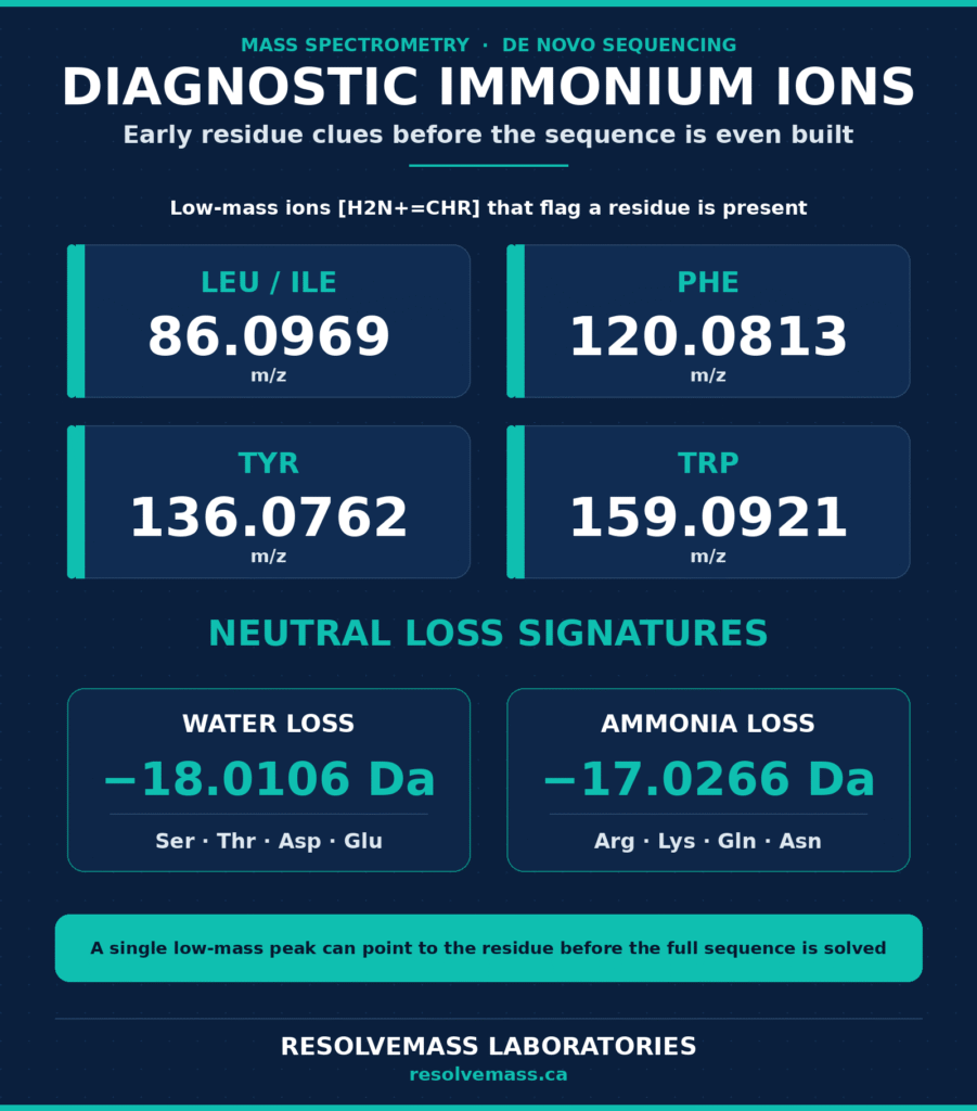
The low-mass region of tandem mass spectra frequently contains diagnostic immonium ions [H2N+=CHR] that provide early evidence for the presence of specific amino acid residues before full sequence reconstruction is completed. Commonly observed immonium ions include those associated with Leucine/Isoleucine (m/z 86.0969), Phenylalanine (m/z 120.0813), Tyrosine (m/z 136.0762), and Tryptophan (m/z 159.0921).
In addition, characteristic neutral loss events occur during peptide fragmentation. Examples include water loss (−18.01056 Da) from residues such as Ser, Thr, Asp, and Glu, and ammonia loss (−17.02655 Da) from residues such as Arg, Lys, Gln, and Asn. These fragmentation patterns provide valuable supplementary information during de novo sequence interpretation.
Evaluating non-covalent complexes or higher-order structures? Learn how Native Mass Spectrometry Services maintain structural integrity during intact mass analysis.
Resolving Isobaric Mass Ambiguities in De Novo Peptide Sequencing by Tandem Mass Spectrometry
Distinguishing between the isobaric amino acids leucine and isoleucine (113.08406 Da) during De Novo Peptide Sequencing by Tandem Mass Spectrometry requires additional side-chain fragmentation information generated through techniques such as EThcD or high-energy CID. These methods produce characteristic d-ions and w-ions that reflect side-chain structure and allow differentiation between the two residues. Conventional tandem MS/MS (MS2) experiments primarily induce backbone cleavage and therefore cannot distinguish leucine from isoleucine based solely on precursor or backbone fragment masses.
Chemistry of Satellite d-Ion and w-Ion Generation
The differentiation of Leucine and Isoleucine relies on inducing fragmentation within their respective aliphatic side chains. When additional collisional energy or radical-mediated activation techniques such as EThcD are applied, cleavage occurs at the Cβ–Cγ bond, generating characteristic satellite ions that reveal side-chain architecture.
- Leucine Side Chain: Leucine possesses an isopropyl group attached to the Cβ carbon. During secondary radical-driven fragmentation of an N-terminal a•-ion or a C-terminal z•-ion, homolytic cleavage results in the loss of an isopropyl radical (•CH(CH3)2, 43.05477 Da), producing a characteristic d-ion or w-ion signature.
- Isoleucine Side Chain: Isoleucine contains a sec-butyl side chain featuring both methyl and ethyl substituents attached to the Cβ carbon. Secondary fragmentation leads to the loss of either an ethyl radical (•CH2CH3, 29.03912 Da) or a methyl radical (•CH3, 15.02347 Da), generating satellite ions with masses that differ from those observed for Leucine.
Targeted Multi-Stage Activation (MS3)
Modern Orbitrap tribrid mass spectrometry platforms employ targeted multi-stage mass spectrometry (MS3) workflows to automatically resolve Leucine/Isoleucine ambiguities. In these approaches, an initial MS2 ETD experiment generates a radical z-ion that contains an unresolved Leu/Ile residue at the N-terminus. This z-ion is subsequently isolated and subjected to higher-energy HCD fragmentation during the MS3 stage. The additional activation promotes Cβ–Cγ side-chain cleavage and produces diagnostic w-ions, enabling definitive differentiation between Leucine and Isoleucine without requiring chemical derivatization or conventional Edman degradation.
Review this comprehensive GLP-1 Analog Peptide Sequencing Workflow to see how multi-stage activation is integrated into routine analysis.
Computational Architectures: From Graph Algorithms to Transformer Deep Learning in De Novo Peptide Sequencing by Tandem Mass Spectrometry
Computational de novo sequencing interprets highly complex tandem mass spectra using either graph-based dynamic programming methods or advanced deep learning transformer architectures that approach spectrum interpretation as a neural machine translation problem. Continuous advancements in computational methodologies have significantly enhanced sequence reconstruction accuracy and enabled automated analysis of increasingly complex datasets.
Classical Graph Theory and Dynamic Programming
Traditional de novo sequencing algorithms such as PEAKS, Novor, pNovo, and Lutefisk convert tandem mass spectra into Directed Acyclic Graphs (DAGs). Spectral peaks that exceed a predefined signal-to-noise threshold are represented as graph nodes, while directed edges connect nodes whose mass differences correspond to the monoisotopic masses of canonical amino acids within a specified mass tolerance window. Dynamic programming algorithms then evaluate all feasible paths through the graph and identify the peptide sequence that most effectively explains the observed fragmentation pattern, including b-ion and y-ion series, while remaining consistent with the precursor ion mass.
Transformer Neural Networks and Sequence-to-Sequence Translation
Contemporary de novo sequencing platforms increasingly utilize Transformer-based deep learning architectures, reformulating peptide sequencing as a sequence-to-sequence translation task. Solutions such as Casanovo, InstaNovo, and π-PrimeNovo convert spectral peak information directly into predicted amino acid sequences.
- Continuous Peak Embedding: Fourier feature-based encoders transform continuous m/z and intensity measurements into high-dimensional vector representations, eliminating the need for coarse m/z binning and preserving detailed spectral information.
- Self-Attention and Contextual Encoders: Transformer encoder layers employ multi-head self-attention mechanisms to analyze contextual relationships among all spectral peaks simultaneously. This enables the recognition of complementary b/y ion pairs, neutral loss events, and long-range spectral dependencies without relying on rigid scoring rules.
- Autoregressive and Non-Autoregressive Decoders: Autoregressive models such as Casanovo generate peptide sequences incrementally using beam-search strategies, while non-autoregressive frameworks such as π-PrimeNovo predict all residue positions simultaneously, substantially reducing inference time during large-scale proteomics workflows.
| Feature / Metric | Classical Graph/DP (PEAKS, Novor, pNovo) | Deep Learning / Transformers (Casanovo, InstaNovo) |
|---|---|---|
| Core Computational Mechanism | Directed Acyclic Graphs (DAGs) combined with Dynamic Programming. | Encoder-decoder Transformer architectures utilizing self-attention mechanisms. |
| Spectral Data Representation | Discretized peak lists matched against theoretical amino acid mass transitions. | Continuous m/z and intensity embeddings generated through Fourier feature projections. |
| Fragmentation Rule Dependency | High dependence on predefined scoring rules for b-, y-, and a-ion series. | Lower dependence on explicit rules; learns spectral relationships directly from training data. |
| Sequencing Accuracy | Moderate performance and increased sensitivity to missing fragment ions. | High accuracy, even when analyzing noisy or partially fragmented spectra. |
| Computational Overhead | Relatively low memory requirements and efficient single-thread performance. | Requires GPU resources for training but supports highly parallelized inference. |
Explore Multi-Attribute Monitoring (MAM) Services for streamlined quality control and sequence confirmation.
Methodological Workflow for De Novo Peptide Sequencing by Tandem Mass Spectrometry
A comprehensive de novo sequencing workflow combines orthogonal enzymatic digestion strategies, high-resolution LC-MS/MS analysis, complementary fragmentation methods, automated computational prediction, and contig-level sequence assembly. Careful execution of each stage maximizes sequence coverage and minimizes gaps within the reconstructed protein sequence.
1. Sample Purification and Reduction/Alkylation
Purified proteins are first denatured using reagents such as 2% sodium deoxycholate or 8 M urea to disrupt higher-order structures. Disulfide bonds are then reduced using reagents such as TCEP or DTT, followed by alkylation with iodoacetamide or iodoacetic acid to permanently cap free cysteine residues and prevent disulfide bond reformation.
2. Orthogonal Multi-Enzyme Digestion
To minimize sequence gaps associated with single-enzyme specificity, multiple parallel digestion strategies are employed using proteases with distinct cleavage preferences:
- Trypsin: Cleaves on the C-terminal side of Lysine and Arginine residues.
- Chymotrypsin: Cleaves preferentially on the C-terminal side of aromatic amino acids such as Tyrosine, Phenylalanine, and Tryptophan, as well as Leucine.
- Pepsin: Performs relatively non-specific cleavage under acidic conditions and is particularly useful for densely folded protein regions.
- Elastase and Lys-C: Generate complementary overlapping peptide fragments that improve coverage across variable and structurally constrained regions.
3. High-Resolution Nano-LC-MS/MS Analysis
The resulting peptide mixtures are separated using reverse-phase nano-UHPLC and analyzed on high-resolution mass spectrometry platforms. Precursor ion scans (MS1) are acquired at resolving powers exceeding 120,000, providing sub-ppm mass accuracy and highly accurate intact mass measurements.
4. Dual Activation Data-Dependent Acquisition
Selected precursor ions are fragmented using complementary activation approaches. HCD generates abundant b-ion and y-ion series, whereas EThcD provides c-ion and z•-ion series together with satellite w-ions that facilitate Leucine/Isoleucine differentiation.
5. De Novo Prediction and Positional Scoring
Acquired spectra are processed using specialized de novo sequencing software to generate candidate amino acid sequences. Positional confidence scores are calculated for each residue, providing quantitative measures of sequence reliability.
6. Contig Sequence Assembly and Mass Validation
Overlapping peptide sequences are assembled into continuous full-length protein contigs using bioinformatics platforms such as Stitch. The reconstructed sequence is subsequently validated by comparing its theoretical intact molecular mass with experimentally measured MS1 proteoform masses.
Access the Peptide Characterization CRO Deliverables Checklist to ensure complete coverage across all regulatory data requirements.
Technical Limitations and Data Deconvolution Challenges in De Novo Peptide Sequencing by Tandem Mass Spectrometry
Despite substantial technological advancements, de novo sequencing remains affected by challenges including incomplete fragmentation, near-isobaric residue masses, and precursor co-isolation events that generate chimeric spectra. Addressing these issues requires both experimental optimization and advanced computational processing.
Missing Fragment Ions and the Proline Effect
Incomplete peptide fragmentation can create sequence gaps when specific backbone bonds fail to undergo cleavage. Low-mass b1 and y1 ions are often absent because of intrinsic ion instability or instrumental low-mass transmission limitations. Additionally, peptides containing Proline exhibit highly asymmetric fragmentation behavior. The rigid cyclic structure of Proline promotes cleavage at N-terminal amide bonds while suppressing fragmentation at neighboring positions. As a result, de novo sequencing algorithms may be forced to interpret combined mass differences corresponding to multiple unresolved amino acid residues.
Assessing secondary structure or aggregation tendencies? Read more about CD Spectroscopy for Peptide Secondary Structure
and Peptide Aggregation Analysis to mitigate structural analytical bottlenecks.
Near-Isobaric Mass Coincidences
In addition to the true isomeric relationship between Leucine and Isoleucine, several amino acid residues and residue combinations exhibit extremely similar monoisotopic masses:
- Lysine (128.09496 Da) vs. Glutamine (128.05858 Da): These residues differ by only 36.38 mDa. Reliable differentiation requires mass accuracy better than 3 ppm or the use of chemical derivatization techniques such as amine dimethylation.
- Glycine-Alanine (G+A = 128.05858 Da) vs. Glutamine (Q): In the absence of an intermediate fragment ion, a Glycine-Alanine dipeptide can become indistinguishable from a single Glutamine residue unless ultra-high-resolution instrumentation is employed.
- Valine-Glycine (V+G = 156.08988 Da) vs. Arginine (R = 156.10111 Da): These masses differ by only 11.23 mDa, necessitating high-field Orbitrap instrumentation and exceptional mass accuracy for confident discrimination.
Chimeric Spectra and Precursor Co-Isolation
When multiple precursor ions possessing similar m/z values are simultaneously isolated within a quadrupole selection window, the resulting tandem mass spectrum contains overlapping fragment ions originating from multiple peptides. These mixed spectra, known as chimeric spectra, complicate sequence interpretation because conventional de novo algorithms generally assume that all fragments originate from a single precursor ion. Advanced spectral deconvolution approaches address this problem by identifying complementary b/y ion relationships and computationally separating mixed spectra into distinct single-peptide fragmentation datasets before sequence reconstruction.
Need assistance navigating IND/NDA requirements? Review the Regulatory Requirements for GLP-1 Peptide Characterization to ensure full compliance for drug substance and drug product specifications.
Conclusion and Strategic Applications of De Novo Peptide Sequencing by Tandem Mass Spectrometry
De Novo Peptide Sequencing by Tandem Mass Spectrometry serves as a powerful and database-independent analytical approach for deciphering complex biological sequences in both proteomics research and biopharmaceutical development. By integrating high-resolution dual-activation mass spectrometry, orthogonal multi-enzyme digestion strategies, and advanced transformer-based deep learning algorithms, researchers can confidently reconstruct previously uncharacterized protein sequences and peptide structures.
As the biopharmaceutical industry increasingly adopts engineered antibody platforms, bispecific therapeutics, antibody-drug conjugates, and novel cyclic peptide modalities, robust sequence characterization becomes essential for structural verification, intellectual property protection, and regulatory compliance. Ongoing advancements in satellite-ion fragmentation methodologies and real-time computational inference are expected to further enhance the analytical power and scope of de novo sequencing technologies.
For organizations seeking specialized expertise in structural characterization, novel peptide identification, or custom proteomics studies, collaboration with experienced CRO laboratories provides access to advanced mass spectrometry instrumentation and validated analytical workflows. To learn more about project-specific solutions or discuss analytical requirements, contact the scientific team at ResolveMass Laboratories Inc.
Frequently Asked Questions (FAQs)
In Collision-Induced Dissociation (CID) and Higher-Energy C-trap Dissociation (HCD), energy is transferred to peptide ions through collisions with inert gas molecules. This process preferentially breaks amide bonds within the peptide backbone, leading to the formation of b-ions and y-ions. These fragment ions are generally stable and abundant, making them highly informative for peptide sequence interpretation and de novo sequencing workflows.
Leucine and Isoleucine possess identical molecular masses, making them indistinguishable through standard precursor mass measurements alone. Advanced fragmentation techniques such as high-energy CID and EThcD generate side-chain-specific fragment ions, including d-ions and w-ions, which provide structural information unique to each residue. These characteristic fragmentation patterns enable confident discrimination between the two amino acids.
Transformer-based deep learning models have significantly enhanced the performance of modern de novo sequencing platforms. These models analyze spectral patterns by learning relationships between fragment ions and amino acid sequences directly from large training datasets. Through self-attention mechanisms, transformers can recognize complex fragmentation signatures, improving sequencing accuracy even when spectra are noisy or partially incomplete.
Relying on a single proteolytic enzyme often produces incomplete sequence coverage because some protein regions may generate peptides that are too long, too short, or difficult to analyze. Using multiple enzymes with different cleavage specificities creates overlapping peptide fragments that cover a larger portion of the protein sequence. This overlapping information improves sequence assembly and increases confidence in the final protein reconstruction.
Post-translational modifications alter the mass of specific amino acid residues and can significantly influence fragmentation behavior. De novo sequencing algorithms identify these modifications by detecting unexpected mass shifts between adjacent fragment ions. Because they do not rely solely on predefined modification libraries, advanced algorithms can also assist in the discovery and localization of previously uncharacterized PTMs.
High-resolution mass spectrometry is essential for reliable de novo sequencing because it enables precise measurement of peptide and fragment ion masses. Resolving powers above 60,000 and often exceeding 120,000 are commonly used to achieve sub-ppm mass accuracy. Such performance is critical for distinguishing closely related residues, reducing assignment errors, and improving overall sequence confidence.
Missing fragment ions are a common challenge in tandem mass spectrometry and can create gaps in peptide sequence information. To address this issue, de novo sequencing algorithms evaluate the total mass difference across the missing region and generate candidate amino acid combinations that fit the observed mass interval. Modern machine learning approaches further improve predictions by incorporating contextual information from neighboring fragment ions.
c-ions and z-ions are fragment ions generated through cleavage of the N–Cα bond within the peptide backbone. These ion types are predominantly produced during electron-based fragmentation techniques such as Electron-Transfer Dissociation (ETD) and Electron-Capture Dissociation (ECD). Because these methods preserve many labile post-translational modifications, c-ions and z-ions are particularly useful for detailed structural and PTM characterization studies.
Chimeric spectra occur when fragments from multiple precursor ions are recorded within a single tandem mass spectrum. Spectral deconvolution algorithms separate these overlapping signals into individual peptide-specific fragmentation patterns before sequence analysis. By reducing interference from co-isolated precursors, deconvolution improves sequence accuracy, minimizes false assignments, and enhances the overall reliability of de novo sequencing results.
References:
- Lebedev, A. T., Damoc, E., Makarov, A. A., & Samgina, T. Y. (2014). Discrimination of leucine and isoleucine in peptides sequencing with Orbitrap Fusion mass spectrometer. Analytical Chemistry, 86(14), 7017–7022. https://doi.org/10.1021/ac501200h
- Eloff, K., Kalogeropoulos, K., Mabona, A., Morell, O., Catzel, R., Rivera-de-Torre, E., Jespersen, J. B., Williams, W., van Beljouw, S. P. B., Skwark, M. J., Laustsen, A. H., Brouns, S. J. J., Ljungars, A., Schoof, E. M., Van Goey, J., auf dem Keller, U., Beguir, K., Carranza, N. L., & Jenkins, T. P. (2025). InstaNovo enables diffusion-powered de novo peptide sequencing in large-scale proteomics experiments. Nature Machine Intelligence, 7(4), 565–579. https://doi.org/10.1038/s42256-025-01019-5
- Yilmaz, M., Fondrie, W. E., Bittremieux, W., Melendez, C. F., Nelson, R., Ananth, V., Oh, S., & Noble, W. S. (2024). Sequence-to-sequence translation from mass spectra to peptides with a transformer model. Nature Communications, 15, 6427. https://doi.org/10.1038/s41467-024-49731-x
- Ebrahimi, S., & Guo, X. (2024). Transformer-based de novo peptide sequencing for data-independent acquisition mass spectrometry (Version 3) [Preprint]. arXiv. https://doi.org/10.48550/arXiv.2402.11363
- Lee, S., & Kim, H. (2024). Bidirectional de novo peptide sequencing using a transformer model. PLoS Computational Biology, 20(2), e1011892. https://doi.org/10.1371/journal.pcbi.1011892
- Gorshkov, V., Kolbeck Hotta, S. Y., Verano-Braga, T., & Kjeldsen, F. (2016). Peptide de novo sequencing of mixture tandem mass spectra. Proteomics, 16(18), 2470–2479. https://doi.org/10.1002/pmic.201500549
Get In Touch With Us


