

Introduction
Endosomal Escape in Peptide Oligonucleotide Conjugates (POC) Therapeutics refers to the physical translocation of synthetic nucleic acid-peptide chimeras from early endosomes, late endosomes, or lysosomal vesicles into the host cell cytoplasm or nucleus. Achieving efficient endosomal release represents the primary quantitative bottleneck limiting therapeutic efficacy, as more than 98% to 99% of endocytosed bioconjugates remain permanently trapped within vesicular compartments destined for enzymatic degradation or exocytosis.
Endosomal Escape in Peptide Oligonucleotide Conjugates (POC) Therapeutics represents the most significant barrier to transforming synthetic nucleic acid constructs into effective extrahepatic medicines. Although chemical modifications, including phosphorothioate (PS) backbones, 2′-O-methoxyethyl (2′-MOE) ribose caps, and Locked Nucleic Acids (LNAs), have largely addressed limitations related to systemic nuclease resistance and plasma half-life, the passage of these therapeutics across the hydrophobic endosomal membrane remains highly inefficient. Peptide-Oligonucleotide Conjugates (POCs) combine the sequence-specific gene silencing, steric blocking, or splice-modulating functions of oligonucleotides with the cell-targeting, membrane-translocating, and endosomolytic properties of functional peptides.
Discover how different structural modifications impact therapeutic efficacy by visiting Types of Peptide-Oligonucleotide Conjugates.
The biophysical challenge associated with POC delivery arises from the disconnect between initial cellular internalisation and subsequent cytosolic availability. Cell-penetrating peptides (CPPs) and receptor-targeted ligands can promote rapid endocytic uptake into early endosomes. However, as these vesicles progressively mature into late endosomes and acidic lysosomes, changes in luminal pH and the presence of hydrolytic enzymes continuously degrade therapeutics that remain trapped within these compartments. Realising the clinical potential of antisense oligonucleotides (ASOs), small interfering RNAs (siRNAs), and splice-switching oligonucleotides (SSOs) requires the development of peptide architectures, bio-responsive linkers, and pharmacological co-therapeutics capable of disrupting endosomal bilayers without inducing systemic cellular toxicity.
Explore targeted delivery strategies for your bioconjugates at Peptide-Oligonucleotide Conjugates Drug Delivery.
Share via:
Article Summary:
- Endosomal escape is the major barrier in peptide oligonucleotide conjugate (POC) therapeutics, with nearly 98–99% of internalized conjugates remaining trapped inside endosomal and lysosomal compartments, limiting their therapeutic activity.
- POCs combine the gene-targeting ability of oligonucleotides with functional peptides that improve cellular uptake, but efficient release into the cytoplasm or nucleus is essential for effective gene silencing and splice-switching applications.
- Several biological mechanisms support endosomal escape, including the proton sponge effect, membrane pore formation, and pH-triggered fusogenic peptides, which destabilize endosomal membranes and promote intracellular cargo release.
- Advanced engineering strategies such as amphipathic peptide architectures, bio-responsive cleavable linkers, and site-specific 1:1 conjugation enhance intracellular delivery while maintaining stability, selectivity, and therapeutic performance.
- Smart linker technologies—including disulfide, acid-sensitive, and enzyme-cleavable linkers—enable controlled cargo release within specific intracellular environments, improving the bioavailability of oligonucleotide therapeutics.
- Small-molecule endosomal modulators can further increase delivery efficiency by altering intracellular trafficking pathways, reducing lysosomal degradation, and significantly improving the functional activity of antisense oligonucleotides and siRNAs.
- Comprehensive analytical characterization using HRMS, IP-RP-HPLC/UPLC, CL-qPCR, and thermal denaturation studies ensures structural integrity, conjugate purity, and intracellular performance, supporting the development of safer and more effective next-generation POC therapeutics.
Biological Mechanisms and Trafficking Bottlenecks of Endosomal Escape in Peptide Oligonucleotide Conjugates (POC) Therapeutics
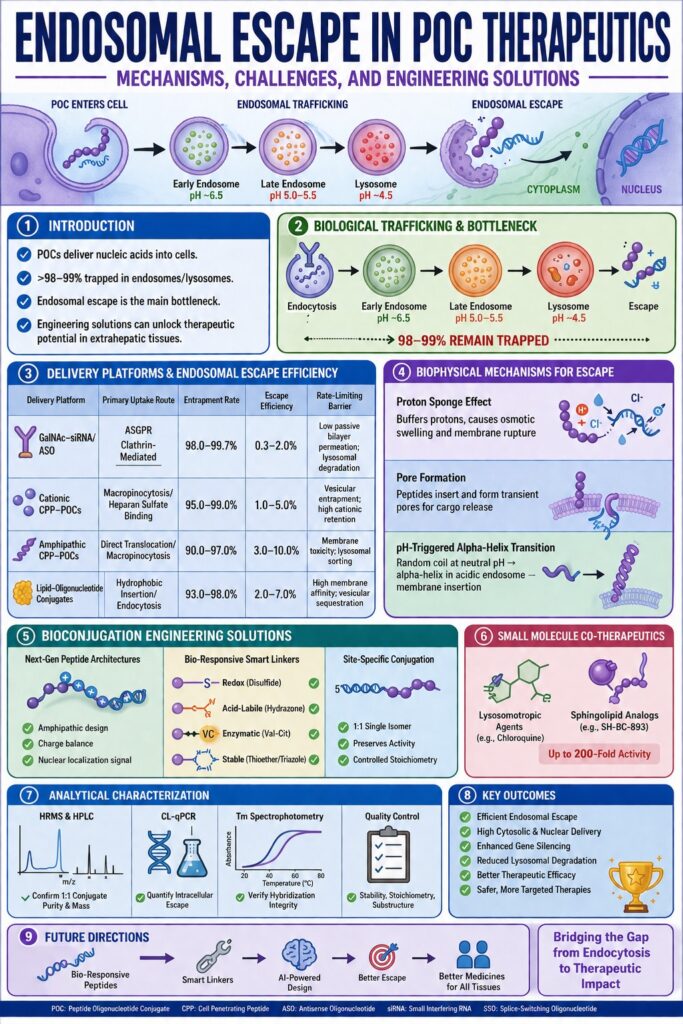
Intracellular trafficking routes POCs through a series of progressive endosomal acidification and maturation stages, during which active sequestration restricts cytosolic delivery. Subcellular quantification has shown that 98.0% to 99.7% of endocytosed material remains entrapped within vesicular compartments, establishing vesicular sorting as the primary bottleneck in intracellular delivery.
Conjugates enter cells through several distinct endocytic pathways, including clathrin-mediated endocytosis (CME), caveolae-mediated endocytosis, and macropinocytosis. The pathway involved is determined primarily by the charge, length, and secondary structure of the peptide moiety. Following internalisation into early endosomes, where the pH is approximately 6.5, conjugates undergo intracellular sorting. A small fraction may recycle back to the plasma membrane, whereas the majority proceeds through the endolysosomal pathway. As vesicles mature, the luminal pH decreases rapidly to approximately 5.0–5.5 in late endosomes and approximately 4.5 in mature lysosomes. This acidification is accompanied by the recruitment of degradative enzymes, including cathepsins and nucleases.
For a detailed breakdown of biological intracellular pathways, visit Peptide-Oligonucleotide Conjugates Mechanism of Action.
Advanced subcellular measurement techniques, including Chemical Ligation Quantitative Polymerase Chain Reaction (CL-qPCR) and Nano-Secondary Ion Mass Spectrometry (NanoSIMS), have generated definitive quantitative insights into intracellular conjugate accumulation. In both hepatocytes and extrahepatic cells, GalNAc- and CPP-conjugated oligonucleotides exhibit escape rates ranging from 0.3% to 2.0%, leaving approximately 98% to 99% of the remaining material trapped within non-productive vesicular compartments. Although endosomal entrapment can create a low-level “depot effect” that gradually releases functional molecules over several months and supports prolonged gene silencing, extrahepatic applications that require high nuclear copy numbers—such as splice switching in neuromuscular disorders, where approximately 100,000 to 700,000 active nuclear molecules may be required per cell—cannot reliably reach therapeutic thresholds without active membrane disruption.
Compare delivery efficiencies and targeting advantages at Peptide vs Antibody Oligonucleotide Conjugates.
| Delivery Platform | Primary Uptake Route | Endosomal Entrapment Rate | Estimated Escape Efficiency | Rate-Limiting Barrier |
|---|---|---|---|---|
| GalNAc-siRNA / ASO | ASGPR Clathrin-Mediated | 98.0% – 99.7% | 0.3% – 2.0% | Low passive bilayer permeation; lysosomal degradation |
| Cationic CPP-POCs | Macropinocytosis / Heparan Sulfate binding | 95.0% – 99.0% | 1.0% – 5.0% | Vesicular entrapment; high cationic membrane retention |
| Amphipathic CPP-POCs | Direct Translocation & Macropinocytosis | 90.0% – 97.0% | 3.0% – 10.0% | Membrane toxicity at high concentration; lysosomal sorting |
| Lipid-Oligonucleotide Conjugates | Hydrophobic Insertion / Endocytosis | 93.0% – 98.0% | 2.0% – 7.0% | High membrane affinity leading to vesicular sequestration |
Biophysical Mechanisms Driving Endosomal Escape in Peptide Oligonucleotide Conjugates (POC) Therapeutics
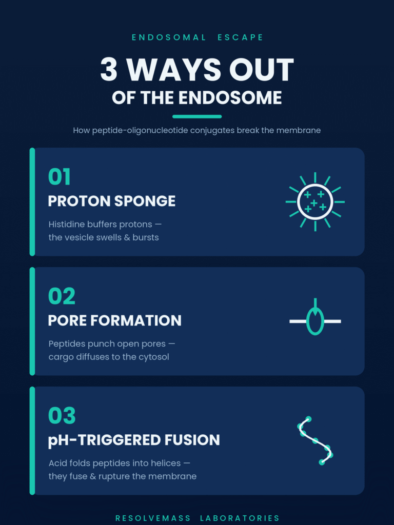
The biophysical mechanisms responsible for endosomal escape include proton sponge buffering, direct lipid bilayer pore formation, and pH-sensitive alpha-helical transitions. These mechanisms destabilise the lipid bilayer during vesicle acidification, thereby enabling the translocation of cytosolic cargo from the endosomal compartment.
Proton Sponge Effect and Histidine-Rich Motifs in Endosomal Escape in Peptide Oligonucleotide Conjugates (POC) Therapeutics
The proton sponge effect relies on weakly basic amino acids to buffer protons within the endosome, promoting osmotic swelling and vesicular membrane rupture. Histidine residues contain an imidazole side chain with a pKa of approximately 6.0 and can therefore undergo selective protonation within acidic endosomes, promoting osmotic destabilisation.
Active proton transport by vacuolar-type H+-ATPase (V-ATPase) pumps progressively lowers the luminal pH during endosomal maturation. When histidine residues are incorporated into cationic peptide structures, they can buffer incoming protons and interfere with the natural acidification process. To maintain electrical neutrality, chloride counter-ions (Cl-) enter the vesicle, accompanied by the passive movement of water. This process increases intra-vesicular osmotic pressure. The resulting mechanical stress can cause transient endosomolysis and promote cargo release. Modifying cationic peptides such as TAT with poly-histidine tracts has been shown to increase genetic cargo transfection efficiency by up to 7,000-fold compared with unmodified counterparts, supporting proton buffering as an important mechanism of endosomal release.
Pore Formation and Membrane Disruption Dynamics
Direct membrane pore formation occurs when amphipathic or cationic peptides insert into the endosomal lipid bilayer and generate transient toroidal or barrel-stave pores. These structural openings allow encapsulated nucleic acid cargo to diffuse directly into the host cell cytoplasm.
Cationic and amphipathic peptides, including melittin variants and gasdermin-D derived sequences, interact directly with negatively charged membrane phospholipids. Electrostatic attraction initially promotes peptide binding to the membrane headgroups, after which hydrophobic peptide surfaces insert into the fatty acid core. During bioconjugate self-assembly or interaction with the endosomal membrane, peptides can undergo a multi-stage binding cascade involving initial electrostatic association within the major groove of the oligonucleotide, minor groove alignment, and subsequent hydrophobic aggregation. This process facilitates bilayer insertion when the conjugate reaches the endosomal membrane.
pH-Triggered Fusogenic Alpha-Helical Transitions
Fusogenic peptides undergo pH-dependent conformational changes, transitioning from random coils at neutral pH to amphipathic alpha-helices within acidic endosomes. This structural transformation promotes insertion into the endosomal membrane and enables localised membrane destabilisation.
Derived from viral envelope proteins such as influenza hemagglutinin-2 (HA2), LK15, or N-E5L, fusogenic motifs remain largely inactive during extracellular circulation at a pH of approximately 7.4. After entering the acidic endosome, where the pH falls below 6.0, protonation of acidic side chains, including glutamic and aspartic acid, reduces internal electrostatic repulsion. This change drives the peptide into a highly structured hydrophobic alpha-helix. The hydrophobic face of the helix inserts into the inner endosomal leaflet, promoting membrane curvature, lipid mixing, and the leakage of cargo into the cytosol.
Learn more about overcoming membrane transport obstacles at Challenges in Peptide-Oligonucleotide Conjugates.
Bioconjugation Engineering Solutions to Optimize Endosomal Escape in Peptide Oligonucleotide Conjugates (POC) Therapeutics
Engineering strategies for improving endosomal escape focus on rational peptide sequence design, bio-responsive cleavable linkers, and chemoselective site-specific conjugation. These approaches are designed to minimise steric interference, preserve systemic stability, and promote controlled cytosolic release of the therapeutic cargo.
Next-Generation Peptide Architectures and Amphipathic Designs
Next-generation peptide designs incorporate amphipathic charge patterning, sequence cyclization, and nuclear localization signals to improve the ability of conjugates to cross endosomal membranes. Architectures such as Pip6a are designed to balance cationic cellular binding with efficient endosomal membrane escape.
Classical CPPs such as penetratin and R9 may show limited performance because of extensive endosomal trapping or rapid systemic clearance. More advanced peptide series, including Pip6a, use hydrophobic core structures flanked by arginine-rich cationic clusters (R8-R10). This configuration provides an optimised charge density that supports both cellular entry and vesicular release in extrahepatic tissues, including muscle and central nervous system tissue. The incorporation of short cationic Nuclear Localization Signals (NLS) further ensures that, after cytosolic entry has been achieved, the cargo can be actively transported into the nucleus to interact with splice-switching machinery.
Review advanced chemical synthesis methodologies at Peptide-Oligonucleotide Conjugate Synthesis Methods.
Bio-Responsive Smart Linkers for Intracellular Cargo Release
Bio-responsive smart linkers are designed to maintain covalent stability during systemic circulation while undergoing cleavage in response to endosomal pH, intracellular redox conditions, or lysosomal enzymes. This cleavage process releases the native oligonucleotide, allowing it to interact more effectively with its target RNA or protein complexes.
Linker selection has a direct influence on both pharmacokinetics and intracellular potency. Disulfide linkers take advantage of the substantial concentration gradient between extracellular glutathione, approximately 2 µM, and cytosolic glutathione, which ranges from 1 to 10 mM. These linkers remain stable in plasma but undergo rapid reduction after entering the cytoplasm. Acid-labile hydrazone or ester linkages can undergo selective hydrolysis within acidic late endosomes, where the pH is below 5.5. Enzyme-cleavable dipeptides, such as Valine-Citrulline, are designed to resist plasma proteases while undergoing rapid cleavage by endolysosomal cathepsin B. This process separates the peptide vector from the oligonucleotide before extensive degradative processing occurs.
Dive into specialized chemistry and bio-responsive bonds at Peptide-Oligonucleotide Conjugate Linker Chemistry.
Site-Specific Conjugation and Stoichiometric Control
Site-specific bioconjugation produces well-defined 1:1 single-isomer conjugates, thereby reducing heterogeneous product populations and helping preserve oligonucleotide functional activity. Conjugation handles are strategically placed at positions that minimise interference with the oligonucleotide, preserving target hybridization and RISC loading.
Non-specific bioconjugation can produce heterogeneous mixtures with variable stoichiometry, resulting in unpredictable pharmacokinetics, altered biodistribution, and reduced target-binding performance. Attaching peptides to the 5′- or 3′-terminus of single-stranded ASOs can preserve base-pairing hybridization affinity. In the case of double-stranded siRNA, conjugation is directed to the 5′-terminus of the sense (passenger) strand, thereby preserving unhindered 5′-phosphorylation and supporting RISC assembly on the antisense (guide) strand. Chemoselective chemistries, including Strain-Promoted Azide-Alkyne Cycloaddition (SPAAC), thiol-maleimide addition, and oxime ligation, provide highly controlled and quantitative site-specific conjugation.
| Linker Mechanism | Chemical Structure / Group | Cleavage Trigger | Subcellular Cleavage Site | Primary Advantage |
|---|---|---|---|---|
| Redox-Sensitive | Disulfide (-S-S-) | High intracellular Glutathione (GSH) | Cytosol / Nucleus | Reversible linkage; releases native unmodified oligonucleotide |
| Acid-Labile | Hydrazone / Ester / Acetal | Low pH (< 5.5) | Late Endosomes | Selective activation in endosomal microenvironments |
| Enzymatic | Valine-Citrulline (Val-Cit) | Cathepsin B cleavage | Endolysosomes | Excellent plasma stability; highly selective enzyme processing |
| Stable Covalent | Thioether / Triazole (Click) | Non-cleavable | N/A (Intact Conjugate) | Maximum systemic stability; ideal for steric-blocking ASOs |
Small Molecule Endolysosomal Modulators and Co-Therapeutics
Small molecule co-therapeutics can improve endosomal escape by regulating intracellular trafficking networks, slowing lysosomal maturation, or increasing the availability of permeable endosomal compartments. The co-administration of synthetic sphingolipid analogs or lysosomotropic compounds can substantially enhance oligonucleotide activity, in some cases by up to 200-fold.
Co-treatment with pharmacological modulators provides an external strategy for recovering the activity of conjugates trapped within intracellular vesicles. Classical lysosomotropic weak bases, such as chloroquine, accumulate within endosomes and promote osmotic swelling and membrane disruption. However, concerns regarding systemic toxicity have limited the clinical application of these compounds.
More recent pharmacological approaches have identified better-tolerated synthetic sphingolipid analogs, including SH-BC-893, which can modify intracellular trafficking without compromising overall membrane integrity. SH-BC-893 simultaneously inhibits ADP-ribosylation factor 6 (ARF6)-dependent endocytic recycling and Phosphoinositide 5-kinase (PIKfyve)-dependent lysosomal fusion. This dual inhibition redirects internalized conjugates away from degradative lysosomes and toward extra-lysosomal endosomal compartments that undergo rapid membrane fusion and fission. These dynamic intermediate vesicles demonstrate increased passive permeability, enhancing ASO and siRNA functional activity by up to 15-fold in vivo across extrahepatic tissues, including the central nervous system and lungs, without inducing measurable tissue toxicity.
Examine absorption, distribution, and stability parameters at Peptide-Oligonucleotide Conjugates Pharmacokinetics.
Analytical Characterization and Quality Control Metrics
Analytical characterization of POCs requires the use of high-resolution mass spectrometry and liquid chromatography to confirm 1:1 structural purity, target-binding affinity, and intracellular escape efficiency. Comprehensive quality control protocols are essential for validating construct stability, stoichiometry, and sub-monomer integrity.
The chemical combination of polyanionic oligonucleotides with polycationic or hydrophobic peptides presents substantial analytical challenges, including non-specific column adsorption, peak broadening, and self-aggregation. High-Resolution Mass Spectrometry (HRMS), using Electrospray Ionization Time-of-Flight (ESI-TOF) or Orbitrap analyzers, enables the confirmation of precise molecular weight, sequence fidelity, and complete deprotection. Ion-Pair Reverse-Phase Liquid Chromatography (IP-RP-HPLC) and Ultra-Performance Liquid Chromatography (UPLC) separate full-length conjugates from unreacted peptides, failure sequences, and aggregation byproducts, supporting the confirmation of purities exceeding 95%.
Ensure construct integrity with quality control solutions at QC Testing for Peptide-Oligonucleotide Conjugates.
Intracellular escape and functional activity can be quantified using Chemical Ligation Quantitative PCR (CL-qPCR), which measures precise oligonucleotide copy numbers in isolated cytosolic and nuclear fractions following free uptake. In addition, Thermal Denaturation (Tm) Spectrophotometry evaluates duplex melting temperatures to confirm that peptide attachment does not compromise hybridization kinetics or the thermodynamic affinity of the target interaction. Advanced bioconjugate research environments, such as ResolveMass Laboratories Inc., use these orthogonal analytical platforms to support the detailed characterization and quality control of custom POC architectures.
Explore detailed analytical characterization workflows at Structural Characterization of Peptide-Oligonucleotide Conjugates.
Conclusion: Future Directions in Endosomal Escape in Peptide Oligonucleotide Conjugates (POC) Therapeutics
Future advances in Endosomal Escape in Peptide Oligonucleotide Conjugates (POC) Therapeutics will depend on the rational integration of bio-responsive peptide motifs, site-specific smart linkers, and predictive machine-learning design frameworks. Together, these integrated strategies have the potential to bridge the gap between inefficient cellular uptake and effective extrahepatic gene silencing.
Achieving efficient Endosomal Escape in Peptide Oligonucleotide Conjugates (POC) Therapeutics remains the primary challenge in expanding nucleic acid medicines toward extrahepatic tissue targets. Overcoming the approximately 99% vesicular entrapment barrier will require a shift beyond simple cell-penetrating peptides toward multifunctional and structurally defined bioconjugates. The combined use of pH-triggered fusogenic transitions, proton-buffering histidine clusters, bio-responsive cleavable linkers, and precise 1:1 bioconjugation strategies provides a realistic pathway for achieving therapeutically relevant nuclear and cytosolic concentrations.
When combined with emerging small-molecule endolysosomal modulators and high-resolution analytical validation, next-generation POC platforms have the potential to redefine treatment modalities for rare genetic disorders, neuromuscular diseases, and oncology. To discuss custom bioconjugation synthesis, analytical testing, or strategic platform development, connect with industry experts through the ResolveMass Contact Page.
Frequently Asked Questions (FAQs)
Histidine contains an imidazole side chain with a pKa close to 6.0, making it particularly responsive to the acidic environment of maturing endosomes. As the endosomal pH decreases, histidine residues become protonated and buffer incoming protons. This process promotes the accumulation of chloride ions and water within the vesicle, increasing osmotic pressure and potentially causing membrane destabilization or lysis. Incorporating histidine-rich sequences can therefore substantially improve intracellular delivery efficiency.
Cationic CPPs, including TAT and polyarginine, primarily depend on a high density of positively charged lysine or arginine residues to interact with negatively charged cell-surface proteoglycans and promote endocytic uptake. Amphipathic CPPs contain both hydrophilic and hydrophobic regions arranged to interact with lipid membranes. These structural features allow them to insert into bilayers, generate transient pores, or undergo pH-triggered alpha-helical transitions that destabilize endosomal membranes. Their distinct architectures therefore influence both cellular uptake and endosomal escape mechanisms.
Cleavable linkers can release the oligonucleotide from its peptide carrier after the conjugate reaches an appropriate intracellular environment. Redox-sensitive disulfides and acid-labile hydrazones, for example, can respond to cytosolic reducing conditions or acidic endosomal compartments. Removing the peptide component can reduce steric interference with target hybridization and intracellular effector systems. This is particularly important for efficient RISC loading in RNAi applications and for the interaction of oligonucleotides with their intended nucleic acid targets.
For many siRNA designs, peptide attachment is directed toward the 5′- or 3′-terminus of the sense (passenger) strand to minimise interference with the functional antisense (guide) strand. Modification of the 5′-terminus of the antisense strand may interfere with essential phosphorylation and recognition events required for RISC assembly. Such interference can substantially reduce the efficiency of RNA interference. Careful selection of the conjugation site is therefore essential for maintaining gene-silencing activity.
Small molecule endolysosomal modulators such as SH-BC-893 can alter intracellular trafficking pathways that normally direct POCs toward degradation. By inhibiting ARF6-mediated endocytic recycling and PIKfyve-mediated lysosomal fusion, these compounds can redirect internalized conjugates toward extra-lysosomal, non-degradative vesicular compartments. These compartments may undergo active membrane fusion and fission, increasing the opportunity for cargo release into the cytosol. Consequently, POC activity can be substantially enhanced, with reported increases reaching up to 200-fold under specific experimental conditions.
High-Resolution Mass Spectrometry (HRMS) is used to determine molecular mass and evaluate sequence and structural integrity of the conjugate. Reverse-Phase Ultra-Performance Liquid Chromatography (RP-UPLC) and Ion-Pair HPLC provide complementary separation of the intended conjugate from free peptide, unreacted oligonucleotide, failure sequences, and other impurities. Together, these techniques help confirm the expected 1:1 stoichiometry and assess overall product purity. Orthogonal analytical methods are particularly valuable for complex POC structures.
The phosphorothioate (PS) backbone replaces a non-bridging oxygen atom with sulfur, increasing the oligonucleotide’s lipophilicity and its ability to bind serum proteins such as albumin. These properties can enhance cell-surface interactions and promote cellular uptake through endocytic pathways. However, the modification does not by itself guarantee efficient endosomal release. Much of the internalized material may remain sequestered within endosomes, creating a prolonged “depot effect”, while direct cytosolic escape generally remains limited to approximately 1% to 2%.
Nuclear Localization Signal (NLS) peptides are short cationic sequences that are recognised by nuclear transport proteins known as importins. After a POC successfully escapes from the endosome and reaches the cytosol, the NLS can facilitate its recognition by the nuclear import machinery. The resulting transport process directs the conjugate through the nuclear pore complex and into the nucleus. This mechanism can support applications requiring nuclear access, including splice-switching and transcriptional targeting.
Scaling up POC bioconjugation requires the integration of chemically distinct components, as hydrophilic nucleic acids and hydrophobic or cationic peptides often have different synthesis and purification requirements. Additional challenges include controlling aggregation during post-synthetic conjugation and maintaining consistent reaction stoichiometry. Purifying the desired conjugate at high yield and achieving purity levels exceeding 95% may require specialised preparative HPLC workflows. Process optimisation is therefore essential for ensuring reproducible quality and scalability during larger-scale manufacturing.
Reference:
- Wickline, S. A., Hou, K. K., & Pan, H. (2023). Peptide-based nanoparticles for systemic extrahepatic delivery of therapeutic nucleotides. International Journal of Molecular Sciences, 24(11), 9455. https://doi.org/10.3390/ijms24119455
- Chahal, G. S., Helbig, K. J., Parton, R. G., & Monson, E. A. (2026). The biology of endosomal escape: Strategies for enhanced delivery of therapeutics. ACS Nano, 20(2), 1789–1813. https://doi.org/10.1021/acsnano.5c18112
- Malinowska, A. L., Huynh, H. L., & Bose, S. (2024). Peptide–oligonucleotide conjugation: Chemistry and therapeutic applications. Current Issues in Molecular Biology, 46(10), 11031–11047. https://doi.org/10.3390/cimb46100655
- Klabenkova, K., Fokina, A., & Stetsenko, D. (2021). Chemistry of peptide-oligonucleotide conjugates: A review. Molecules, 26(17), 5420. https://doi.org/10.3390/molecules26175420
- Hou, K. K., Pan, H., Schlesinger, P. H., & Wickline, S. A. (2015). A role for peptides in overcoming endosomal entrapment in siRNA delivery—A focus on melittin. Biotechnology Advances, 33(6), 931–940. https://doi.org/10.1016/j.biotechadv.2015.05.005
- Finicle, B. T., Eckenstein, K. H., Revenko, A. S., Anderson, B. A., Wan, W. B., McCracken, A. N., Gil, D., Fruman, D. A., Hanessian, S., Seth, P. P., & Edinger, A. L. (2023). Simultaneous inhibition of endocytic recycling and lysosomal fusion sensitizes cells and tissues to oligonucleotide therapeutics. Nucleic Acids Research, 51(4), 1583–1599. https://doi.org/10.1093/nar/gkad023
Get In Touch With Us
About The Author


