Introduction
Peptide-oligonucleotide conjugates (POCs) demand tightly controlled chemical, thermal, and physical conditions to preserve their structural integrity and biological functionality because the peptide and nucleic acid components exhibit markedly different stability characteristics. Establishing stringent protocols for the handling and storage of peptide-oligonucleotide conjugates is therefore essential to minimize degradation, maintain target-binding affinity, and ensure consistent analytical as well as therapeutic performance. As a recognized leader in high-resolution bioanalytical workflows, ResolveMass Laboratories Inc. has developed these best practices to assist researchers in overcoming the unique challenges associated with handling these sophisticated hybrid macromolecules. Since POCs integrate highly charged, hydrophilic oligonucleotides with chemically diverse and frequently hydrophobic peptide sequences, conventional storage approaches designed for individual biomolecules are insufficient. As a result, a comprehensive understanding of degradation mechanisms, reconstitution behavior, and vessel surface interactions is necessary to minimize aggregation, prevent chemical modifications, and reduce sample loss.
To learn more about the specific structural challenges involved, explore our overview on the challenges in peptide-oligonucleotide conjugates.
Article Summary:
- Peptide-oligonucleotide conjugates (POCs) require carefully controlled handling and storage because their peptide and nucleic acid components have different stability profiles, making them vulnerable to degradation if improperly managed.
- Multiple degradation mechanisms, including nuclease activity, protease digestion, depurination, oxidation, deamidation, and unstable linker cleavage, can reduce conjugate integrity, highlighting the need for optimized pH, temperature, and chemical conditions.
- Buffer selection and reconstitution methods play a critical role in maintaining stability. Neutral to slightly alkaline TE buffer is generally preferred, while hydrophobic, acidic, or specialized POCs may require tailored solvents to prevent aggregation and improve solubility.
- Proper storage practices such as keeping samples lyophilized at low temperatures, preparing single-use aliquots, avoiding repeated freeze-thaw cycles, and using freeze-stable buffers significantly extend shelf life and preserve biological activity.
- Reducing sample loss is equally important. Using ultra-low binding or siliconized laboratory consumables minimizes adsorption of POCs to container surfaces, ensuring higher recovery and more reliable experimental results.
- Advanced analytical techniques, including high-resolution LC-MS/MS with ETD, ECD, and UVPD fragmentation, enable precise characterization of POCs by confirming conjugation sites, detecting degradation products, and verifying structural integrity.
- A comprehensive workflow combining optimized handling, stabilized linker chemistry, appropriate storage conditions, and rigorous analytical characterization ensures maximum stability, reproducibility, and performance of peptide-oligonucleotide conjugates in both research and therapeutic applications.
Chemical Architecture and Hybrid Degradation Pathways
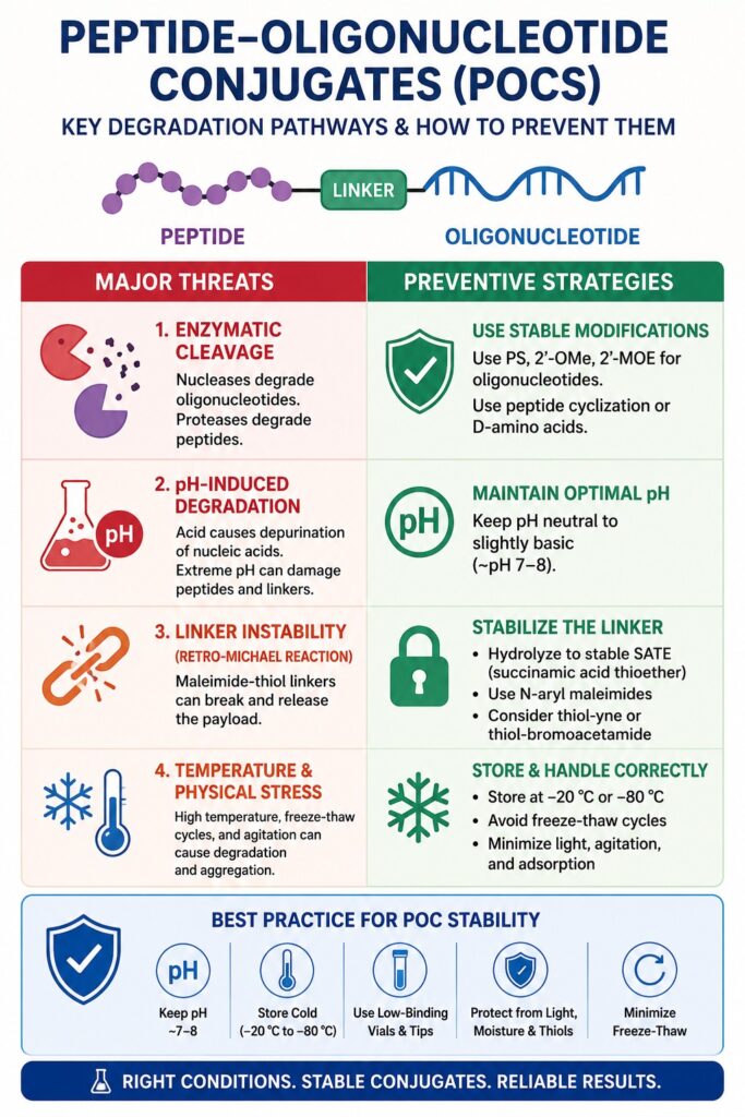
Peptide-oligonucleotide conjugates undergo degradation through several concurrent mechanisms, including enzymatic cleavage of both phosphodiester and amide backbones, acid-induced depurination, and cleavage of chemically sensitive linkers. A thorough understanding of these degradation pathways is critical for developing appropriate handling, reconstitution, and stabilization strategies that effectively preserve the integrity of both the peptide and oligonucleotide components.
For an in-depth look at how these molecules interact with biological systems, visit our page on peptide-oligonucleotide conjugates mechanism of action.
Enzymatic Vulnerability and Chemical Hydrolysis
Native oligonucleotides are highly susceptible to rapid degradation by serum nucleases, whereas peptide sequences are readily broken down by proteolytic enzymes. Although structural modifications such as replacing conventional phosphodiester linkages with phosphorothioate (PS) bonds or incorporating 2′-O-methyl (2′-OMe) and 2′-O-methoxyethyl (2′-MOE) sugar modifications significantly improve metabolic stability, these modifications do not completely eliminate enzymatic susceptibility. Likewise, peptide stability can be enhanced through cyclization or by incorporating unnatural D-amino acids. However, even these stabilized peptides remain vulnerable when exposed to environments contaminated with active proteases.
The rate of chemical hydrolysis is strongly influenced by both pH and temperature. Under acidic conditions (pH < 5.0), the glycosidic bonds connecting purine nucleobases (adenine and guanine) to the deoxyribose or ribose backbone undergo rapid acid-catalyzed cleavage through a process known as depurination. This reaction produces unstable apurinic sites that are highly susceptible to subsequent β-elimination, ultimately resulting in irreversible strand cleavage. In contrast, highly basic environments may damage peptide sequences that contain chemically sensitive amino acid residues or labile linkers. These considerations emphasize the importance of maintaining a carefully controlled neutral-to-slightly-basic pH throughout sample handling and storage procedures.
Discover more about ensuring long-term integrity through peptide-oligonucleotide conjugate stability.
Linker Instability and Maleimide-Thiol Retro-Michael Reactions
The stability of the chemical linker plays a decisive role in determining the long-term integrity of peptide-oligonucleotide conjugates because unstable linkers can undergo premature cleavage, separating the peptide from its attached oligonucleotide payload. Maleimide-thiol linkages are particularly susceptible to retro-Michael reactions when exposed to thiol-rich biological environments, resulting in the release of the oligonucleotide and increasing the likelihood of off-target payload redistribution.
The conventional thiol-maleimide conjugation reaction initially generates a succinimide thioether (SITE) intermediate. Under physiological conditions, or in the presence of competing endogenous thiols such as glutathione or serum albumin, this SITE intermediate can undergo a retro-Michael β-elimination reaction. During this process, the regenerated maleimide functionality becomes available to react with alternative thiol-containing molecules within the surrounding biological environment. This unwanted exchange leads to payload migration and significantly reduces targeting specificity.
To minimize degradation caused by retro-Michael reactions, post-conjugation ring-opening hydrolysis is commonly employed. Following conjugation, exposing the thiosuccinimide adduct to carefully controlled, mildly basic conditions promotes hydrolysis of the succinimide ring, generating a highly stable succinamidic acid thioether (SATE). The SATE derivative exhibits exceptional resistance to retro-Michael reactions and thiol-exchange processes, with reported physiological half-lives extending beyond two years. Formation of this stabilized structure can be further accelerated using several complementary strategies:
- N-Aryl substitutions: Introducing electron-withdrawing substituents, including N-phenyl or N-fluorophenyl groups, onto the maleimide nitrogen promotes faster hydrolysis of the succinimide ring.
- Basic amino group engineering: Positioning a basic amine in close proximity to the maleimide serves as an intramolecular catalyst, accelerating hydrolysis under neutral pH conditions.
- Alternative chemistries: Employing thiol-yne coupling or thiol-bromoacetamide reactions generates stable vinyl sulfide or irreversible thioether linkages that are inherently resistant to retro-Michael cleavage.
The following table summarizes the major degradation mechanisms affecting peptide-oligonucleotide conjugates along with the corresponding preventive strategies.
| Degradation Pathway | Primary Targets | Chemical Mechanism | Prevention / Mitigation Strategy |
|---|---|---|---|
| Nuclease Cleavage | Oligonucleotide backbone | Enzymatic hydrolysis of phosphodiester bonds | Phosphorothioate (PS) modifications; sterile, nuclease-free storage. |
| Protease Cleavage | Peptide domain | Enzymatic hydrolysis of peptide amide bonds | Peptide cyclization; incorporation of D-amino acids; sterile handling. |
| Depurination | Adenine and Guanine bases | Acid-catalyzed cleavage of β-N-glycosidic bonds | Reconstitute at pH 7.5–8.0; avoid acidic deprotection conditions and acidic buffers. |
| Retro-Michael Cleavage | Thiosuccinimide linkages | β-elimination and thiol exchange with glutathione (GSH) or albumin | Perform post-conjugation ring-opening hydrolysis to generate SATE. |
| Oxidation | Cys, Met, and Trp residues | Atmospheric oxygen-induced chemical modification | Purge storage buffers with argon or nitrogen and store samples in airtight containers. |
| Deamidation | Asn and Gln residues | Hydrolytic conversion of side chains into carboxylic acids | Maintain samples in the lyophilized state whenever possible and avoid prolonged aqueous storage. |
Review the engineering principles behind these stable connections in our guide on peptide-oligonucleotide conjugate linker chemistry.
Linker Chemistry and the Dynamics of Thiol-Maleimide Conjugates
Thiol-maleimide linkages are susceptible to retro-Michael reactions under both in vitro and in vivo conditions. This instability can be substantially reduced by performing post-conjugation ring-opening hydrolysis, which converts the succinimide intermediate into the more stable succinamidic acid thioether (SATE). Formation of the SATE structure chemically stabilizes the conjugate, preventing payload migration and minimizing unwanted exchange reactions with endogenous thiols such as glutathione.
For a broader understanding of how these are built, see our detailed peptide-oligonucleotide conjugate synthesis methods.
pH Dependence of Maleimide Reactions
The thiol-maleimide coupling reaction demonstrates excellent chemoselectivity toward thiol groups only within a relatively narrow pH window. At pH values between 6.5 and 7.5, maleimides react with thiols at rates approximately 1,000 times greater than with primary amines, including the ε-amino groups present on lysine residues. However, once the pH rises above 7.5, this selectivity decreases significantly because free primary amines begin to compete for reaction with the maleimide double bond. In addition, elevated pH conditions accelerate hydrolysis of the maleimide ring itself, producing inactive maleamic acid before conjugation can occur. This undesirable side reaction substantially decreases conjugation efficiency and can dramatically reduce overall coupling yields.
Rearrangement and Thiazine Formation
When peptides containing an N-terminal cysteine are conjugated to maleimide-functionalized oligonucleotides, an often-overlooked side reaction involves the transformation of the initially formed succinimidyl thioether into a six-membered thiazine ring structure. The extent and rate of this thiazine rearrangement depend heavily on several factors, including the pH of the reaction medium, the chemical nature of the linker, and the amino acid residues immediately adjacent to the conjugation site. Under neutral or alkaline conditions, the N-terminal amine becomes sufficiently deprotonated, enabling it to perform an intramolecular nucleophilic attack on the carbonyl group of the succinimide ring. This intramolecular rearrangement proceeds rapidly at physiological or elevated pH values. In contrast, conducting the conjugation reaction under mildly acidic conditions, such as pH 5.0, maintains the N-terminal amine in its protonated, non-nucleophilic state, thereby effectively suppressing thiazine formation.
The sequence-dependent and pH-dependent characteristics of this rearrangement are illustrated below using a model tripeptide system (Cys-Xxx-Phe-OH).
| Peptide Conjugate | Thiazine % at pH 5.0 (336 hours) | Thiazine % at pH 7.3 (1 hour) | Thiazine % at pH 8.4 (1 hour) |
|---|---|---|---|
| CGF-MPA (Gly adjacent) | 1.1% | 6.1% | 17.6% |
| CEF-MPA (Glu adjacent) | 1.0% | 3.4% | 8.4% |
| CKF-MPA (Lys adjacent) | 0.5% | 8.7% | 16.8% |
| CSF-MPA (Ser adjacent) | 0.9% | 4.8% | 11.2% |
| CLF-MPA (Leu adjacent) | 0.3% | 4.3% | 6.6% |
Operational Guidelines: Reconstitution of Peptide-Oligonucleotide Conjugates
Successful reconstitution of peptide-oligonucleotide conjugates requires the use of carefully selected buffer systems that preserve both peptide and oligonucleotide stability. Sterile Tris-EDTA (TE) buffer maintained at pH 7.5 to 8.0 is generally preferred because it minimizes depurination while simultaneously protecting the conjugate against enzymatic degradation through the chelation of essential metal ions. Reconstitution procedures should always be customized according to the isoelectric point and hydrophobic characteristics of the peptide component in order to minimize precipitation, reduce aggregation, and maximize solution stability.
Buffer Selection and the Chelating Role of EDTA
For optimal sample preparation, sterile TE buffer consisting of 10 mM Tris-HCl and 1 mM EDTA at pH 8.0, prepared with nuclease-free water, is strongly recommended. The mildly alkaline pH helps preserve the phosphodiester backbone of the oligonucleotide by reducing susceptibility to acid-catalyzed depurination. The inclusion of EDTA is particularly important for maintaining long-term stability in solution because EDTA functions as a hexadentate chelating agent that effectively binds divalent metal ions such as Mg²⁺ and Ca²⁺. These metal ions serve as essential catalytic cofactors for the majority of DNA and RNA nucleases. By removing these cofactors from solution, EDTA inhibits nuclease activity and significantly reduces enzymatic degradation. If downstream biological assays or PCR applications cannot tolerate chelating agents, sterile 10 mM Tris-Cl buffer may be used as an alternative. However, storing peptide-oligonucleotide conjugates in water alone should be limited exclusively to short-term applications involving single-use aliquots.
Learn about the various forms available for research in our article on types of peptide-oligonucleotide conjugates.
Sequence-Specific Buffers for Handling and Storage for Peptide-Oligonucleotide Conjugates
The amino acid composition of the peptide component plays a major role in determining solubility characteristics and therefore influences the selection of the most appropriate reconstitution solvent. Although highly charged and hydrophilic peptide-oligonucleotide conjugates generally dissolve readily in standard TE buffer, conjugates containing substantial proportions of hydrophobic or electrically neutral amino acid residues require a more carefully controlled reconstitution strategy.
Acidic Sequences: Peptide segments enriched with aspartic acid (Asp) and glutamic acid (Glu) possess an overall negative charge. These conjugates should initially be dissolved in a small volume of a basic solution, such as 0.1% aqueous ammonia, followed by gradual dilution with sterile water or TE buffer until the desired working concentration is achieved.
Basic Sequences: Peptides containing high levels of arginine (Arg) and lysine (Lys) carry a net positive charge and generally exhibit excellent solubility under neutral conditions. When supplied as trifluoroacetate (TFA) salts, reconstitution in neutral phosphate-buffered saline (PBS) or TE buffer is typically sufficient to achieve complete dissolution.
Hydrophobic Sequences: Peptides rich in alanine (Ala), valine (Val), leucine (Leu), isoleucine (Ile), phenylalanine (Phe), or tryptophan (Trp) display pronounced hydrophobic characteristics. To minimize aggregation and precipitation, these conjugates should first be dissolved in a minimal volume of a dry, sterile, biocompatible organic solvent such as dimethyl sulfoxide (DMSO) or dimethylformamide (DMF). TE buffer should then be added gradually while maintaining the final concentration of organic solvent below 10% (v/v) to preserve compatibility with downstream biological applications.
Specialized Modality Solvents: Peptide-RNA chimeras must always be reconstituted using certified RNase-free water to eliminate the risk of RNA degradation. For peptide-PNA (peptide nucleic acid) and peptide-PMO (phosphorodiamidate morpholino oligomer) conjugates, supplementing the stock solution with formamide is recommended to promote complete dissolution while minimizing the formation of secondary structures.
| Sequence Class | Dominant Amino Acids / Modalities | Recommended Reconstitution Vehicle | Potential Storage Risk |
|---|---|---|---|
| Acidic POCs | Asp, Glu | 0.1% aqueous ammonia, followed by dilution with sterile water or TE buffer | Poor solubility under acidic conditions; precipitation. |
| Basic POCs | Arg, Lys | Neutral TE buffer (pH 7.5–8.0) or sterile PBS | High hygroscopicity; deliquescence upon air exposure. |
| Hydrophobic POCs | Val, Leu, Ile, Phe, Trp | Minimal volume of anhydrous DMSO or DMF (<10%), followed by gradual dilution | Extensive aggregation and sample precipitation. |
| Labile POCs | Cys, Met, Trp | Oxygen-free, degassed TE buffer purged with argon | Rapid oxidation and disulfide crosslinking. |
| PNA-Peptides | Peptide Nucleic Acids | TE buffer supplemented with formamide | Intramolecular hybridization and limited solubility. |
| RNA-Peptides | siRNA, microRNA | RNase-free sterile buffer (pH 7.5) | Rapid degradation caused by ubiquitous RNase enzymes. |
Mitigation of Vessel Adsorption and Mechanical Product Loss
Non-specific adsorption of peptide-oligonucleotide conjugates can be significantly reduced by using pre-siliconized or certified ultra-low binding (ULB) laboratory plasticware. These specialized surfaces minimize both electrostatic and hydrophobic interactions with container walls, thereby improving sample recovery. Because cationic peptides interact strongly with the negatively charged surfaces of conventional polypropylene containers, effective surface deactivation is essential for maintaining accurate sample concentrations throughout analytical workflows.
Electrostatic and Hydrophobic Wall Binding
Highly charged cell-penetrating peptides (CPPs) contain substantial numbers of positively charged arginine and lysine residues. Standard laboratory tubes manufactured from untreated polypropylene or glass possess negatively charged silanol groups or other polar surface functionalities. This difference in surface charge promotes rapid and high-affinity electrostatic adsorption of the positively charged peptide domains onto the walls of the storage vessel.
Similarly, peptide domains possessing strong hydrophobic characteristics readily associate with untreated polymer surfaces through hydrophobic interactions. When peptide-oligonucleotide conjugates are handled at low working concentrations within the micromolar or nanomolar range, this non-specific surface adsorption can substantially reduce the concentration of active material remaining in solution. The resulting sample loss may produce significant analytical errors and introduce considerable bias into screening experiments.
To eliminate losses caused by surface adsorption, all procedures involving reconstitution, dilution, or aliquoting should be performed exclusively using pre-siliconized microcentrifuge tubes or certified ultra-low retention laboratory consumables. Siliconization creates a thin, chemically inert organosiloxane coating on glass or polymer surfaces, effectively masking both charged and hydrophobic binding sites that would otherwise promote sample adsorption.
For advanced microscopy applications or imaging chamber preparations, glass surfaces should be pre-coated with a 5% bovine serum albumin (BSA) solution. This treatment blocks potential adsorption sites, minimizes surface wetting, and reduces the likelihood of peptide-oligonucleotide conjugates or nanoparticles adhering to the substrate.
Understand how these delivery vehicles function in our overview of peptide-oligonucleotide conjugates drug delivery.
Thermal Stabilization, Lyophilization, and Freeze-Thaw Integrity
Maximum long-term thermal stability of peptide-oligonucleotide conjugates is achieved by storing the material in a lyophilized state at temperatures of −20°C or below. Once reconstituted, solutions should be divided immediately into single-use aliquots and rapidly frozen using liquid nitrogen or dry ice. This approach minimizes repeated freeze-thaw cycles, reduces degradation associated with fluctuating microenvironmental pH, and helps preserve the structural and functional integrity of both the peptide and oligonucleotide components during long-term storage.
Dynamics of Lyophilization and Cake Formulation
In aqueous solution, peptide-oligonucleotide conjugates exhibit limited stability because water functions as a nucleophile that promotes hydrolytic cleavage of both the phosphodiester backbone and chemically sensitive linker structures. Consequently, removing water through lyophilization is one of the most effective strategies for significantly extending product shelf life. Although freeze-drying greatly improves long-term stability, the freezing and drying stages expose the conjugate to considerable physical and thermodynamic stress. During ice crystal formation, dissolved solutes are excluded from the growing ice lattice, resulting in cryoconcentration and the formation of localized microenvironments with elevated ionic strength. These conditions can promote peptide denaturation, increase intermolecular interactions, and initiate physical aggregation.
To preserve the structural and functional integrity of peptide-oligonucleotide conjugates throughout the lyophilization process, carefully selected lyoprotective excipients should be incorporated into the formulation.
Cryoprotectants (Sucrose/Trehalose): These disaccharides undergo vitrification during freeze-drying, producing a rigid amorphous glassy matrix that stabilizes the conjugate by preventing molecular mobility, self-association, and structural collapse. In addition to providing physical stabilization, they establish hydrogen-bonding interactions with peptide amide groups and the oligonucleotide backbone, effectively replacing the protective hydration shell that is removed during dehydration.
Bulking Agents (Mannitol/Glycine): Crystalline bulking agents provide the mechanical framework required to support the lyophilized cake. They improve structural integrity, minimize cake collapse during primary and secondary drying, and facilitate rapid, uniform, and complete reconstitution after storage.
Non-ionic Surfactants (Polysorbate 20): These surfactants reduce surface tension at the ice-water interface during the freezing stage, thereby minimizing surface-induced denaturation of peptide domains and improving overall product stability throughout the lyophilization cycle.
Avoiding the Sodium Phosphate Freeze-Induced pH Shift
Repeated freeze-thaw cycling of reconstituted peptide-oligonucleotide conjugate solutions can significantly compromise molecular integrity. One frequently underestimated mechanism responsible for chemical degradation during slow freezing is the substantial pH shift associated with sodium phosphate buffer systems.
As sodium phosphate buffer solutions are cooled, dibasic sodium phosphate (Na₂HPO₄), which represents the alkaline component of the buffer, exhibits substantially lower solubility at reduced temperatures than monobasic sodium phosphate (NaH₂PO₄), the acidic component. As a result, the dibasic salt preferentially precipitates from solution during freezing. The corresponding loss of alkaline buffering capacity causes the localized pH of the remaining unfrozen liquid phase to decrease dramatically, often falling from approximately pH 7.0 to highly acidic conditions ranging between pH 3.5 and 4.0. Under these acidic microenvironmental conditions, the oligonucleotide component undergoes rapid depurination, while acid-sensitive linker chemistries become increasingly susceptible to hydrolytic cleavage, resulting in substantial degradation of the conjugate.
To minimize degradation associated with freeze-induced pH changes, the following best practices should be implemented:
Utilize Freeze-Stable Buffers: Reconstitute and store peptide-oligonucleotide conjugates in buffer systems that maintain stable pH values during freezing, including Tris-HCl, histidine, or potassium phosphate buffers. Potassium phosphate systems are particularly advantageous because their monobasic and dibasic salts possess comparable solubility characteristics and therefore precipitate at similar rates, minimizing significant pH fluctuations.
Flash-Freezing: Samples should always be frozen rapidly by immersion in liquid nitrogen or a dry ice/ethanol bath. Rapid freezing promotes instantaneous vitrification, immobilizing dissolved solutes before phase separation and salt crystallization can occur, thereby preventing the pH shifts responsible for degradation.
Strict Aliquoting: Following reconstitution, stock solutions should be divided immediately into small, single-use aliquots. This practice eliminates repeated freeze-thaw cycles and ensures that each aliquot experiences only one freezing event and one thawing event before use, thereby preserving molecular integrity.
Learn how to validate the quality of your stored samples with QC testing for peptide-oligonucleotide conjugates.
Advanced High-Resolution Characterization of Peptide-Oligonucleotide Conjugates
Comprehensive characterization of peptide-oligonucleotide conjugates depends on advanced high-resolution liquid chromatography-mass spectrometry (LC-MS) combined with non-ergodic fragmentation techniques capable of accurately evaluating structural heterogeneity and confirming conjugation integrity. These sophisticated analytical methods enable clear discrimination between intact hybrid conjugates, partially degraded species, linker cleavage products, truncated sequences, and unconjugated starting materials, providing detailed structural confirmation throughout product development and quality control.
Exploiting Mass Defects for Analytical Identification
High-resolution mass spectrometry (HRMS) provides exceptional analytical capability for characterizing peptide-oligonucleotide conjugates by exploiting the distinct mass defect characteristics of peptide and nucleic acid components. Peptides generally contain relatively high proportions of elements such as nitrogen and hydrogen, both of which contribute positive mass defects. Conversely, oligonucleotides are enriched in oxygen and phosphorus, elements that exhibit negative mass defects. Modern high-resolution mass spectrometers readily resolve these subtle differences with exceptional accuracy. By analyzing these characteristic mass defect signatures, analysts can rapidly identify heteroconjugates, distinguish them from homopolymeric impurities, and verify the precise stoichiometric relationship between peptide and oligonucleotide components, including confirmation of a 1:1 conjugation ratio.
Non-Ergodic Fragmentation for Linker Mapping
Accurate determination of the conjugation site within complex peptide-oligonucleotide conjugates requires advanced fragmentation strategies during LC-MS/MS analysis. Conventional collision-induced dissociation (CID) is classified as an ergodic fragmentation technique because it distributes collision energy throughout the molecule before fragmentation occurs. As a consequence, CID preferentially cleaves the weakest chemical bonds present within the conjugate. In peptide-oligonucleotide hybrids, this typically results in cleavage of the relatively labile peptide-oligonucleotide linker, producing two independent molecular fragments and preventing precise localization of the conjugation site.
To overcome these limitations, non-ergodic fragmentation methods are routinely employed because they preserve the integrity of the covalent linker while generating highly informative fragmentation patterns.
Electron Transfer Dissociation (ETD) and Electron Capture Dissociation (ECD): These radical-mediated fragmentation techniques selectively cleave peptide amide bonds to generate c-type and z-type fragment ions while simultaneously fragmenting the oligonucleotide backbone without disrupting the chemically sensitive covalent linker. This preservation of linker integrity enables detailed structural characterization and direct confirmation of the conjugation site.
Ultraviolet Photodissociation (UVPD): UVPD employs high-energy photons to induce extensive fragmentation across both peptide and oligonucleotide domains simultaneously. The resulting complementary fragmentation patterns provide high-resolution sequence information and facilitate comprehensive structural mapping of the intact hybrid conjugate.
Chromatographic Coupling: To maximize analytical performance, high-resolution mass spectrometers are commonly coupled with Ion-Pair Reverse-Phase Liquid Chromatography (IP-RPLC) or Hydrophilic Interaction Liquid Chromatography (HILIC). These chromatographic techniques provide highly efficient separation of complex conjugate mixtures, enabling sensitive detection of trace degradation products, unconjugated precursors, structural variants, and other low-abundance impurities before mass spectrometric analysis.
Explore the techniques used to verify these molecules at our resource on structural characterization of peptide-oligonucleotide conjugates.
Actionable Recommendations for Handling and Storage for Peptide-Oligonucleotide Conjugates
Successful management of peptide-oligonucleotide conjugates depends on implementing rigorous cold-chain procedures, utilizing validated ultra-low binding laboratory consumables, and selecting buffer systems that remain stable during freezing and long-term storage. Adhering to these practical recommendations helps preserve biological activity, maintain chemical integrity, and maximize the long-term stability of custom peptide-oligonucleotide conjugates.
The workflow below summarizes the comprehensive operational strategy recommended by ResolveMass Laboratories Inc. for the handling, reconstitution, and storage of these advanced hybrid bioconjugates.
For researchers moving toward clinical trials, we provide essential guidance on peptide-oligonucleotide conjugates in IND submissions.
To ensure these recommendations are implemented consistently in laboratory practice, personnel should follow the operational procedures outlined below.
1. Shipment and Receipt Protocols
Dry Ice Verification: Upon receiving a shipment containing a custom peptide-oligonucleotide conjugate, confirm that the material has been transported on dry ice whenever required. Although standard catalog products and certain custom orders may tolerate brief shipment at ambient temperature, they should be transferred to appropriate cold storage immediately upon arrival to preserve molecular stability.
Immediate Storage: Place lyophilized conjugate vials directly into an ultra-low temperature freezer maintained at −20°C or preferably −80°C. Storage in frost-free freezers should be strictly avoided because these systems undergo periodic warming cycles that cause repeated micro-melting events, increasing the risk of sample degradation.
2. Microenvironmental Reconstitution Workflow
Aqueous TE Buffer Reconstitution: Prepare a stock solution at a concentration of 100 µM by adding sterile TE buffer (10 mM Tris-HCl, 1 mM EDTA, pH 8.0). The volume of buffer added, expressed in µL, should equal ten times the number of nanomoles of peptide-oligonucleotide conjugate contained within the vial.
Organic Solvent Pre-dissolution: Highly hydrophobic conjugates should first be dissolved in 100% anhydrous DMSO that has been dried over 3 Å molecular sieves. Following complete dissolution, sterile TE buffer should be added gradually while ensuring that the final concentration of organic solvent remains below 10% of the total solution volume to maintain compatibility with downstream biological applications.
Gentle Hydration: Introduce the reconstitution solvent slowly along the inner wall of the siliconized vial to minimize localized concentration gradients. Allow the sample to hydrate undisturbed at room temperature for approximately 10 to 15 minutes before gently mixing by inversion. Vigorous vortexing should be avoided because excessive mechanical agitation may promote aggregation or structural disruption.
3. Surface Chemistry and Consumables Control
Vessel Standardization: All procedures involving pipetting, dilution, aliquoting, and storage should be performed exclusively using certified ultra-low binding (ULB) polypropylene microcentrifuge tubes or pre-siliconized laboratory vessels. These specialized consumables substantially reduce non-specific adsorption and improve sample recovery.
Contact Minimization: Untreated glass containers, conventional polypropylene laboratory plates, and standard pipette tips should be avoided whenever possible. These untreated surfaces readily adsorb positively charged peptide domains through electrostatic interactions, resulting in rapid sample loss and reduced analytical accuracy.
4. Thermal Preservation Strategy
Flash-Freezing in an Inert Atmosphere: For extended storage of reconstituted peptide-oligonucleotide conjugate solutions, purge the headspace of each vial with dry argon or nitrogen gas to minimize oxidation of methionine (Met), cysteine (Cys), and tryptophan (Trp) residues. Following inert gas purging, immediately flash-freeze sealed aliquots using either a dry ice-ethanol bath or liquid nitrogen to prevent phase separation and freeze-induced degradation.
Potassium Phosphate Substitution: Whenever phosphate-based buffer systems are required, potassium phosphate should be selected in place of conventional sodium phosphate buffers. Potassium phosphate exhibits significantly greater pH stability during freezing and therefore minimizes the acidic pH shifts responsible for depurination and hydrolytic degradation.
Before beginning your experimental process, you can review our peptide-oligonucleotide conjugates preclinical services to ensure your workflow is optimized for success.
Conclusion
Implementing comprehensive protocols for the handling and storage of peptide-oligonucleotide conjugates is essential for preserving the molecular integrity, biological performance, and therapeutic potential of these sophisticated hybrid biomolecules. Careful control of storage conditions, thermal management, reconstitution procedures, and surface chemistry substantially improves sample stability, minimizes analytical variability, and supports reliable translation from research to therapeutic development.
Key best practices include using validated ultra-low retention laboratory consumables to reduce non-specific surface adsorption, avoiding sodium phosphate buffer systems to eliminate freeze-induced acidic depurination, and stabilizing maleimide-thiol linkages through post-conjugation ring-opening conversion to the succinamidic acid thioether (SATE) form. Together, these strategies significantly improve long-term conjugate stability while maintaining structural integrity throughout analytical and biological applications.
ResolveMass Laboratories Inc. provides specialized expertise in high-resolution LC-MS/MS characterization, mass defect analysis, and non-ergodic fragmentation sequencing using ETD and ECD technologies. These advanced analytical capabilities enable comprehensive structural confirmation, precise quality assessment, and robust metabolic characterization, helping ensure that custom peptide-oligonucleotide conjugate programs achieve exceptional analytical accuracy and reliable biological performance.
For specialized assistance with custom peptide-oligonucleotide synthesis, analytical characterization, quality control validation, or high-resolution mass spectrometry profiling, consult a ResolveMass scientific expert through the ResolveMass Contact Page.
Frequently Asked Questions:
Dry peptide-oligonucleotide conjugates should be maintained in their lyophilized form inside tightly sealed, moisture-resistant containers with an appropriate desiccant. For optimal long-term preservation, they should be stored at −20°C or, preferably, −80°C. Keeping the material dry and at ultra-low temperatures minimizes hydrolysis, oxidation, and other degradation pathways. Under these controlled conditions, lyophilized conjugates can remain stable for several years.
A sterile Tris-EDTA (TE) buffer containing 10 mM Tris-HCl and 1 mM EDTA at pH 7.5–8.0 is widely recommended for reconstitution. The mildly alkaline pH helps protect the oligonucleotide from acid-induced depurination, while EDTA chelates divalent metal ions such as Mg²⁺ and Ca²⁺ that are required for nuclease activity. This combination enhances solution stability and reduces the risk of enzymatic degradation during handling and storage.
Repeated freeze-thaw cycles expose peptide-oligonucleotide conjugates to mechanical stress caused by ice crystal formation and changes in solute concentration. These conditions can promote peptide aggregation, destabilize sensitive linker chemistries, and negatively affect the structural integrity of the hybrid molecule. Frequent temperature fluctuations may also reduce biological activity and increase the likelihood of irreversible degradation. Using single-use aliquots is the most effective way to eliminate these risks.
During freezing, the alkaline component of sodium phosphate buffer, Na₂HPO₄, precipitates more readily than the acidic component, NaH₂PO₄. This imbalance causes the pH of the remaining liquid phase to decrease significantly, often reaching highly acidic conditions. Such an acidic environment accelerates depurination of nucleic acid bases and increases the hydrolysis of acid-sensitive linkers. Selecting freeze-stable buffers can help prevent this form of degradation.
The reconstitution buffer should be added slowly along the inner wall of the vial to allow gradual hydration of the lyophilized material. After the solution has rested for sufficient hydration, gentle inversion or slow mixing is preferred to achieve complete dissolution. Vigorous shaking or vortexing should be avoided because excessive mechanical force can induce aggregation and damage sensitive hybrid structures. Careful handling preserves both molecular integrity and sample recovery.
Allowing sealed vials to reach room temperature inside a desiccator prevents atmospheric moisture from condensing onto the cold lyophilized powder. Because these conjugates are highly hygroscopic, immediate exposure to humid air can result in rapid moisture absorption and partial dissolution of the dry cake. This unwanted hydration may initiate hydrolytic degradation and compromise accurate sample weighing. Controlled equilibration therefore helps maintain sample quality before reconstitution.
The surface properties of laboratory containers have a significant impact on sample recovery, particularly for conjugates containing positively charged peptide sequences. Untreated glass and standard polypropylene surfaces can promote electrostatic and hydrophobic adsorption, leading to measurable sample loss. Using certified ultra-low binding plasticware or pre-siliconized tubes minimizes these interactions and improves recovery. Selecting appropriate consumables is especially important when working with low-concentration samples.
Following thiol-maleimide conjugation, the initially formed succinimide thioether (SITE) intermediate can undergo retro-Michael reactions, resulting in linker instability and payload migration. Promoting ring-opening hydrolysis under mildly basic conditions converts this intermediate into the more stable succinamidic acid thioether (SATE). The SATE structure is highly resistant to thiol exchange and significantly reduces the likelihood of retro-Michael cleavage. This stabilization strategy greatly improves the long-term integrity of thiol-maleimide conjugates.
Reference:
- de Vries, R. H., & Steendam, R. (2021). Improving the stability of maleimide–thiol conjugation for drug targeting. ChemBioChem, 22(2), 307–321. https://doi.org/10.1002/cbic.202000474
- Russo, S. (2019, December 18). Importance of EDTA buffer pH to resuspend oligos? ResearchGate. ResearchGate
- Green, N. M., & Sambrook, J. (2010). Molecular cloning: Laboratory protocols for DNA and RNA storage and handling. Cold Spring Harbor Protocols, 2010(10), pdb.top79. https://doi.org/10.1101/pdb.top79
- Klabenkova, K., Fokina, A., & Stetsenko, D. (2021). Chemistry of peptide-oligonucleotide conjugates: A review. Molecules, 26(17), 5420. https://doi.org/10.3390/molecules26175420
- Oliveira, S. (2017, February 2). Does freeze thaw effect will have an impact on the peptides stability? ResearchGate. ResearchGate
- Krivos, K. L., & Limbach, P. A. (2010). Sequence analysis of peptide:Oligonucleotide heteroconjugates by electron capture dissociation and electron transfer dissociation. Journal of the American Society for Mass Spectrometry, 21(8), 1387–1397. https://doi.org/10.1016/j.jasms.2010.04.011

