


Introduction
Peptide-oligonucleotide conjugates gain entry into cells mainly through energy-dependent endocytosis or energy-independent direct plasma membrane translocation, with the dominant mechanism determined by peptide chemistry, overall charge, and secondary structural characteristics. Understanding how do peptide-oligonucleotide conjugates enter cells? is essential for overcoming the hydrophobic barrier of cellular membranes and improving the delivery of targeted nucleic acid therapeutics. Peptide-Oligonucleotide Conjugates (POCs) integrate the sequence-specific gene-silencing or splice-modulating functions of synthetic oligonucleotides with functional peptides that promote cellular uptake, tissue-selective delivery, and efficient intracellular trafficking.
Learn more about comprehensive analytical testing and candidate evaluation through ResolveMass Preclinical Services for Peptide-Oligonucleotide Conjugates.
Unconjugated therapeutic oligonucleotides—including antisense oligonucleotides (ASOs), small interfering RNAs (siRNAs), peptide nucleic acids (PNAs), and phosphorodiamidate morpholino oligomers (PMOs)—are limited by significant pharmacokinetic challenges. Their relatively high molecular weight (>5–15 kDa) together with the highly polyanionic phosphorothioate or phosphodiester backbone generates strong electrostatic repulsion from the negatively charged plasma membrane, resulting in extremely poor passive diffusion through the lipid bilayer. Although chemical modifications such as 2′-O-methoxyethyl (2′-MOE) sugar substitutions and Locked Nucleic Acids (LNAs) enhance nuclease stability and extend systemic circulation time, insufficient cellular uptake continues to represent one of the most important quantitative barriers restricting therapeutic performance beyond hepatic tissues.
Compare the structural differences and advantages between modalities at Peptide vs. Antibody-Oligonucleotide Conjugates.
Covalent attachment of functional peptides transforms nucleic acid delivery from an inefficient passive process into an actively facilitated mechanism of cellular internalization. Cell-penetrating peptides (CPPs), receptor-targeting ligands, and endosomal escape peptides (EEPs) each contribute unique strategies for crossing biological membranes and improving intracellular delivery. Comprehensive characterization of these sophisticated bioconjugates requires advanced analytical methodologies. Liquid chromatography-mass spectrometry (LC-MS) services at ResolveMass Laboratories Inc. provide detailed structural confirmation, purity assessment, and conjugate stoichiometry verification, enabling researchers to correlate physicochemical properties with cellular uptake efficiency.
Explore the functional mechanisms of action driving uptake at Peptide-Oligonucleotide Conjugates Mechanism of Action.
Share via:
Article Summary:
- Peptide-oligonucleotide conjugates (POCs) improve intracellular delivery by combining therapeutic oligonucleotides with functional peptides that enhance membrane penetration, targeted uptake, and gene modulation efficiency.
- Cellular entry occurs through two primary mechanisms: direct membrane translocation, which delivers cargo directly into the cytoplasm, and energy-dependent endocytosis, which internalizes conjugates through membrane-bound vesicles.
- Multiple endocytic pathways—including macropinocytosis, clathrin-mediated endocytosis, and caveolae-mediated endocytosis—are influenced by peptide structure, charge, receptor interactions, and membrane composition.
- Endosomal trapping remains the biggest delivery challenge, as the majority of internalized conjugates become confined within intracellular vesicles, limiting the amount of therapeutic cargo reaching its biological target.
- Advanced escape strategies, such as proton sponge mechanisms and pH-responsive fusogenic peptides, help release conjugates from endosomes before degradation, significantly improving therapeutic performance.
- Chemical engineering approaches, including site-specific conjugation, cleavable linkers, and protease-resistant peptide designs, enhance stability, cellular uptake, and controlled intracellular release of oligonucleotide therapeutics.
- Comprehensive LC-MS and mass spectrometry characterization verifies conjugate identity, purity, molecular integrity, and conjugation efficiency, ensuring consistent quality and supporting the successful development of peptide-oligonucleotide therapeutics.
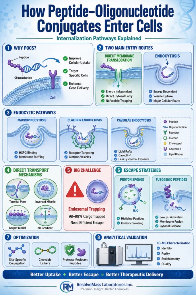
Primary Internalization Pathways Governing How Do Peptide-Oligonucleotide Conjugates Enter Cells
Peptide-oligonucleotide conjugates access target cells through two principal mechanisms: energy-dependent endocytosis, in which conjugates are enclosed within membrane-bound vesicles, and energy-independent direct membrane translocation, which delivers the conjugates into the cytosol without vesicular transport. The predominance of either pathway is influenced by peptide charge density, amphipathic character, conjugate stoichiometry, and the structural properties of the attached oligonucleotide backbone.
Discover the structural varieties and classification of conjugates at Types of Peptide-Oligonucleotide Conjugates.
The route of cellular entry is governed by complex biophysical interactions involving the peptide component, the nucleic acid cargo, and the plasma membrane. Strongly basic, arginine-rich cationic peptides initially associate with negatively charged molecules present on the cell surface, including heparan sulfate proteoglycans (HSPGs) and anionic phospholipid headgroups. Amphipathic peptides possess distinct hydrophobic and hydrophilic surfaces, allowing them to combine electrostatic membrane association with direct insertion into the hydrophobic core of the lipid bilayer.
The physicochemical characteristics of the attached nucleic acid also play a major role in determining the preferred internalization pathway. Electrically neutral oligonucleotide analogues such as PNAs and PMOs preserve both the positive charge and structural flexibility of conjugated CPPs, thereby promoting direct membrane translocation even at relatively low concentrations. In contrast, negatively charged ASOs and double-stranded siRNAs partially offset the positive charges of cationic peptides, making energy-dependent endocytic pathways the predominant mechanism of cellular uptake.
| Internalization Pathway | Energy Requirement | Primary Biophysical Trigger | Intracellular Destination | Typical Trapping Rate | Representative Peptide Class |
|---|---|---|---|---|---|
| Direct Translocation | Energy-Independent (4°C active) | Local charge accumulation, transient pore formation | Cytosol / Direct cytoplasmic access | 0% (Bypasses vesicles) | Polyarginine (R₈/R₉), Tat, amphipathic CPPs |
| Macropinocytosis | Energy-Dependent (37°C) | HSPG cross-linking, actin-driven membrane ruffling | Early / Late Endosomes | 90.0%–99.0% | Cationic & Amphipathic CPPs (e.g., Pip6a, Penetratin) |
| Clathrin-Mediated Endocytosis | Energy-Dependent (37°C) | Specific receptor-ligand binding, clathrin coat assembly | Early Endosomes → Lysosomes | 98.0%–99.7% | Receptor ligands (e.g., RGD, GalNAc, Transferrin mimics) |
| Caveolae-Mediated Endocytosis | Energy-Dependent (37°C) | Lipid raft dynamics, caveolin-1 oligomerization | Caveosomes / Endoplasmic Reticulum | 90.0%–95.0% | Hydrophobic motifs, lipid-modified peptides |
Energy-Independent Direct Plasma Membrane Translocation
Direct plasma membrane translocation allows peptide-oligonucleotide conjugates to move across the lipid bilayer and enter the cytosol without requiring metabolic energy or vesicular trafficking through endosomal compartments. This energy-independent mechanism generally occurs when conjugates accumulate at sufficiently high local concentrations, leading to temporary disruption of membrane architecture that facilitates cytoplasmic delivery.
Toroidal Pore Formation and pH Gradient Drives
Toroidal pore formation is initiated when cationic peptide residues destabilize the plasma membrane, causing phospholipid headgroups to bend inward and generate continuous lipid-lined aqueous pores. Transmembrane pH gradients further promote this transport process by regulating electrostatic interactions between peptide guanidinium groups and membrane-associated fatty acids.
Arginine-rich cell-penetrating peptides are particularly effective at initiating pore formation because their guanidinium side chains establish strong bidentate hydrogen bonds with the phosphate headgroups of membrane phospholipids. As peptide molecules accumulate on the membrane surface, electrostatic attraction generates increasing mechanical stress within the outer lipid leaflet. This membrane strain induces the bilayer to curve inward, creating a toroidal pore in which both peptide molecules and phospholipid headgroups collectively form the pore lining.
Transmembrane pH differences further facilitate this mode of cellular entry. At the extracellular physiological pH (approximately pH 7.4), guanidinium groups interact strongly with fatty acid carboxylates located within the outer membrane leaflet. As the peptide-oligonucleotide conjugate progresses toward the lower pH environment at the cytosolic membrane interface, altered protonation states weaken these ionic interactions. This disruption releases the conjugate into the cytoplasm while allowing the transient toroidal pore to reseal, thereby restoring membrane integrity after successful cargo delivery.
Inverted Micelle and Carpet Model Internalization
Inverted micelle-mediated transport encloses peptide-oligonucleotide conjugates within lipid-encapsulated hydrophilic cores that traverse the membrane through inversion, whereas the carpet model promotes membrane permeabilization through extensive peptide accumulation across the membrane surface. Both mechanisms effectively circumvent endosomal sequestration and deliver conjugated cargo directly into the cytosolic environment.
The inverted micelle mechanism begins when positively charged peptide residues bind electrostatically to negatively charged membrane lipids, triggering localized membrane invagination. Membrane phospholipids subsequently reorganize into a spherical inverted micelle in which hydrophobic fatty acid tails remain oriented toward the lipid bilayer, while hydrophilic phospholipid headgroups surround the peptide-oligonucleotide conjugate within an internal aqueous compartment. The resulting micellar structure then inverts across the membrane core before releasing the conjugate into the cytoplasm following fusion with the inner membrane leaflet.
Within the carpet model, amphipathic peptides align parallel to the plasma membrane, progressively coating the external lipid surface. Their positively charged amino acid residues interact extensively with negatively charged membrane components, modifying membrane fluidity while simultaneously increasing local surface tension. Once peptide accumulation reaches a critical threshold, lipid packing becomes destabilized, producing temporary membrane thinning or detergent-like disruption. These transient structural changes permit direct diffusion of the peptide-oligonucleotide conjugate into the cytoplasm while avoiding vesicular internalization.
Energy-Dependent Endocytic Entry Routes
Energy-dependent endocytosis is the predominant mechanism through which most peptide-oligonucleotide conjugates are internalized under physiological conditions and therapeutically relevant concentrations. This ATP-dependent process transports conjugates into membrane-enclosed vesicles through pathways including macropinocytosis, clathrin-mediated endocytosis, and caveolae-mediated uptake.
Macropinocytosis and Heparan Sulfate Proteoglycan Interactions
Macropinocytosis facilitates the uptake of peptide-oligonucleotide conjugates through actin-mediated membrane ruffling initiated by electrostatic interactions with cell-surface heparan sulfate proteoglycans (HSPGs). During this process, large quantities of extracellular fluid together with membrane-bound bioconjugates are engulfed into sizeable endocytic vesicles known as macropinosomes.
The process is initiated when positively charged peptide domains interact with HSPGs expressed on the plasma membrane. This binding activates signalling molecules, including the small GTPases Rac1 and Cdc42, together with p21-activated kinase 1 (PAK1). These signalling pathways stimulate localized actin polymerization, resulting in the formation of membrane protrusions known as lamellipodia. The membrane ruffles subsequently fold back and fuse with the plasma membrane, entrapping extracellular fluid and concentrated POC-HSPG complexes within macropinosomes measuring approximately 0.2 μm to 5.0 μm in diameter. Although macropinocytosis efficiently internalizes substantial quantities of peptide-oligonucleotide conjugates, the cargo remains confined within fluid-filled vesicles that progressively mature into degradative compartments unless effective endosomal escape mechanisms are activated.
Clathrin-Mediated and Receptor-Targeted Endocytosis
Clathrin-mediated endocytosis transports receptor-bound peptide-oligonucleotide conjugates into inwardly budding clathrin-coated pits that are released as intracellular vesicles through the activity of the GTPase dynamin. Receptor-targeting peptides, including integrin-binding RGD motifs and ASGPR-binding ligands, exploit this pathway to achieve selective uptake by specific tissues and cell types.
Conjugation of targeting peptides to therapeutic oligonucleotides enables high-affinity recognition of specific membrane receptors. For instance, cyclic Arg-Gly-Asp (cRGD) peptide conjugates preferentially bind αvβ3 integrins expressed on tumour endothelial cells, whereas GalNAc-peptide conjugates selectively recognize the asialoglycoprotein receptor located on hepatocytes. Following ligand-receptor engagement, adaptor protein complexes (AP-2) are recruited to the cytoplasmic surface of the plasma membrane, promoting the assembly of clathrin triskelions into an organized lattice structure. Progressive polymerization of this lattice drives membrane invagination and the formation of a clathrin-coated pit. Dynamin subsequently assembles around the neck of the developing vesicle and hydrolyses GTP to sever the vesicle from the plasma membrane. After internalization, the vesicle rapidly loses its clathrin coat before fusing with early endosomal compartments.
Explore targeted strategies for intracellular payload release at Peptide-Oligonucleotide Conjugates Drug Delivery Methods.
Caveolae-Mediated Endocytosis and Lipid Raft Dynamics
Caveolae-mediated endocytosis directs hydrophobic or lipid-modified peptide-oligonucleotide conjugates through cholesterol-rich lipid raft domains stabilized by caveolin-1 oligomers. Compared with clathrin-mediated uptake, this pathway reduces rapid lysosomal trafficking by routing internalized cargo through relatively non-acidic intracellular membrane compartments.
Peptides containing hydrophobic regions or lipid modifications display strong affinity for sphingolipid- and cholesterol-rich microdomains present within the plasma membrane. Caveolae appear as characteristic flask-shaped invaginations approximately 50–80 nm in diameter and are coated by oligomerized caveolin-1 proteins. Interaction of hydrophobic peptide-oligonucleotide conjugates with these membrane domains activates localized Src kinase signalling, leading to caveolin-1 phosphorylation and dynamin-dependent vesicle scission. Rather than entering highly acidic endolysosomal compartments immediately, caveolar vesicles typically develop into caveosomes or traffic through neutral regions of the endoplasmic reticulum and Golgi apparatus, thereby protecting therapeutic oligonucleotides from premature nuclease-mediated degradation.
Endosomal Trapping and Biophysical Escape Mechanics
Endosomal sequestration remains the most significant quantitative limitation affecting the therapeutic efficiency of peptide-oligonucleotide conjugates, with more than 98% to 99% of internalized material becoming confined within intracellular vesicles. Successful therapeutic delivery therefore depends on efficient disruption of the endosomal membrane before progressive acidification and lysosomal enzymes degrade the conjugated payload.
Understand key design principles for bio-responsive escape at Endosomal Escape in Peptide-Oligonucleotide Conjugates.
Proton Sponge Effect and Histidine-Rich Motifs
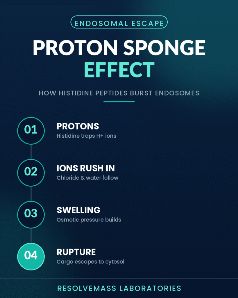
The proton sponge mechanism exploits the buffering capacity of histidine-rich peptides, whose imidazole side chains absorb accumulating protons during endosomal maturation. Continuous proton buffering stimulates the influx of chloride counterions and water, ultimately producing osmotic swelling that ruptures the endosomal membrane.
As early endosomes mature into late endosomes, vacuolar ATPases (V-ATPases) actively transport protons into the vesicular lumen, reducing the internal pH from approximately 6.5 to between 5.0 and 5.5. Incorporation of histidine residues, which possess an imidazole side chain with a pKa of approximately 6.0, enables the peptide sequence to function as an intracellular proton buffer. These residues bind incoming protons and delay the expected decline in luminal pH. To preserve electrical neutrality, V-ATPases continue pumping additional protons into the vesicle while chloride ions (Cl⁻) simultaneously enter through passive transport mechanisms. The increasing ionic concentration elevates osmotic pressure within the endosome, drawing water into the compartment. Progressive swelling eventually generates sufficient hydrostatic pressure to rupture the endosomal membrane, releasing trapped peptide-oligonucleotide conjugates into the cytoplasm.
pH-Triggered Fusogenic Transitions and Membrane Disruption
Fusogenic peptides undergo pH-dependent structural transitions, converting from flexible random conformations into amphipathic α-helices upon exposure to the acidic environment of endosomes (pH < 6.0). The newly formed hydrophobic face of the α-helix inserts into the endosomal membrane, destabilizing the lipid bilayer and promoting release of the therapeutic cargo into the cytosol.
Amphipathic endosomal escape peptides (EEPs) and peptide derivatives originating from viral fusion proteins, including HA2 and Pip6a, exploit the natural acidification that accompanies endosomal maturation. Under physiological extracellular conditions (pH 7.4), negatively charged glutamic acid and aspartic acid residues generate electrostatic repulsion that prevents stable α-helical formation. As the conjugates enter the acidic endosomal lumen (pH 5.0–5.5), these acidic residues become protonated and electrically neutral. This change drives the peptide into a stable amphipathic α-helical conformation. The hydrophobic surface of the helix subsequently penetrates the inner endosomal membrane, inducing localized membrane curvature, transient pore formation, and lipid bilayer fusion. These structural alterations permit peptide-oligonucleotide conjugates to escape from the endosome into the cytoplasm before lysosomal degradation can occur.
| Delivery Platform | Primary Cell Entry Route | Endosomal Entrapment Rate | Estimated Escape Rate | Primary Intracellular Bottleneck |
|---|---|---|---|---|
| Unconjugated ASO / siRNA | Gymnosis / Fluid-phase Endocytosis | 99.0%–99.9% | 0.1%–1.0% | Poor membrane affinity; lysosomal degradation |
| GalNAc-Oligonucleotide | ASGPR Clathrin-Mediated Endocytosis | 98.0%–99.7% | 0.3%–2.0% | Extensive endosomal recycling; minimal passive leakage |
| Cationic CPP-POCs | Macropinocytosis / HSPG Binding | 95.0%–99.0% | 1.0%–5.0% | Endosomal membrane sequestration |
| Amphipathic CPP-POCs | Direct Translocation & Macropinocytosis | 90.0%–97.0% | 3.0%–10.0% | Concentration-dependent toxicity; lysosomal sorting |
| Endosomal Escape Vehicle (EEV)-POCs | Receptor Endocytosis & pH-Disruption | 85.0%–95.0% | 5.0%–15.0% | Linker cleavage efficiency; cytosolic clearance kinetics |
Chemical Optimization and Analytical Validation of Conjugate Transport
Enhancing how peptide-oligonucleotide conjugates enter cells requires carefully optimized site-specific conjugation chemistry, peptide architectures with increased resistance to proteolytic degradation, and bio-responsive linker systems. Chemoselective conjugation strategies generate highly homogeneous products, ensuring consistent cellular uptake, predictable biological performance, and minimal toxicity.
Review the essential linker chemistries supporting selective intracellular cleavage at Peptide-Oligonucleotide Conjugate Linker Chemistry.
Key engineering approaches that improve cellular internalization include:
- Chemoselective Conjugation Chemistries: Employing copper-catalysed azide-alkyne cycloaddition (CuAAC), strain-promoted azide-alkyne cycloaddition (SPAAC), or thiol-maleimide coupling enables precise 1:1 molar conjugation between defined functional groups on both the peptide and oligonucleotide, resulting in highly uniform bioconjugates. To explore synthetic protocols, see Peptide-Oligonucleotide Conjugate Synthesis Methods.
- Bioreversible Linker Architectures: Incorporating intracellularly cleavable linkers—including glutathione-sensitive disulfide bonds, cathepsin-B-cleavable Val-Cit dipeptides, or acid-labile hydrazone linkages—allows rapid peptide release following cytosolic entry. This separation enables the liberated oligonucleotide to interact efficiently with its intended mRNA or pre-mRNA target without steric interference.
- Proteolytic Scaffolding Modifications: Introducing D-amino acids, non-canonical amino acids, or peptide cyclization strategies significantly enhances resistance to plasma and endosomal proteases. These structural modifications preserve transporter integrity and sustain cellular delivery activity under physiological conditions. For critical stability considerations, check Peptide-Oligonucleotide Conjugate Stability.
Comprehensive characterization of conjugate integrity and product purity is critical for establishing reproducible cellular uptake profiles. Advanced LC-MS and mass spectrometry platforms available at ResolveMass Laboratories Inc. verify molecular weight, conjugation efficiency, sequence identity, and overall product quality, supporting the successful development of peptide-oligonucleotide therapeutics for clinical applications.
Read about resolving common synthetic and stability barriers at Challenges in Peptide-Oligonucleotide Conjugates.
Conclusion
Understanding how do peptide-oligonucleotide conjugates enter cells? reveals that successful intracellular delivery depends on a coordinated balance between direct membrane translocation and multiple endocytic uptake pathways. Improving endosomal escape through rational molecular engineering, together with comprehensive analytical characterization, remains a fundamental requirement for maximizing the clinical potential of nucleic acid therapeutics.
Examine analytical methodologies used to confirm target structure and purity at Structural Characterization of Peptide-Oligonucleotide Conjugates.
By combining cell-penetrating or receptor-targeting peptides with carefully engineered bio-cleavable linkers, peptide-oligonucleotide conjugates harness endogenous cellular uptake pathways while incorporating specialized mechanisms that facilitate efficient endosomal escape. Detailed structural characterization using mass spectrometry and LC-MS services at ResolveMass Laboratories Inc. verifies product purity, confirms conjugate stoichiometry, and ensures consistent biological performance throughout development. To learn more about bioconjugate characterization and customized analytical support, visit the ResolveMass Laboratories Inc. Contact Page.
Frequently Asked Questions
Following cellular internalization, the majority of peptide-oligonucleotide conjugates become confined within endosomal vesicles instead of reaching the cytoplasm. As these vesicles mature, they progress toward lysosomal compartments where acidic conditions and degradative enzymes can damage the therapeutic cargo. Consequently, only a small fraction of the administered conjugates successfully escape into the cytosol, making endosomal escape one of the most critical determinants of therapeutic effectiveness.
Cell-penetrating peptides, particularly those enriched with positively charged amino acids such as arginine, interact strongly with negatively charged heparan sulfate proteoglycans on the cell surface. This interaction activates intracellular signalling molecules, including Rac1 and PAK1, which promote actin cytoskeleton rearrangement and membrane ruffling. The resulting membrane folds engulf extracellular fluid together with the peptide-oligonucleotide conjugates, producing large vesicles known as macropinosomes.
The proton sponge effect improves cytosolic delivery by using histidine-rich peptide sequences that buffer protons as endosomes become increasingly acidic. Continuous proton accumulation triggers the influx of chloride ions and water, causing osmotic swelling within the vesicle. Eventually, the increased internal pressure disrupts the endosomal membrane, releasing the trapped peptide-oligonucleotide conjugates into the cytoplasm before degradation can occur.
Fusogenic peptides respond to the acidic pH of the endosomal environment by undergoing a structural transition into amphipathic α-helices. This conformational change exposes hydrophobic surfaces that insert into the endosomal membrane and destabilize the surrounding lipid bilayer. The temporary membrane disruption allows therapeutic conjugates to exit the endosome and gain access to their intracellular RNA targets.
Several highly selective conjugation strategies are employed to generate stable peptide-oligonucleotide conjugates with defined structures. Frequently used approaches include copper-catalyzed azide-alkyne cycloaddition (CuAAC), strain-promoted azide-alkyne cycloaddition (SPAAC), thiol-maleimide coupling, and thioether bond formation. These chemistries provide precise attachment sites, high reaction efficiency, and excellent product reproducibility for therapeutic development.
Energy-dependent uptake relies on cellular ATP and typically occurs at physiological temperatures through endocytic mechanisms such as macropinocytosis, clathrin-mediated endocytosis, or caveolae-mediated uptake. In contrast, energy-independent uptake allows peptide-oligonucleotide conjugates to cross the plasma membrane directly without requiring metabolic energy or vesicle formation. The preferred mechanism depends on factors including peptide composition, charge distribution, and conjugate concentration.
Although receptor-targeted peptide conjugates improve delivery to specific cell types by promoting receptor-mediated internalization, they do not completely eliminate endosomal sequestration. Most receptor-bound conjugates still enter cells through clathrin-mediated endocytosis and become enclosed within endosomal vesicles. For optimal intracellular delivery, these systems are commonly combined with endosomal escape mechanisms that facilitate release into the cytoplasm.
The carpet model describes a mechanism in which amphipathic peptides align parallel to the plasma membrane and progressively accumulate across its surface. As peptide density increases, membrane organization becomes destabilized, leading to localized thinning and disruption of lipid packing. These transient structural changes permit peptide-oligonucleotide conjugates to cross the membrane directly without requiring vesicular transport.
Mass spectrometry plays a central role in confirming the structural quality of peptide-oligonucleotide conjugates throughout development. It accurately verifies molecular mass, conjugation stoichiometry, sequence integrity, and overall product purity while identifying residual starting materials or unwanted by-products. These analytical data help ensure batch consistency and establish reliable relationships between conjugate structure and biological performance.
Reference:
- Klabenkova, K., Fokina, A., & Stetsenko, D. (2021). Chemistry of peptide-oligonucleotide conjugates: A review. Molecules, 26(17), 5420. https://doi.org/10.3390/molecules26175420
- Okafor, M., Schmitt, D., Ory, S., Gasman, S., Hureau, C., Faller, P., & Vitale, N. (2025). The different cellular entry routes for drug delivery using cell penetrating peptides. Biology of the Cell, 117(6), e70012. https://doi.org/10.1111/boc.70012
- Malinowska, A. L., Huynh, H. L., & Bose, S. (2024). Peptide-oligonucleotide conjugation: Chemistry and therapeutic applications. Current Issues in Molecular Biology, 46(10), 11031–11047. https://doi.org/10.3390/cimb46100655
- Bianchi, A., Cazzola, H., Malaguti, M., & Corradini, R. (2023). Peptide-oligonucleotide conjugates: Current strategies and applications in medicine. Biomedicines, 11(2), 494. https://doi.org/10.3390/biomedicines11020494
- Juliano, R. L., Ming, X., Nakagawa, O., Xu, R., Yoo, H., & Leong, K. W. (2008). Intracellular delivery of an anionic antisense oligonucleotide via receptor-mediated endocytosis. Nucleic Acids Research, 36(12), 4158–4171. https://doi.org/10.1093/nar/gkn315
- Dastpeyman, M., Sharifi, R., Amin, A., Karas, J. A., Cuic, B., Pan, Y., Nicolazzo, J. A., Turner, B. J., & Shabanpoor, F. (2021). Endosomal escape cell-penetrating peptides significantly enhance pharmacological effectiveness and CNS activity of systemically administered antisense oligonucleotides. International Journal of Pharmaceutics, 599, 120398. https://doi.org/10.1016/j.ijpharm.2021.120398
Get In Touch With Us
About The Author


