


Introduction
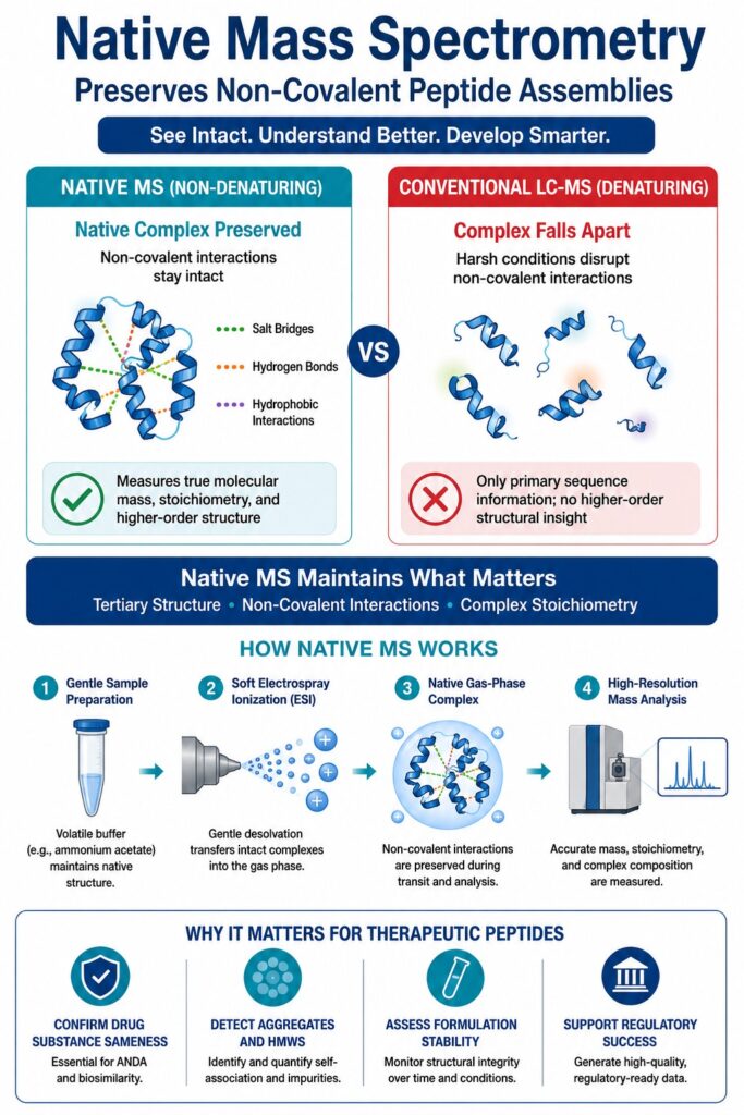
The structural characterization of biomolecular assemblies increasingly depends on Native Mass Spectrometry for Therapeutic Peptide Characterization, as it enables the direct preservation and quantitative assessment of tertiary structures, non-covalent interactions, and stoichiometric relationships of peptide therapeutics in their biologically active forms. Unlike denaturing analytical methods, native mass spectrometry maintains intact molecular assemblies throughout the analysis, providing valuable higher-order structural information that conventional peptide-focused proteomic approaches are unable to obtain. Retaining these native structural arrangements is essential because the therapeutic performance, biological activity, and pharmacokinetic behavior of modern peptide-based drugs are closely associated with their higher-order conformations and self-association characteristics.
Biotherapeutic peptides, including long-acting glucagon-like peptide-1 (GLP-1) receptor agonists and various lipopeptides, are intentionally designed to undergo self-association or interact with carrier proteins such as human serum albumin. These molecular interactions reduce renal elimination and significantly extend the circulating half-life of the therapeutic agent. Conventional denaturing liquid chromatography-mass spectrometry (LC-MS) methods disrupt these delicate assemblies through the use of strong acids and organic solvents, preventing accurate evaluation of the peptide’s authentic assembly state. To overcome this analytical limitation, specialized contract research organizations (CROs) and contract development and manufacturing organizations (CDMOs), including ResolveMass Laboratories Inc., utilize high-resolution native mass spectrometry to verify drug substance equivalence, evaluate formulation stability, and demonstrate regulatory compliance. As a Health Canada licensed and USFDA-registered facility, ResolveMass Laboratories Inc. provides comprehensive, traceable, and scientifically robust biophysical datasets that support submissions for investigational new drug (IND) applications and abbreviated new drug (ANDA) filings in accordance with international regulatory expectations.
To accelerate your drug development pipeline, explore our specialized CRO for GLP-1 Peptide Characterization services to ensure absolute structural accuracy and validation.
Share via:
Article Summary:
- Native mass spectrometry (Native MS) enables therapeutic peptides to be analyzed in their intact, biologically relevant form by preserving non-covalent interactions and higher-order structures that are typically disrupted by conventional denaturing LC-MS methods.
- Soft electrospray ionization (ESI) and volatile buffer systems allow peptide assemblies to transition into the gas phase without significant structural disruption, making it possible to accurately evaluate molecular mass, oligomeric state, and complex stoichiometry.
- Advanced mass spectrometers such as Orbitrap UHMR, Q-TOF, and FT-ICR provide the high mass range, resolution, and accuracy needed to characterize intact peptide complexes, detect subtle structural variations, and support detailed higher-order structural analysis.
- Charge reduction strategies using reagents like triethylammonium acetate (TEAA), ethylenediamine diacetate (EDDA), or gas-phase proton transfer improve the stability of fragile peptide complexes, reduce spectral congestion, and enhance data interpretation.
- Native MS plays a vital role in studying therapeutic lipopeptides, including GLP-1 receptor agonists, by identifying self-associated oligomers, monitoring aggregation behavior, distinguishing intact molecules from degradation products, and assessing product quality.
- The technique also enables real-time investigation of dynamic biological interactions, including peptide self-assembly, reversible oligomerization, metal-ion coordination, and membrane-associated peptide complexes under conditions that closely resemble their native environment.
- For biopharmaceutical development, Native MS provides essential analytical evidence for demonstrating molecular integrity, manufacturing consistency, formulation stability, and regulatory compliance, making it a valuable tool for peptide characterization throughout research, development, and quality control.
Molecular Forces Stabilizing Gas-Phase Peptide Assemblies
The structural integrity of gas-phase peptide assemblies within a mass spectrometer is maintained through a carefully balanced combination of electrostatic salt bridges, hydrogen bonding, and van der Waals interactions. Following desolvation, where bulk water molecules are removed, electrostatic interactions become substantially stronger because of the dramatic reduction in the dielectric constant. This strengthening of electrostatic attraction enables native-like tertiary conformations and non-covalent interfaces originally present in solution to remain intact throughout the millisecond-scale transit of the ions inside the mass analyzer.
The successful transfer of a solvated peptide complex into the gas phase relies on the gentle desolvation characteristics of electrospray ionization (ESI). In contrast to denatured or unfolded peptides, which generally acquire numerous charge carriers, natively folded peptide molecules possess compact surface geometries that produce a narrow distribution of lower charge states at higher mass-to-charge (m/z) values. As solvent evaporates from the electrospray droplets, repeated Coulombic fission events occur at the Rayleigh limit until the intact, fully desolvated peptide assembly is released into the gas phase. The behavior of individual non-covalent interactions during this transition ultimately determines whether the resulting gas-phase ion accurately reflects the native solution structure.
Electrostatic Interactions and Salt Bridges: In aqueous solution, charged amino acid residues are surrounded by water molecules that reduce electrostatic attraction through solvation. Once transferred into the vacuum environment of the mass spectrometer (ε = 1), these shielding effects disappear, causing salt bridges to become considerably stronger. These reinforced electrostatic interactions serve as structural anchors that preserve the integrity of the peptide complex and stabilize its quaternary architecture throughout analysis.
Hydrophobic Forces: Although the entropic contribution responsible for hydrophobic clustering in solution no longer exists after transition into the gas phase, the hydrophobic cores of acylated peptides generally remain kinetically trapped and structurally intact, provided excessive vibrational activation is avoided during ion transmission.
Hydrogen Bonding and Van der Waals Forces: These short-range intermolecular interactions play a vital role in preserving secondary structural elements, including α-helices and β-sheets. Their continued stability prevents collapse of the peptide backbone and minimizes random structural rearrangements during gas-phase analysis.
Complement your mass spectrometry data and confirm secondary conformations using our advanced platform for CD Spectroscopy for Peptide Secondary Structure Characterization.
Implementing Native Mass Spectrometry for Therapeutic Peptide Characterization on Advanced Instrument Platforms
Performing high-resolution Native Mass Spectrometry for Therapeutic Peptide Characterization requires advanced mass analyzers capable of operating across extended mass-to-charge ranges. Commonly employed platforms include Quadrupole Time-of-Flight (Q-TOF), Orbitrap Ultra-High Mass Range (UHMR), and Fourier Transform Ion Cyclotron Resonance (FT-ICR) mass spectrometers. These sophisticated analytical systems efficiently transmit, trap, and analyze large intact non-covalent peptide assemblies while maintaining the resolving power necessary to distinguish subtle molecular mass differences. Through precise optimization of collision cell pressures and electrical potentials, these instruments preserve higher-order structural organization during ion transmission and detection.
Historically, Q-TOF instruments represented the preferred choice for native mass spectrometry because of their excellent transmission efficiency for high-m/z ions. However, recent developments in Orbitrap technology equipped with the High Mass Range (HMRn) option have significantly advanced native MS by providing exceptional resolving power (>100,000 at m/z 4000) together with outstanding mass accuracy. These capabilities are particularly valuable for characterizing complex proteoforms and target-bound peptide assemblies. FT-ICR mass spectrometers operating under high magnetic field strengths, such as 15 T, currently represent the highest level of native mass spectrometric characterization. These systems enable simultaneous top-down sequencing through electron-capture dissociation (ECD) while also providing intact mass analysis of macromolecular assemblies extending to several hundred kilodaltons. Furthermore, the integration of native Mass Spectrometry Imaging (native MSI) with liquid extraction surface analysis (LESA) or nano-desorption electrospray ionization (nano-DESI) on these high-performance platforms enables visualization of the spatial distribution of intact folded peptide assemblies directly within biological tissue sections.
For multi-domain therapeutic molecules, map out your sequence precision by reviewing our optimized GLP-1 Analog Peptide Sequencing Workflow.
Methodological Workflows and Buffers for Native Mass Spectrometry for Therapeutic Peptide Characterization
Successful implementation of Native Mass Spectrometry for Therapeutic Peptide Characterization depends on replacing non-volatile physiological salts with volatile, mass spectrometry-compatible buffer systems such as ammonium acetate. This buffer exchange process is commonly performed immediately before electrospray ionization using size-exclusion spin columns or microdialysis techniques to preserve native conformational integrity. Since non-volatile salts generate substantial peak broadening through random adduct formation, maintaining highly purified volatile buffer conditions is essential for acquiring high-quality native mass spectra.
| Buffer Component / Parameter | Standard Operating Range | Analytical Function and Relevance |
|---|---|---|
| Ammonium Acetate (NH₄CH₃COO) | 50 mM to 200 mM | A volatile, MS-compatible buffering system that reproduces physiological ionic strength and pH while minimizing ion suppression effects. |
| Solution pH | 6.7 to 8.0 | Maintained close to physiological neutrality to preserve the native tertiary and quaternary structures of therapeutic peptides. |
| Organic Co-solvents | < 5% | Restricted to very low concentrations to avoid premature hydrophobic denaturation and structural unfolding of peptide molecules. |
| Glycerol / Non-volatile Additives | 0% (Strictly Avoided) | Completely excluded because they produce severe peak broadening and significant signal deterioration during mass spectrometric analysis. |
| Micro Bio-Spin Columns | 6 kDa molecular weight cut-off | Utilized for rapid, high-efficiency buffer exchange immediately before nanoelectrospray ionization. |
To achieve effective desolvation while preventing thermal dissociation of peptide assemblies, instrument parameters such as source backing pressure and collisional activation voltage require careful optimization. Standard high-vacuum instrument configurations are modified to sustain elevated pressures, often reaching several mbar during the early stages of vacuum generation. Increased gas pressure enhances collisional cooling, reducing the kinetic energy of heavy peptide complexes and improving their transmission efficiency into the mass analyzer. When residual solvent or salt adducts remain associated with peptide ions, operators apply a carefully controlled potential drop at the ion funnel exit to remove solvent shells selectively without disrupting the fundamental non-covalent interactions responsible for maintaining structural integrity.
Access Analytical Guidelines: Download the comprehensive Peptide Characterization CRO Deliverables Checklist to ensure your structural data meets all essential laboratory benchmarks.
Charge Reduction Chemistry and Gas-Phase Stability
Lowering the overall charge carried by electrosprayed peptide complexes enhances their structural stability in the gas phase by minimizing destabilizing Coulombic repulsion. This objective can be achieved by incorporating solution additives such as triethylammonium acetate (TEAA) or by exposing electrospray droplets to organic solvent vapors, including acetonitrile, within the ion source housing. Reducing the number of charge carriers present on the peptide surface decreases electrostatic stress, thereby preserving fragile non-covalent assemblies that might otherwise dissociate during gas-phase analysis.
Throughout the electrospray ionization process, peptide ions possessing high charge states are vulnerable to Coulombic unfolding and asymmetric monomer dissociation. Employing charge-reducing reagents shifts the observed charge-state distribution toward higher m/z values, thereby increasing the separation between neighboring spectral peaks and substantially improving spectral clarity and resolution. The selection of an appropriate charge-reducing strategy significantly influences both the gas-phase conformation and the stability of peptide complexes.
Triethylammonium Acetate (TEAA) and Ethylenediamine Diacetate (EDDA): These basic buffer additives function as proton-transfer reagents during droplet evaporation, lowering the net charge of emerging peptide ions and promoting the preservation of compact native conformations.
Solvent Vapor Exposure (Acetonitrile): Introducing neutral acetonitrile vapor into the atmospheric region of the electrospray ionization source decreases the average charge state of peptide assemblies without requiring chemical modification of the solution. This strategy also reduces the occurrence of common alkali metal adducts that may interfere with spectral interpretation.
Proton Transfer Charge Reduction (PTCR): Within the mass spectrometer, multiply charged peptide ions are exposed to perfluorinated anions, including species generated from perfluorodecylamine. Gas-phase proton transfer reactions reduce the charge state of these ions, effectively separating overlapping ion populations and improving the interpretation of highly congested native mass spectra.
Stoichiometric Deconvolution of GLP-1 Receptor Agonists and Lipopeptide Assemblies
Native mass spectrometry enables the resolution of co-existing lipopeptide oligomers by separating individual quaternary assembly states within the m/z domain while accurately determining their molecular masses and relative abundances. This analytical capability is particularly important for acylated therapeutics such as liraglutide and semaglutide, which naturally undergo self-association to generate complex and dynamic higher-order structures in solution. Measuring the intact mass of these assemblies under native conditions allows researchers to differentiate biologically functional oligomeric drug species from degraded or potentially immunogenic high-molecular-weight species (HMWS).
Therapeutic lipopeptides including semaglutide (4113.58 Da) and tirzepatide (4813.53 Da) contain hydrophobic fatty acid side chains that promote the formation of non-covalent oligomeric assemblies within pharmaceutical formulations. Using denaturing size-exclusion chromatography coupled with high-resolution mass spectrometry (dSEC-HRMS), investigators can effectively separate, identify, and quantify these high-molecular-weight species. This high-resolution analytical strategy also permits detection of truncated peptide variants together with non-dissociable covalent impurities at concentrations as low as 0.1%, making it an essential tool for confirming product purity, consistency, and safety throughout release testing.
| Peptide Formulation / Species | Theoretical Mass (Da) | Observed Mass (Da) | Relative Abundance (%) | Proposed Structural / Compositional Assignment |
|---|---|---|---|---|
| Semaglutide Monomer | 4113.58 | 4113.58 | >97.0 | Intact acylated therapeutic peptide monomer. |
| Semaglutide HMWS1 | 8227.16 | 8227.16 | 2.60 | Non-dissociable covalent dimer. |
| Semaglutide HMWS2-1 | 12340.74 | 11649.28 | 0.17 | Significantly truncated, non-dissociable trimer (Δmass = -691.46 Da). |
| Tirzepatide Monomer | 4813.53 | 4813.53 | >99.8 | Intact dual GIP/GLP-1 receptor agonist monomer. |
| Tirzepatide HMWS1a | 9627.06 | 9627.06 | 0.06 | Non-dissociable covalent dimer. |
| Tirzepatide HMWS1b | 9627.06 | 9627.06 | 0.07 | Structurally distinct or conformational covalent dimer. |
| Tirzepatide HMWS1a-1 | 9627.06 | 7903.16 | Low Abundance | Truncated, non-dissociable dimer (Δmass = -1723.86 Da). |
Track In-Depth Oligomer Profiles: To accurately profile high-molecular-weight species and structural degradants, leverage our specialized GLP-1 Peptide Impurity Characterization and sequence mapping workflows.
Time-Course Kinetics and Anisotropic Assembly Pathways
The oligomerization behavior of acylated peptides such as liraglutide represents a dynamic, time-dependent process that progresses through an anisotropic assembly pathway before forming stable higher-order molecular architectures. During the earliest stages, small and short-lived intermediates are generated, which gradually reorganize into a “fuzzy oil drop” configuration that effectively sequesters hydrophobic fatty acid chains away from the surrounding polar solvent. Native ion mobility-mass spectrometry (nIM-MS), combined with molecular dynamics (MD) simulations, provides real-time visualization of this structural evolution and reveals the fundamental mechanisms responsible for peptide self-association.
Liraglutide (4113.0 Da) undergoes a sequential and pH-dependent transition from low-order oligomeric species into large macromolecular assemblies. When prepared at a concentration of 1 mg·mL⁻¹ in 20 mM ammonium acetate at pH 6.7, the peptide exhibits multiple co-existing structural populations. Within approximately 10 minutes following preparation, monomeric molecules together with low-order oligomers (n = 2-8) dominate the m/z 2000-3500 region of the native mass spectrum. As incubation progresses over approximately 270 minutes, these smaller intermediates gradually reorganize into larger assemblies, with oligomeric species ranging from n = 13-16 (m/z 3500-5000) becoming the predominant structural population. After 24 hours, the charge states associated with the n = 13-16 assemblies decrease, while a highly stable n = 17 oligomer carrying 14 positive charges ([17]¹⁴⁺) becomes the dominant species.
[Monomers / Extended States] (t = 0)
       │
       ▼ (t < 10 min)
[Transient Oligomers (n = 2–8)] (Hydrophobic acylation clustering)
       │
       ▼ (t = 30–90 min)
[Intermediate Assemblies (n = 13–16)] (Fuzzy oil drop condensation)
       │
       ▼ (t = 270 min – 24 h)
[Stable Quaternary Complexes (n = 17)] (Reversible, histidine-protonation dependent)
Importantly, this self-assembly process remains fully reversible. Increasing the solution pH from 6.7 to 8.1 decreases the relative abundance of the n = 13-17 oligomeric complexes by nearly 50% within only 10 minutes, resulting in the rapid re-emergence of smaller oligomeric species ranging from n = 2-8. This rapid structural interconversion is governed by changes in the protonation state of the N-terminal histidine residues, which directly regulate the rate of monomer dissociation, the primary rate-limiting step controlling oligomer transformation. Structural characterization through electron-capture dissociation (ECD) demonstrates restricted fragmentation within the C-terminal region, evidenced by reduced production of z-ions. These observations confirm that the C-terminus remains deeply buried inside the compact oligomeric core. Furthermore, advanced single-molecule native mass spectrometry techniques, including Direct Mass Technology (DMT), have identified exceptionally large liraglutide assemblies ranging from n = 25-62. These massive structures are stabilized through external hydrophilic interactions that surround and reinforce previously formed hydrophobic cores.
Discover how to track and limit multi-mer formation using our targeted Peptide Aggregation Analysis workflows.
Metal-Peptide Coordination Stoichiometry via Soft Ionization
Native electrospray ionization allows direct observation and quantitative characterization of metal-peptide coordination complexes in solution without disrupting their native structural organization. Unlike conventional denaturing mass spectrometry methods, native ionization preserves biologically relevant metal-binding interactions throughout analysis. Since numerous biological and therapeutic peptides depend upon coordination with essential metal ions, including zinc (Zn²⁺), to achieve their active conformations, evaluating these interactions under non-denaturing conditions is critical for confirming structural integrity and biological functionality. The exceptional mass accuracy provided by native mass spectrometry enables simultaneous identification, resolution, and quantification of multiple co-existing metallo-peptide species within highly complex mixtures.
A representative example involves titration of the histidine-rich peptide HRGP330, a 35-amino acid peptide containing 17 histidine residues together with four glutamic/aspartic acid residues, using increasing concentrations of zinc acetate. Native mass spectra reveal a highly dynamic coordination landscape characterized by multiple metal-binding stoichiometries.
1:1 Molar Ratio: At stoichiometric equivalence (1:1), native nanoESI-MS produces nearly identical signal intensities for both the unbound (apo) peptide and the mono-zinc complex (Zn₁-HRGP330), while smaller populations corresponding to the di-zinc (Zn₂) complex are also observed.
2 Molar Equivalents: Following the addition of two molar equivalents of zinc, the Zn₂-HRGP330 species becomes the dominant complex, whereas the signal corresponding to the apo peptide nearly disappears.
5 Molar Equivalents: At five molar equivalents of zinc, native mass spectra display a broad and heterogeneous distribution of zinc-bound complexes. The tetra-zinc (Zn₄) species represents the predominant population, followed by Zn₅ and Zn₃ complexes, while lower-intensity signals corresponding to Zn₆ and Zn₂ remain detectable.
Assuming that each Zn²⁺ ion requires coordination by four ligands, the 35-residue sequence of HRGP330 provides a total of 21 potential donor residues, theoretically supporting coordination of as many as five zinc ions through mono-dentate binding interactions. Detection of the Zn₆ species under elevated zinc concentrations indicates that peptide coordination likely involves bridging bidentate aspartate/glutamate carboxylate groups or occupation of lower-affinity coordination sites requiring fewer than four peptide-derived ligands. This highly detailed speciation profile represents a unique analytical advantage of native mass spectrometry because conventional denaturing techniques remove coordinated metal ions from the peptide backbone, leaving only the mass of the apo peptide available for analysis.
For highly structured or tied configurations, view our platforms for Cyclic Peptide Characterization to preserve complex geometries during soft ionization.
Advanced Reconstitution in Membrane-Mimetic Systems
Characterization of membrane-active peptides together with their receptor complexes requires their incorporation into membrane-mimetic carriers such as detergent micelles, nanodiscs, or peptidiscs before electrospray ionization. These biomimetic systems stabilize hydrophobic transmembrane domains within aqueous solution while allowing selective removal of the surrounding carrier during mass spectrometric analysis through collisional activation. This strategy enables detailed investigation of membrane-associated peptide interactions under native-like conditions while preserving biologically relevant lipid interactions and target-binding characteristics.
Detergent micelles generated using compounds such as n-dodecyl-β-D-maltoside (DDM) or lauryldimethylamine N-oxide (LDAO) are commonly employed to stabilize membrane-associated complexes in volatile ammonium acetate solutions. During transmission through the mass spectrometer, collision-induced dissociation supplies sufficient activation energy to vaporize the surrounding detergent micelle, thereby releasing the intact and folded peptide complex directly into the mass analyzer. Although this technique is highly effective, the elevated activation energy required for detergent removal may occasionally induce thermal denaturation or remove essential regulatory lipid molecules associated with the target complex.
To reduce the likelihood of thermal stress, peptidiscs provide a gentler, detergent-free alternative for membrane protein reconstitution. Constructed from bi-helical peptide segments possessing carefully optimized hydrophobic and hydrophilic surfaces, peptidiscs surround the hydrophobic transmembrane regions of membrane complexes and effectively prevent aggregation in aqueous buffer systems. Comparative native mass spectrometry studies involving membrane proteins, including the 30 kDa helical AceI-Bril complex, have demonstrated efficient gas-phase release from both detergent micelles and peptidisc environments.
LDAO Micelles: Gas-phase ejection produces a well-resolved monomeric protein ion with an observed molecular mass of 30421 ± 0.5 Da.
Peptidiscs: Reconstituted complexes generate an essentially identical observed mass of 30418 ± 3 Da, confirming complete preservation of structural integrity throughout sample preparation and mass spectrometric analysis.
Although releasing the target complex from a peptidisc requires a higher activation energy (250 V) than that needed for conventional detergent micelles (120 V), the peptidisc platform offers a significantly more stable and non-denaturing solution environment. This enhanced stability substantially reduces the likelihood of structural collapse, thermal damage, or premature dissociation of protein subunits during sample preparation while preserving the native architecture of membrane-associated peptide complexes.
Learn how to isolate conformation and solution states simultaneously with 2D NMR for Peptide Characterization.
Technical Performance of ResolveMass Laboratories in Complex Biotherapeutic Workflows
ResolveMass Laboratories Inc. plays a significant role in supporting modern biopharmaceutical development through the implementation of high-resolution native mass spectrometry workflows within a rigorously regulated quality framework. Operating as an ISO 9001:2015 certified, USFDA-registered, and Health Canada GMP licensed contract research organization (CRO), the laboratory generates accurate, traceable, and regulatory-ready structural datasets that support both investigational new drug (IND) and abbreviated new drug (ANDA) submissions. By employing advanced Orbitrap and Q-TOF mass spectrometry platforms, ResolveMass Laboratories Inc. performs comprehensive characterization of peptide therapeutics and polymer-based drug products while maintaining stringent quality assurance and quality control standards throughout every stage of analysis.
For organizations developing peptide therapeutics, demonstrating drug substance “sameness” for generic ANDA applications or establishing structural biosimilarity presents a highly demanding analytical challenge. Conventional quality control methodologies frequently fail to detect dynamic self-association behavior, non-covalent aggregation, and low-level chemical modifications that may influence product quality, stability, or therapeutic performance. ResolveMass Laboratories Inc. addresses these analytical limitations by integrating high-resolution native mass spectrometry with advanced liquid chromatography techniques, enabling detailed structural characterization and molecular mapping that align with the scientific and regulatory expectations established by international health authorities.
Review the crucial guidelines for marketing approval with our deep dive on Regulatory Requirements for GLP-1 Peptide Characterization.
Beyond the evaluation of tertiary and quaternary molecular structures, the laboratory possesses extensive expertise in custom organic synthesis, characterization of controlled-release polymer systems including PLGA and PLA matrices, and comprehensive impurity assessment. These analytical capabilities include highly sensitive quantification of trace nitrosamines together with per- and polyfluoroalkyl substances (PFAS). Through this broad range of scientific services, ResolveMass Laboratories Inc. provides an integrated analytical platform that supports therapeutic developers throughout the complete product lifecycle, from early-stage research and development to GMP-compliant release testing and regulatory submission.
Track critical product traits via high-throughput automation using Multi-Attribute Monitoring (MAM) for Peptide Characterization.
Conclusion
The incorporation of Native Mass Spectrometry for Therapeutic Peptide Characterization into biopharmaceutical development delivers an exceptional level of structural insight into the stoichiometry, stability, and higher-order organization of complex peptide therapeutics. This advanced analytical approach enables developers to confidently demonstrate drug substance sameness, evaluate aggregation behavior, and verify manufacturing consistency across the product development process. By preserving fragile non-covalent interactions and directly measuring intact molecular assemblies under native conditions, native mass spectrometry provides critical biophysical information that cannot be obtained using conventional denaturing analytical techniques. These capabilities make it an indispensable technology for the successful development, characterization, and regulatory evaluation of safe, effective, and high-quality peptide therapeutics.
Set up a detailed study blueprint and view our comprehensive options for Peptide Physicochemical Characterization Services to securely guide your molecule to market.
Organizations seeking advanced biophysical characterization or regulatory-compliant analytical support for peptide therapeutics can benefit from the specialized CRO and CDMO services offered by ResolveMass Laboratories Inc. The company’s scientific expertise encompasses high-resolution native mass spectrometry, custom synthesis, impurity profiling, and comprehensive analytical characterization designed to meet the requirements of modern biopharmaceutical development. Researchers and pharmaceutical developers interested in discussing project requirements or arranging a technical consultation regarding custom synthesis or high-resolution mass spectrometry services can contact ResolveMass Laboratories Inc. through the Contact Us page.
Frequently Asked Questions
Native Mass Spectrometry for Therapeutic Peptide Characterization preserves non-covalent interactions by employing soft electrospray ionization (ESI) together with volatile buffers that closely resemble physiological conditions. During ionization, peptide assemblies are transferred into the gas phase with minimal structural disruption. As solvent molecules are removed, electrostatic interactions, hydrogen bonds, and salt bridges become sufficiently stable to maintain the native-like architecture of the peptide complex throughout mass analysis.
Ammonium acetate is widely used because it is a volatile buffer that evaporates completely during the electrospray ionization process, leaving minimal background contamination. This property helps generate clean mass spectra with excellent signal quality and mass accuracy. Unlike non-volatile salts such as phosphate or sodium chloride, ammonium acetate does not produce excessive adduct formation or peak broadening that can interfere with accurate structural analysis.
Native mass spectrometry establishes subunit stoichiometry by accurately measuring the intact molecular mass of the complete peptide assembly under non-denaturing conditions. The measured mass is then compared with the known masses of individual monomeric components or smaller sub-complexes. This comparison enables researchers to determine the exact number and arrangement of subunits within the native complex while preserving its biological organization.
Acylated GLP-1 receptor agonists such as liraglutide and semaglutide often exist as mixtures of monomers and multiple oligomeric species in solution. Native mass spectrometry separates these different assemblies according to their mass-to-charge (m/z) ratios without disrupting their native structures. This approach allows simultaneous identification, mass determination, and relative quantification of individual oligomeric populations, providing a detailed picture of formulation heterogeneity.
The “fuzzy oil drop” model explains how liraglutide molecules self-assemble into stable higher-order oligomers. In this structural arrangement, the hydrophobic fatty acid chains cluster together to form a protected internal core, while the hydrophilic peptide regions remain exposed to the surrounding solvent. This organization enhances structural stability and helps explain the peptide’s self-association behavior under physiological conditions.
N-terminal cyclization is a process that may occur during the manufacturing or storage of liraglutide, often promoted by trace levels of formaldehyde contamination. This modification produces a measurable mass increase of 12 Da because of cyclization involving the N-terminal histidine residue. High-resolution liquid chromatography coupled with mass spectrometry enables accurate detection, identification, and monitoring of this impurity during quality control testing.
Charge-reducing reagents such as triethylammonium acetate (TEAA) or imidazole decrease the number of charges acquired by peptide ions during electrospray ionization. Lower charge states reduce internal Coulombic repulsion, helping preserve compact native conformations throughout gas-phase analysis. As a result, fragile non-covalent peptide assemblies remain intact and produce clearer, more interpretable mass spectra with improved resolution.
Yes. Native mass spectrometry is well suited for monitoring dynamic structural changes and reversible oligomerization processes in real time. For example, altering the pH of a liraglutide solution can rapidly shift the equilibrium between larger oligomeric assemblies and smaller peptide species. These structural transitions can be directly observed and quantified, providing valuable insights into peptide stability and self-association mechanisms.
Native mass spectrometry imaging (native MSI) combines techniques such as liquid extraction surface analysis (LESA) and nanoelectrospray ionization (nanoESI) with high-resolution mass spectrometers to analyze biological tissues. This approach preserves intact peptide complexes while simultaneously determining their spatial distribution within tissue sections. Because the analysis does not require fluorescent or chemical labeling, it provides highly accurate localization of native peptide therapeutics in biological samples.
Reference:
- Boeri Erba, E., & Petosa, C. (2015). The emerging role of native mass spectrometry in characterizing the structure and dynamics of macromolecular complexes. Protein Science, 24(8), 1176–1192. https://doi.org/10.1002/pro.2661
- Gavriilidou, A. F. M., Sokratous, K., Yen, H.-Y., & De Colibus, L. (2022). High-throughput native mass spectrometry screening in drug discovery. Frontiers in Molecular Biosciences, 9, Article 837901. https://doi.org/10.3389/fmolb.2022.837901
- Kaltashov, I. A., Bobst, C. E., & Abzalimov, R. R. (2012). Advanced mass spectrometry-based methods for the analysis of conformational integrity of biopharmaceutical products. Pharmaceutical Research, 29(4), 995–1007. https://doi.org/10.1007/s11095-011-0657-8
- Zhang, H., Cui, W., Gross, M. L., & Blankenship, R. E. (2013). Native mass spectrometry of photosynthetic pigment-protein complexes. FEBS Letters, 587(8), 1012–1020. https://doi.org/10.1016/j.febslet.2013.01.005
- Reading, E., Keener, J. E., Liko, I., Allison, T. M., & Hopper, J. T. S. (2025). Native mass spectrometry of membrane proteins reconstituted in peptidiscs. Chemical Biology, 21(2), 115–127. https://doi.org/10.1039/D5CB00236B
- Morgan, E. M., Smith, J. H., Keane, F. D. L., Sobott, F., Blindauer, C. A., & Stewart, A. J. (2018). Native electrospray mass spectrometry approaches to probe the interaction between zinc and an anti-angiogenic peptide from histidine-rich glycoprotein. Scientific Reports, 8, Article 8647. https://doi.org/10.1038/s41598-018-26924-1
Get In Touch With Us
About The Author


