

Introduction
NMR-Based Structural Elucidation provides a definitive analytical approach for determining the three-dimensional architecture, solution-state dynamics, and covalent linkage geometry of peptide-oligonucleotide conjugates (POCs) with atomic-level resolution. These chimeric molecules combine the sequence-selective targeting functions of therapeutic nucleic acids, including antisense oligonucleotides (ASOs), small interfering RNAs (siRNAs), microRNAs, and aptamers, with functional peptides that can provide membrane penetration, receptor recognition, or selective organ targeting. Through this combination, POCs can address several longstanding limitations associated with nucleic acid drug delivery. Determining how the peptide and oligonucleotide portions affect one another’s native conformations, binding thermodynamics, and conformational stability requires detailed structural information under solution conditions that closely resemble their functional environment. Consequently, high-resolution nuclear magnetic resonance (NMR) spectroscopy is an essential analytical technology for the development and characterization of these biopharmaceuticals.
You can explore the core types of peptide-oligonucleotide conjugates and their therapeutic applications.
The integration of two fundamentally different biomolecular components into one conjugate creates substantial analytical complexity. Oligonucleotides contain polyanionic phosphodiester or phosphorothioate backbones together with stacked, hydrophobic nucleobases, while peptides may contain combinations of hydrophobic, amphiphilic, and polycationic amino acid sequences. NMR-Based Structural Elucidation enables non-destructive characterization in solution while providing simultaneous information about intra-domain folding, tertiary interactions between domains, and the detailed spatial arrangement of the covalent junction connecting the peptide and nucleic acid components.
Discover the primary mechanisms for cellular entry of peptide-oligonucleotide conjugates in biopharmaceutical development.
Share via:
Quick Summary:
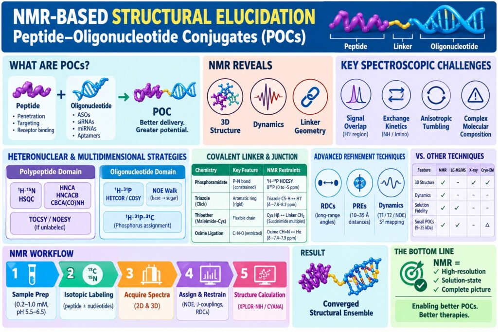
- NMR-based structural elucidation provides atomic-level insight into the three-dimensional structure, solution-state dynamics, and covalent linkage geometry of peptide–oligonucleotide conjugates (POCs).
- POCs combine peptides and nucleic acids to support membrane penetration, receptor recognition, and selective organ targeting, while addressing challenges in nucleic acid drug delivery.
- Major spectroscopic challenges include overlapping proton signals, rapid amide exchange, and anisotropic molecular tumbling, requiring specialized multidimensional NMR methods.
- Heteronuclear NMR strategies such as HSQC, HNCA, HNCACB, TOCSY, NOESY, and phosphorus-based experiments enable reliable assignment of peptide and oligonucleotide domains.
- Covalent linker characterization uses inter-domain NOEs, heteronuclear interactions, and specialized experiments such as 1H^{1}H1H-31P^{31}P31P HOESY to define junction geometry and flexibility across phosphoramidate, triazole, thioether, and oxime linkages.
- Advanced refinement techniques including RDCs, PREs, and relaxation measurements provide long-range structural restraints and reveal domain orientation, rigidity, and flexible hinge-like motion.
- A complete NMR workflow combines sample preparation, isotopic labeling, multidimensional data acquisition, resonance assignment, restraint generation, and molecular dynamics calculations to produce a converged structural ensemble—making NMR a powerful tool for understanding and developing complex POC therapeutics.
Spectroscopic Challenges in the NMR-Based Structural Elucidation of Biomolecular Chimeras
The NMR-based structural elucidation of peptide-oligonucleotide conjugates presents several spectroscopic difficulties arising from extensive chemical shift overlap between ribose and amino acid protons, differences in solvent exchange behavior, and anisotropic rotational tumbling. Resolving these highly congested signals requires specialized heteronuclear and multidimensional NMR acquisition strategies specifically adapted to hybrid biomolecular systems.
A major characterization challenge is the substantial redundancy of proton (¹H) chemical shifts across the two conjugated domains. Ribose sugar ring protons (H2′, H3′, H4′, H5′, H5″) of nucleic acids generally appear within the 3.5–5.0 ppm region, where they overlap with polypeptide backbone alpha-protons (Hα) and numerous aliphatic side-chain resonances. In addition, aromatic base protons (H6/H8/H2) exhibit extensive overlap with aromatic side-chain resonances originating from phenylalanine, tyrosine, and tryptophan residues, particularly within the 6.5–8.5 ppm range.
Peptide Domain                        Covalent Linker                       Oligonucleotide Domain
[Amphiphilic / Cationic] <=======> [Amide / Triazole / Thioether] <=======> [Polyanionic Phosphorothioate]
   - Fast Amide Exchange              - Local Flexibility Constraints          - Hydrogen-Bonded Base Pairs
   - Broad Chemical Shift             - Inter-domain NOE Network               - Narrow Sugar Proton Dispersion
Differences in proton exchange kinetics introduce another layer of complexity during spectrum interpretation. Peptide backbone amide protons (NH) can exchange rapidly with bulk aqueous solvent under physiological pH conditions. Therefore, lower temperatures (278–298 K) and mildly acidic buffer conditions (pH 5.5–6.5) may be required to preserve observable resonances. By comparison, Watson-Crick and Hoogsteen base-pairing imino protons (T-H3, G-H1) are strongly dependent on the stability of the corresponding base pairs and local secondary structure. Their exchange behavior can consequently differ substantially between flexible terminal regions and more rigid internal duplex segments.
Read more about structural characterization of peptide-oligonucleotide conjugates using advanced analytical techniques.
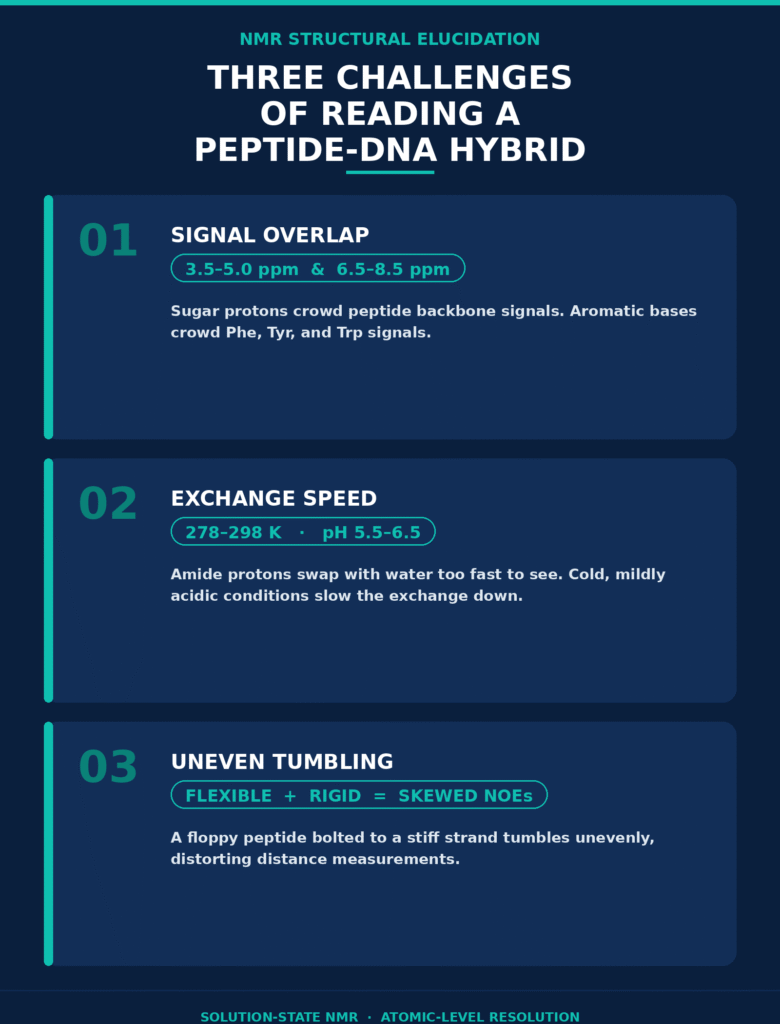
Anisotropic rotational correlation times (τm) create an additional physical limitation. When flexible peptide tails are covalently connected to rigid double-stranded or hairpin nucleic acid structures, the resulting conjugate can exhibit complicated, non-isotropic rotational behavior in solution. Such anisotropic tumbling affects nuclear dipolar relaxation and can interfere with conventional Nuclear Overhauser Effect (NOE) intensity-to-distance relationships. Accurate structural interpretation therefore requires advanced computational refinement approaches that account for these non-uniform motional properties.
Review the key analytical and synthetic challenges in POC development during drug design.
Heteronuclear Multidimensional Assignment Strategies
Heteronuclear multidimensional assignment strategies reduce spectral congestion by distributing proton resonances across additional isotopic dimensions involving carbon (¹³C), nitrogen (¹⁵N), and phosphorus (³¹P). This multidimensional dispersion facilitates reliable sequential assignment of both the polypeptide backbone and the nucleic acid phosphodiester or phosphorothioate chain.
Pulse Sequences for the Polypeptide Domain
Assignment of polypeptide backbone and side-chain resonances is typically performed using 2D ¹H-¹⁵N HSQC/HMQC together with 3D triple-resonance pulse sequences, including HNCA, HNCACB, and CBCA(CO)NH. These heteronuclear experiments establish correlations between amide nitrogen and proton resonances and their associated intra- and inter-residue carbon atoms, thereby generating reliable sequential assignment pathways.
For unlabeled or selectively labeled peptide domains, homonuclear 2D ¹H-¹H TOCSY (Total Correlation Spectroscopy) and 2D ¹H-¹H NOESY experiments can be used to identify intra-residue spin systems and sequential Hαᵢ → NHᵢ₊₁ connectivities. Uniform ¹³C/¹⁵N labeling, however, provides a major advantage by enabling 3D triple-resonance spectroscopy. These experiments reduce the impact of proton chemical shift overlap by correlating nitrogen and carbon resonances through scalar J-coupling pathways. Scalar J-couplings (³JHNα) obtained from HNHA experiments can further provide backbone dihedral angle (φ) restraints through domain-specific Karplus relationships. These restraints help establish whether conjugation promotes or disrupts an α-helix, β-sheet, or random coil conformation within the peptide domain.
Check out modern synthesis methods for peptide-oligonucleotide conjugates used in structural studies.
Multidimensional NMR Methods for Oligonucleotide Backbones
Oligonucleotide backbone assignments depend on sequential nuclear Overhauser effect (NOE) connectivity between base and sugar protons, supported by heteronuclear ¹H-³¹P correlations. Together, these spectral pathways enable tracing of the phosphodiester chain and provide evidence for base-pairing patterns and secondary structures present in solution.
The conventional sequential assignment approach, commonly referred to as the “NOE walk,” follows correlations between aromatic base protons and sugar protons belonging to the same nucleotide as well as the adjacent 5′-neighbor:
H6/H8ᵢ → H1′ᵢ → H6/H8ᵢ₊₁ → H1′ᵢ₊₁
Within defined secondary structures, intra-strand and inter-strand NOE cross-peaks involving imino protons (G-H1 and T/U-H3) provide evidence for duplex or hairpin formation. Because phosphodiester and phosphorothioate backbones substantially influence the three-dimensional geometry of nucleic acids, heteronuclear ¹H-³¹P HETCOR, 2D ¹H-³¹P COSY, and 3D ¹H-¹³C-³¹P experiments can be applied to assign phosphorus resonances and follow their positions along the phosphodiester chain. Phosphorothioate (PS) modifications create chiral phosphorus centers, represented by Rp and Sp diastereomers. These stereochemical configurations can produce distinct ³¹P chemical shift resonances, offering sensitive indicators of stereochemical purity and local backbone conformation.
See effective sequence confirmation strategies for POCs for quality control.
NMR-Based Structural Elucidation of Covalent Linker Geometry and Junction Dynamics
Determination of covalent linker geometry requires the observation of inter-domain nuclear Overhauser contacts and scalar heteronuclear interactions that extend across the chemical coupling junction. Such measurements provide direct information regarding the relative spatial orientation and conformational mobility of the peptide and nucleic acid domains.
The covalent junction may be generated through direct phosphoramidate coupling, copper-catalyzed azide-alkyne cycloaddition (triazole click chemistry), oxime ligation, or maleimide-thiol thioether coupling. The selected chemistry has a direct influence on how the two molecular domains are positioned relative to one another. Structural characterization of the junction therefore relies on identifying unambiguous inter-domain NOE cross-peaks (d < 5 Å) between terminal amino acid side chains and nucleotide sugars or bases located near the attachment site. Specialized heteronuclear Overhauser spectroscopy, including ¹H-³¹P HOESY, can provide direct information about spatial proximity between peptide protons and the terminal ³¹P nucleus associated with the linkage region.
Dive into the details of linker chemistry in peptide-oligonucleotide conjugates to optimize stability.
| Conjugation Chemistry | Linker Structural Features | Key Inter-Domain NMR Restraints | Diagnostic Chemical Shift Handles |
|---|---|---|---|
| Phosphoramidate Linkage | Direct P-N covalent bond; highly constrained junction | ¹H-³¹P HOESY (NHlinker ↔ P) | ³¹P shift displacement (0 to -5 ppm) |
| Triazole (Click Chemistry) | Aromatic 1,2,3-triazole ring; rigid planar core | Triazole C5-H ↔ Sugar H1′ NOEs | Heteroaromatic singlet (¹H approximately 7.8–8.2 ppm) |
| Thioether (Maleimide-Cys) | Flexible aliphatic chain with succinimide ring | Cys Hβ ↔ Linker CH₂ NOEs | Succinimide CH-CH₂ multiplet dispersion |
| Oxime Ligation | Restricted rotation around the C=N-O double bond | Oxime CH=N ↔ Peptide Hα NOEs | Aldoxime proton singlet (¹H approximately 7.4–7.9 ppm) |
Refinement Techniques: Residual Dipolar Couplings, PREs, and Solution Dynamics
High-resolution refinement of peptide-oligonucleotide chimeric structures can incorporate Residual Dipolar Couplings (RDCs) and Paramagnetic Relaxation Enhancements (PREs) to introduce long-range angular and distance restraints. These advanced NMR parameters supplement local NOE-derived distance restraints and improve the accuracy of global domain positioning and alignment.
Conventional NOE distance restraints (d ∝ I⁻¹⁄⁶) are primarily local and generally describe interproton distances below approximately 5–6 Å. For extended biomolecular conjugates, the accumulation of small local structural uncertainties can result in substantial global deviations, making the relative orientation of the peptide and oligonucleotide domains difficult to define. RDC measurements can address this limitation by analyzing the conjugate in a weakly aligning liquid crystalline environment, such as Pf1 bacteriophage, neutral polyacrylamide strained gels, or phospholipid bicelles.
In an anisotropic medium, incomplete molecular tumbling results in residual dipolar couplings (DIJ) that remain observable rather than averaging completely to zero. The heteronuclear dipolar interaction between nuclei I and J is determined by their internuclear separation (rIJ) and by the orientation angle (θ) of the internuclear vector relative to the principal axes of the alignment tensor (A):
DIJ(θ, φ) = -[μ0 γI γJ ħ / (4π² rIJ³)]
            × [Azz(3cos²θ - 1) + (3/2) Axx-yy sin²θ cos(2φ)]
Measurements of ¹DNH and ¹DCH within the peptide backbone, together with ¹DC-H and ¹DP-H measurements in the nucleic acid backbone, provide global angular restraints relative to a common molecular alignment frame. These restraints are particularly useful for establishing the relative orientation of the two domains within the complete conjugate.
Paramagnetic Relaxation Enhancement (PRE) measurements provide a complementary source of long-range structural information, typically covering distances of approximately 10–35 Å. A site-specific paramagnetic nitroxide spin label, such as MTSL, or a lanthanide chelate containing Gd³⁺ can be attached to one molecular domain. The resulting paramagnetic center increases the transverse relaxation rate (R2) of nuclear spins in the other domain in an r⁻⁶-dependent manner. This pronounced distance dependence allows spatial contacts between domains separated by relatively large distances to be identified.
To determine whether conjugation changes the dynamic behavior of either domain, ¹⁵N and ¹³C spin-lattice (T1) and spin-spin (T2) relaxation measurements can be combined with heteronuclear Overhauser effects (¹H-{¹⁵N} NOE). Lipari-Szabo ModelFree analysis can then be applied to derive the generalized order parameter (S²). This parameter describes the degree of local structural restriction over nanosecond-to-picosecond timescales, where S² = 1 represents highly restricted or rigid motion and S² = 0 corresponds to complete disorder. Mapping S² values across the conjugate can therefore reveal regions of enhanced rigidity as well as flexible hinge-like motion around the covalent bridge.
Understand key parameters surrounding peptide-oligonucleotide conjugate stability under physiological conditions.
Comparative Analytical Modalities for Chimera Characterization
Solution-state NMR spectroscopy provides distinctive advantages over complementary analytical technologies because it can characterize structural ensembles and molecular dynamics under aqueous conditions that closely resemble the native environment. In contrast to mass spectrometry and X-ray crystallography, NMR can simultaneously investigate solution-phase conformations, inter-domain interactions, and internal molecular flexibility.
High-resolution mass spectrometry (HRMS) is highly effective for confirming molecular weight, stoichiometry, and covalent purity, but it does not directly establish three-dimensional solution folds, tertiary interactions between domains, or dynamic conformational equilibria. X-ray Crystallography presents another limitation because successful analysis generally requires formation of well-ordered crystals from sufficiently rigid samples. Flexible peptide tails, mobile linkers, and disordered oligonucleotide loops can make crystallization particularly challenging. Cryo-Electron Microscopy (Cryo-EM) is highly valuable for large macromolecular assemblies (>50 kDa), but the comparatively small size of many therapeutic POCs (5–25 kDa) can limit image contrast and make high-resolution characterization more difficult.
Compare structural properties of peptide vs. antibody-oligonucleotide conjugates in targeted therapy.
| Characterization Parameter | NMR Spectroscopy | High-Resolution LC-MS/MS | X-Ray Crystallography | Cryo-EM |
|---|---|---|---|---|
| Structural Resolution | Atomic (<1.0 Å ensemble) | Chemical composition / sequence | Atomic (1.0–2.5 Å) | Medium-to-High (2.0–4.0 Å) |
| Dynamic Ensemble Data | Full solution dynamics (ps-μs-ms) | Static gas-phase snapshot | Crystal packing static state | Static freeze-frame snapshot |
| Solvent/Solution Fidelity | Physiological aqueous buffers | Desalting / gas-phase ion traps | Dehydrated crystal lattice | Vitrified ice film |
| Linker Geometry Mapping | Explicit inter-domain NOE networks | Fragment ion localization (MSⁿ) | Density map interpretation | Electron density modeling |
| Sample Molecular Weight | Ideal for 5–50 kDa systems | Unlimited range | Unlimited range | Unfavorable for <50 kDa |
Methodological Workflow for Complete Structural Resolution
A comprehensive NMR workflow for peptide-oligonucleotide conjugates combines sample optimization, multidimensional spectral acquisition, resonance assignment, structural restraint generation, and molecular dynamics calculations. Performing these stages systematically allows the generation of a converged structural ensemble with a high degree of confidence.
The standard experimental protocol can be organized into the following sequence:
- Sample Preparation and Buffer Optimization: Prepare the conjugate at concentrations ranging from 0.2 mM to 1.0 mM in 90% H₂O / 10% D₂O when exchangeable protons are being investigated, or in 99.9% D₂O when the analysis focuses on non-exchangeable ribose and aromatic protons. Physiological or mildly acidic conditions should be maintained using 10–50 mM phosphate buffer at pH 5.5–6.5 with 50–150 mM NaCl. These conditions can help reduce rapid amide proton exchange and improve the observability of exchangeable resonances.
- Isotopic Enrichment Strategy: Prepare the peptide domain using uniform ¹³C,¹⁵N labeling through solid-phase peptide synthesis (SPPS) with isotopically labeled Fmoc amino acids. This labeling approach can be combined with either natural-abundance nucleotides or selectively ¹³C/¹⁵N-labeled nucleotides for the nucleic acid component, depending on the desired assignment strategy and spectral complexity.
- Multidimensional Data Acquisition: Acquire 2D ¹H-¹⁵N HSQC, ¹H-¹³C HSQC, 3D HNCA, 3D HNCACB, and 3D CBCA(CO)NH spectra using modern high-field NMR spectrometers operating at approximately 600–900 MHz and equipped with cryogenic triple-resonance probes. Additional ¹H-¹H NOESY, ¹H-³¹P HETCOR, and ¹H-³¹P HOESY experiments should be collected to provide information needed for resolving the geometry and interactions at the conjugation junction.
- Inter- and Intra-Domain Assignment Pathways: Conduct sequential assignment pathways along both the oligonucleotide backbone and peptide chain. Spatial cross-peaks associated with the conjugation region should be identified to establish connectivity between terminal peptide residues and the 3′ or 5′ oligonucleotide attachment site.
- Restraint Generation and Ensemble Calculation: Convert integrated NOE peak volumes into interproton distance restraints categorized as strong (1.8–2.8 Å), medium (2.0–4.0 Å), or weak (3.0–5.0 Å). Dihedral angle restraints (φ, ψ) can be obtained from scalar J-coupling measurements. Distance, dihedral, and RDC restraints are subsequently combined during simulated annealing calculations using structural calculation software such as XPLOR-NIH or CYANA. The resulting calculations generate low-energy structural ensembles that can be assessed for convergence and consistency with the experimentally measured NMR parameters.
Review protocols on storage stability and handling of peptide-oligonucleotide conjugates for laboratory testing.
Conclusion
NMR-Based Structural Elucidation serves as a gold-standard analytical strategy for defining the three-dimensional solution structures, domain dynamics, and linker topologies of peptide-oligonucleotide conjugates. As biopharmaceutical development increasingly incorporates chimeric biomolecules to address sophisticated drug-delivery requirements, advanced NMR workflows provide the detailed structural characterization and quality-control information needed to understand these complex therapeutic systems.
Learn about preparing peptide-oligonucleotide conjugates for IND submissions and establishing regulatory specifications.
High-field multidimensional NMR is uniquely positioned to provide atomic-resolution information regarding solution-state secondary structures, dynamic orientations between molecular domains, and the precise geometry of covalent linkers within a single non-destructive analytical workflow. To learn how advanced high-field NMR platforms and customized structural characterization strategies can support and accelerate therapeutic biomolecular conjugate programs, consult the analytical experts at ResolveMass Laboratories Inc. through their direct portal: ResolveMass Contact Services.
Frequently Asked Questions
Phosphorus-31 NMR directly monitors the chemical environment of phosphorus atoms within phosphodiester and phosphorothioate (PS) backbones. Conversion of a non-bridging oxygen to sulfur creates a chiral phosphorus center, producing Rp and Sp diastereomers with distinguishable ³¹P resonances. These spectral differences can be used to assess stereochemical composition, stereopurity, and local backbone conformational changes.
Exchangeable proton measurements, including amide NH and nucleobase imino or amino protons, are generally performed in 90% H₂O / 10% D₂O with solvent suppression methods such as WATERGATE. Lower temperatures of approximately 278–288 K can help reduce proton exchange with the solvent. Non-exchangeable ribose, base H6/H8/H2, and amino acid aliphatic side-chain protons are commonly analyzed in 99.9% D₂O to minimize interference from the water signal.
Generalized order parameters (S²) obtained from NMR relaxation measurements describe internal molecular mobility on picosecond-to-nanosecond timescales. Higher S² values, such as those above 0.8, indicate restricted motion and may reflect increased structural organization within a conjugated peptide. Conversely, lower values below 0.4 are consistent with greater flexibility or random coil-like behavior, helping reveal how ASO conjugation affects CPP dynamics.
Highly charged alignment environments can interact strongly with polyanionic nucleic acid domains and potentially cause precipitation or structural perturbation. Neutral or appropriately compatible alignment systems, including Pf1 bacteriophage, neutral polyacrylamide strained gels, and alkylpoly(ethylene glycol)/alcohol liquid crystals, can provide suitable conditions for RDC measurements. These media help establish orientational restraints while minimizing unwanted electrostatic interactions with the conjugate.
Zero-linker phosphoramidate conjugates can produce substantial resonance overlap between terminal peptide residues and the nucleotide attachment region. Although 1D ¹H NMR may provide preliminary qualitative evidence of conjugation, definitive assignment is more challenging. Heteronuclear 2D ¹H-³¹P Spin-Echo Difference or 2D ¹H-³¹P HETCOR experiments provide improved resolution for identifying trans-junction connectivity and characterizing scalar coupling pathways.
Higher magnetic fields generally improve chemical shift dispersion and can help separate overlapping resonances within crowded ribose H2′/H3′/H4′ and peptide Hα regions. However, increased field strength can also enhance Chemical Shift Anisotropy (CSA)-related relaxation effects, contributing to broader ³¹P and ¹³C resonances in larger conjugates. Transverse Relaxation-Optimized Spectroscopy (TROSY) approaches can help reduce relaxation-related line broadening under suitable experimental conditions.
Temperature-dependent NMR experiments performed from approximately 278 K to 338 K can monitor chemical shift temperature coefficients (Δδ/ΔT) for peptide amide (NH) and nucleobase imino (NH) protons. Protons involved in stable hydrogen bonds or well-defined secondary structures generally display smaller temperature-dependent chemical shift changes. Larger shifts and faster exchange behavior are more characteristic of solvent-exposed or weakly protected sites.
Some hydrophobic peptide-oligonucleotide conjugates can form insoluble nanoparticles, lipid-associated complexes, or gel-like formulations intended for depot delivery. Solution NMR requires sufficient molecular mobility and rapid isotropic tumbling, which may not be available in these systems. High-resolution Magic Angle Spinning (MAS) solid-state NMR can instead investigate dipolar couplings and chemical shift anisotropies directly in solid or gel-phase materials, providing information about molecular organization and packaging.
NOESY cross-peak intensity (I) has an approximate inverse sixth-power relationship with internuclear distance (I ∝ r⁻⁶), making peak intensities useful for generating structural distance restraints. Calibration can be performed using known internal reference distances, such as the approximately 2.45 Å H5-H6 separation in cytosine or established aromatic proton distances within Tyr/Phe residues. Inter-domain NOE signals are then interpreted using conservative distance limits that account for differences in molecular motion and anisotropic tumbling.
Reference:
- Saurí, J. (2026). The evolving landscape of NMR structural elucidation. Molecules, 31(5), 888. https://doi.org/10.3390/molecules31050888
- Wagstaff, J. L., Rowe, M. L., Hsieh, S.-J., DiCara, D., Marshall, J. F., Williamson, R. A., & Howard, M. J. (2012). NMR relaxation and structural elucidation of peptides in the presence and absence of trifluoroethanol illuminates the critical molecular nature of integrin αvβ6 ligand specificity. RSC Advances, 2(29), 11019–11028. https://doi.org/10.1039/C2RA21655H
- Klabenkova, K., Fokina, A., & Stetsenko, D. (2021). Chemistry of peptide-oligonucleotide conjugates: A review. Molecules, 26(17), 5420. https://doi.org/10.3390/molecules26175420
- Malinowska, A. L., Huynh, H. L., & Bose, S. (2024). Peptide-oligonucleotide conjugation: Chemistry and therapeutic applications. Current Issues in Molecular Biology, 46(10), 11031–11047. https://doi.org/10.3390/cimb46100655
- Gras, M., Smietana, M., & Adler, P. (2025). Peptide–oligonucleotide conjugates: Catalytic preparation in aqueous solution or on-column. Current Protocols, 5(6), e70154. https://doi.org/10.1002/cpz1.70154
- Kummerlöwe, G., & Luy, B. (2009). Residual dipolar couplings as a tool in determining the structure of organic molecules. TrAC Trends in Analytical Chemistry, 28(4), 483–493. https://doi.org/10.1016/j.trac.2008.11.016
- Klabenkova, K., Fokina, A., & Stetsenko, D. (2021). Chemistry of peptide-oligonucleotide conjugates: A review. Molecules, 26(17), 5420. https://doi.org/10.3390/molecules26175420

