

Introduction
Peptide-Oligonucleotide Conjugate Sequence Confirmation Strategies integrate multi-stage tandem mass spectrometry (MS/MS) with targeted enzymatic digestion to achieve comprehensive primary sequence verification and accurate conjugation site mapping across hybrid biopolymer backbones. By combining complementary gas-phase fragmentation mechanisms with selective proteolytic and nucleolytic cleavage approaches, analytical scientists can systematically characterize structural heterogeneity within complex chimeric constructs.
For a broader review of manufacturing and sequence verification workflows, explore our insights on peptide-oligonucleotide conjugate synthesis methods.
Peptide-Oligonucleotide Conjugates (POCs) represent a therapeutic class that combines the cell-penetrating, tissue-homing, and endosomal-escape properties of peptides with the gene-silencing or splice-modulating activities of antisense oligonucleotides (ASOs), small interfering RNAs (siRNAs), and phosphorodiamidate morpholino oligomers (PMOs). However, establishing the primary structure of these chimeric molecules presents significant analytical challenges. Peptides and oligonucleotides exhibit fundamentally different physicochemical characteristics, including contrasting solution charge states, distinct gas-phase dissociation behaviors, and different liquid chromatography retention mechanisms. Cationic peptides ionize efficiently in positive electrospray ionization mode (ESI+) and generate characteristic b/y or c/z fragment ions, whereas polyanionic nucleic acids preferentially ionize in negative mode (ESI−) and produce a-, w-, d-, and z-type fragment ions.
When these two structurally distinct biopolymers are covalently connected through synthetic linkers, such as maleimide-thiol succinimide adducts, stable triazole rings generated through click chemistry, or oxime bonds, conventional single-stage mass spectrometry approaches often fail to provide adequate structural information. Collisional activation can result in energy dissipation throughout the relatively fragile phosphodiester or phosphorothioate backbone, leaving the peptide chain substantially intact and limiting accurate conjugation site identification. Therefore, robust analytical frameworks that combine tandem mass spectrometry with enzymatic digestion are necessary to verify primary sequence fidelity, support ICH Q6B regulatory expectations, characterize post-synthetic modifications, and confirm batch-to-batch structural consistency throughout preclinical and clinical development.
To understand the core obstacles in analyzing these molecules, see our guide on the challenges in peptide-oligonucleotide conjugates.
Share via:
Quick Summary:
- POC sequence confirmation combines high-resolution MS/MS with targeted enzymatic digestion to verify peptide and oligonucleotide sequences and accurately map conjugation sites.
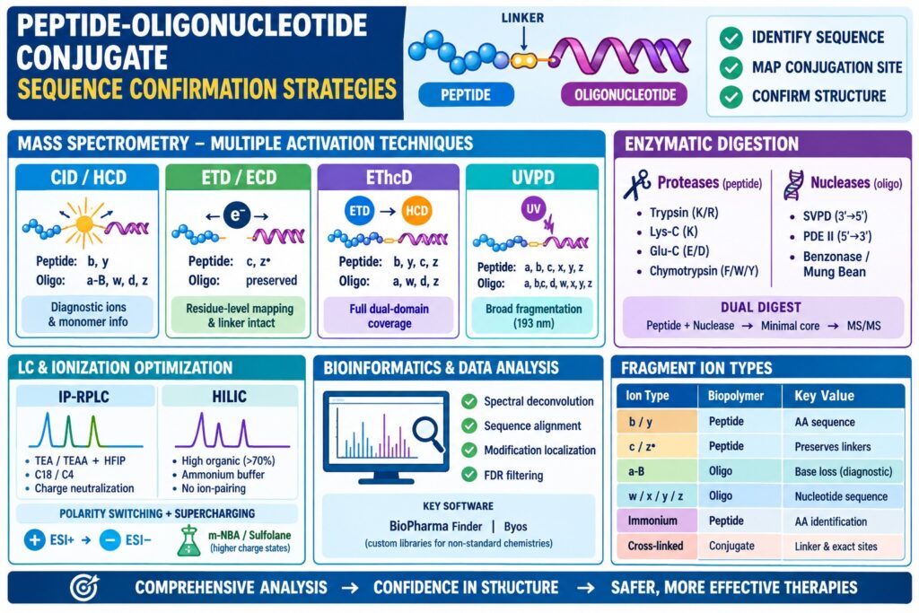
- Multiple MS/MS techniques—CID/HCD, ETD/ECD, EThcD, and UVPD—provide complementary fragmentation and improve coverage across both biopolymer domains.
- ETD and EThcD are especially valuable for preserving fragile linkers and modifications while enabling residue-level conjugation-site mapping.
- Protease and nuclease digestion simplify complex conjugates; trypsin, Lys-C, Glu-C, chymotrypsin, SVPD, PDE II, and other nucleases provide targeted sequence information.
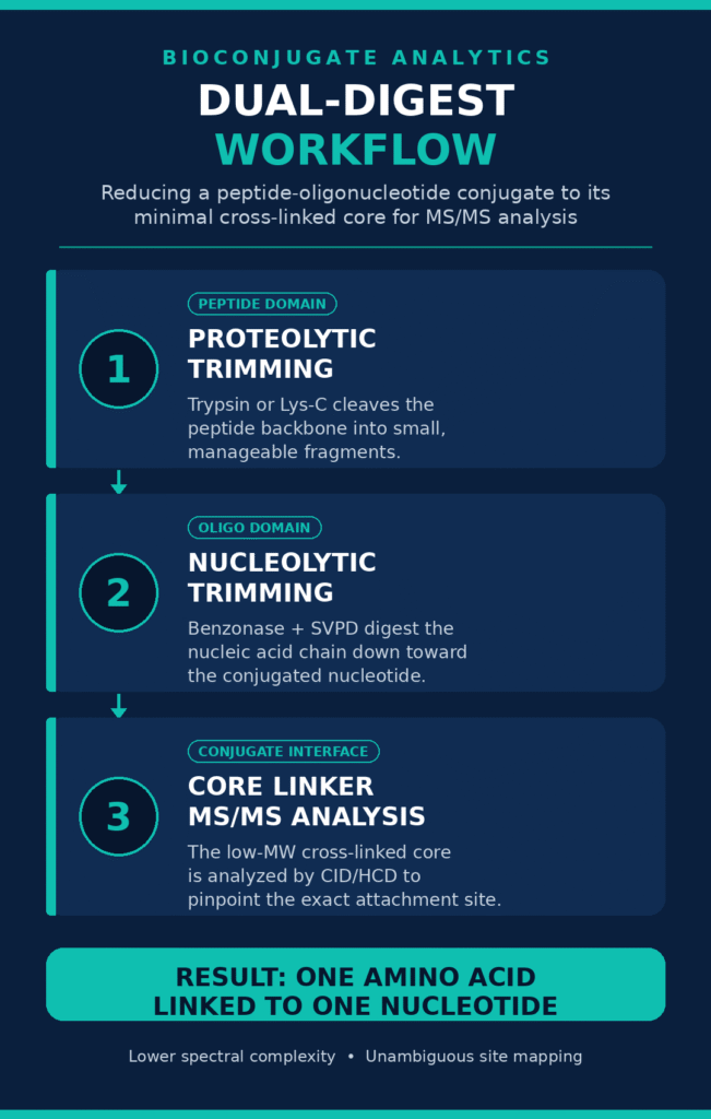
- Dual-digest workflows trim both peptide and oligonucleotide regions toward a minimal cross-linked core, enabling clearer MS/MS identification of exact attachment points.
- LC-MS optimization using IP-RPLC or HILIC, polarity switching, and charge-state supercharging improves chromatographic separation, ionization, and fragmentation efficiency.
- Bioinformatics and spectral deconvolution match experimental fragment ions to theoretical sequences, localize modifications, and confirm structural integrity, supporting regulatory expectations and batch-to-batch consistency.
Tandem Mass Spectrometry (MS/MS) Peptide-Oligonucleotide Conjugate Sequence Confirmation Strategies
Tandem mass spectrometry sequence confirmation strategies for peptide-oligonucleotide conjugates involve the combined use of collision-based and radical-driven fragmentation mechanisms to disrupt both the peptide amide backbone and the nucleic acid phosphodiester framework. Appropriate selection of the gas-phase dissociation technique minimizes asymmetric energy absorption and promotes comprehensive sequence coverage across both molecular domains.
Collision-Induced Dissociation (CID) and Higher-Energy Collisional Dissociation (HCD)
Collision-Induced Dissociation (CID) and Higher-energy Collisional Dissociation (HCD) generate vibrational excitation through collisions between gas-phase precursor ions and inert gas molecules. These processes preferentially promote cleavage of phosphodiester bonds in oligonucleotides and amide bonds in isolated peptides. In intact heteroconjugates, CID and HCD can produce low-mass diagnostic reporter ions and base-loss fragments that provide evidence for monomer composition.
During resonant-excitation CID, precursor ions collide with helium or nitrogen buffer gas, progressively accumulating internal vibrational energy until the lowest-energy chemical bonds undergo cleavage. Although CID of unmodified peptides typically produces predictable N-terminal b-ions and C-terminal y-ions, applying CID to intact peptide-oligonucleotide conjugates generally promotes preferential cleavage along the nucleic acid phosphodiester or phosphorothioate backbone. This process produces abundant nucleobase-loss (a-B) and sugar-phosphate cleavage fragments, including w, d, and z ions, while leaving much of the attached peptide chain unfragmented.
Beam-type collision dissociation, known as Higher-energy Collisional Dissociation (HCD), introduces higher kinetic energy within a dedicated collision cell before transferring the resulting fragment ions to a high-resolution mass analyzer. HCD can overcome some of the energy-dissipation limitations by generating intense low-mass diagnostic ions, including amino acid immonium ions and free nucleobase ions (A−, C−, G−, T−). These diagnostic species can support confirmation of modified residues, including 2′-O-methoxyethyl (2′-MOE) ribose sugars and phosphorothioate linkages. However, HCD alone remains inadequate for comprehensive de novo sequence mapping of the attached peptide domain.
To evaluate environmental vulnerability and stability parameters under stress testing conditions, review our analysis on peptide-oligonucleotide conjugate stability.
Electron Transfer Dissociation (ETD) and Electron Capture Dissociation (ECD)
Electron Transfer Dissociation (ETD) and Electron Capture Dissociation (ECD) introduce electrons into multiply protonated conjugate precursor ions, promoting non-ergodic N-Cα bond cleavage along the peptide backbone while maintaining fragile modifications and nucleotide linkers. This radical-mediated fragmentation pathway avoids extensive vibrational energy redistribution and therefore helps preserve labile bioconjugate linkages.
Unlike collisional activation techniques, ETD does not depend primarily on thermal energy distribution among the vibrational modes of the precursor ion. Instead, a multiply charged positive precursor ion [M + nH]n+ reacts with a radical anion, such as fluoranthene or azobenzene, resulting in electron transfer and formation of an unstable hypervalent radical intermediate. This intermediate rapidly undergoes homolytic cleavage along peptide N-Cα bonds, generating complementary c-type and z•-type fragment ion series.
Because ETD promotes peptide backbone cleavage independently of the relative thermodynamic bond energies, it can preserve labile post-translational modifications, phosphorothioate linkages, and covalent linker bridges. As a result, ETD can provide residue-level characterization of the bioconjugation site. Nevertheless, ETD performance decreases when low precursor charge states (< +3) are analyzed because strong intramolecular non-covalent interactions can prevent effective separation of the resulting c and z fragment ions.
Hybrid Activation Regimes: EThcD and UVPD in Peptide-Oligonucleotide Conjugate Sequence Confirmation Strategies
Hybrid Electron Transfer Higher-energy Collisional Dissociation (EThcD) and Ultraviolet Photodissociation (UVPD) enable broad dual-backbone fragmentation by combining radical-driven cleavage with additional collisional or photon-based excitation within a single MS/MS analysis. These advanced approaches can generate simultaneous b, y, c, and z peptide ions together with a, w, d, and z nucleic acid fragment series.
During an EThcD experiment, precursor ions initially undergo an ETD reaction within an ion trap, producing radical N-Cα peptide bond cleavage products. The resulting primary fragment ions and charge-reduced species are subsequently transferred to the HCD collision cell, where secondary collisional activation occurs. This additional HCD pulse supplies sufficient internal energy to disrupt non-covalent hydrogen bonding and electrostatic interactions that can keep ETD-generated fragments associated, while also promoting cleavage of remaining phosphodiester and peptide bonds.
Ultraviolet Photodissociation (UVPD) employs high-energy ultraviolet lasers, typically 193 nm ArF excimer lasers, to deliver photon energy directly to the conjugate ion backbone on a nanosecond timescale. The rapid energy deposition promotes non-selective cleavage throughout both peptide and oligonucleotide domains. This generates a, b, c, x, y, and z fragment ions together with base-loss species while maintaining the central bioconjugate linker.
For additional pharmacokinetic characterization and in vivo half-life optimization data, consult our findings on peptide-oligonucleotide conjugates pharmacokinetics.
| Fragmentation Technique | Primary Gas-Phase Mechanism | Dominant Fragment Ion Types Produced | Primary Application Domain in Bioconjugates | Key Advantage | Key Limitation |
|---|---|---|---|---|---|
| Collision-Induced Dissociation (CID) | Low-energy vibrational excitation via gas collisions | Peptide: b, yOligo: a-B, w, d, z | Digested peptide fragments; short oligomer ladders | Standard availability on entry-level MS instrumentation | Preferential phosphodiester cleavage suppresses peptide fragmentation |
| Higher-energy Collisional Dissociation (HCD) | Beam-type collisional activation in dedicated cell | Peptide: b, y, immoniumOligo: a, w, base ions | Nucleobase modification screening; reporter ion detection | High-resolution, high-accuracy spectra in Orbitrap analyzers | Over-fragmentation of nucleic acids; incomplete peptide coverage |
| Electron Transfer Dissociation (ETD) | Radical electron transfer inducing non-ergodic cleavage | Peptide: c, z•Oligo: Preserved backbone | Residue-level conjugation site mapping; labile linker preservation | Preserves fragile modifications and bioconjugate linkers | Requires high precursor charge states (> +3); subject to charge reduction |
| Hybrid EThcD | ETD reaction followed by secondary HCD pulse | Dual-backbone:Peptide (b, y, c, z)Oligo (a, w, d, z) | Intact heteroconjugate full-length sequence confirmation | Simultaneous, dual-domain sequence coverage | Slower spectral acquisition rate; requires high-end mass spectrometers |
| Ultraviolet Photodissociation (UVPD) | High-energy photon absorption (193 nm laser) | Comprehensive:a, b, c, d, w, x, y, z | Complex multi-domain bioconjugates; top-down mapping | Unique fragmentation pathways independent of charge state | High instrument complexity; dense, complex fragment spectra |
Enzymatic Digestion Protocols for Peptide-Oligonucleotide Conjugate Sequence Confirmation Strategies
Enzymatic digestion protocols simplify complex peptide-oligonucleotide conjugates into smaller, better-defined fragments by employing targeted proteases and nucleases. Lowering molecular weight and reducing charge heterogeneity through controlled enzymatic digestion can improve sequence coverage beyond what top-down mass spectrometry can provide independently.
Proteolytic Cleavage Workflows for Conjugated Peptides
Proteolytic cleavage employs site-specific endopeptidases to break the peptide domain into discrete fragments while maintaining the attached oligonucleotide payload intact. Characterization of these conjugated digestion products helps localize the bioconjugation site to a defined amino acid sequence.
Trypsin remains the standard enzyme for bottom-up proteomic workflows because it cleaves peptide amide bonds primarily at the C-terminal side of lysine and arginine residues. In peptide-oligonucleotide conjugate mapping, tryptic digestion generates unmodified reference peptides from unconjugated regions as well as a conjugated peptide fragment associated with the intact oligonucleotide payload.
However, bioconjugation commonly occurs at lysine primary amines or cysteine thiols. Covalent modification of a lysine residue can sterically interfere with tryptic recognition and result in missed cleavage at the conjugation site. To address this limitation, analytical workflows can incorporate complementary proteases:
- Endoproteinase Lys-C: Cleaves C-terminal to lysine residues and retains activity under harsh denaturing conditions, making it useful for hydrophobic cell-penetrating peptides.
- Endoproteinase Glu-C: Cleaves C-terminal to glutamic acid and aspartic acid residues, providing an alternative cleavage pattern when basic residues are blocked through conjugation.
- Chymotrypsin: Cleaves C-terminal to aromatic residues, including phenylalanine, tryptophan, and tyrosine, making it useful for characterizing highly basic peptide domains enriched in arginine and lysine.
Sample preparation protocols require careful control of reaction conditions. Disulfide reduction with dithiothreitol (DTT) or TCEP and alkylation with iodoacetamide (IAM) should be performed under optimized pH and temperature conditions to minimize unwanted alkylation side reactions involving oligonucleotide nucleobases or maleimide-thiol exchange reactions.
To learn how cellular internalization mechanisms influence intracellular routing, read our overview on peptide-oligonucleotide conjugates drug delivery.
Nuclease Digestion and Exonuclease Laddering Strategies
Nuclease digestion uses target-specific exonucleases and endonucleases to sequentially cleave phosphodiester or phosphorothioate bonds, generating single-nucleotide stepwise ladders for sequence confirmation. Exonuclease laddering can determine the nucleic acid sequence from both the 3′- and 5′-termini and can proceed directly toward the conjugated peptide linker.
Exonucleases sequentially remove nucleotide monophosphates from a nucleic acid chain. Timed partial exonuclease digestions generate a nested series of truncated conjugate species. LC-MS analysis of the resulting digestion mixture reveals sequential mass differences corresponding to specific nucleotide monophosphates:
- Deoxyadenosine monophosphate (Δm = 313.06 Da)
- Deoxycytidine monophosphate (Δm = 289.05 Da)
- Deoxyguanosine monophosphate (Δm = 329.05 Da)
- Thymidine monophosphate (Δm = 304.06 Da)
The principal nucleases used in sequence confirmation workflows include:
- Snake Venom Phosphodiesterase (SVPD / Phosphodiesterase I): A 3’→5′ exonuclease that sequentially cleaves single-stranded oligonucleotides from the 3′-terminus until steric hindrance near the bioconjugate linker or modified sugar backbones prevents further digestion.
- Bovine Spleen Phosphodiesterase (PDE II): A 5’→3′ exonuclease that provides complementary sequence confirmation beginning at the 5′-terminus.
- Benzonase and Mung Bean Nuclease: Non-specific endonucleases that cleave internal phosphodiester bonds and reduce large ASO or siRNA domains into shorter fragments.
Integrated Dual-Digest Workflows
Integrated dual-digest workflows combine proteases and nucleases within a single or sequential incubation strategy to reduce a bioconjugate to a minimal cross-linked core. Trimming both molecular domains to a single amino acid connected through the linker to a single nucleoside helps isolate the chemical structure of the bioconjugation site.
When characterizing high-molecular-weight bioconjugates, such as antibody-oligonucleotide conjugates or long peptide-PMO constructs, individual enzyme digests may produce fragments that remain too large or structurally complex for clean MS/MS fragmentation. Dual-digest protocols address this challenge through a structured workflow:
- Step 1 (Proteolytic Trimming): The conjugate is incubated with trypsin or Lys-C to reduce the peptide backbone into smaller, more manageable peptide fragments.
- Step 2 (Nucleolytic Trimming): The sample is treated with an exonuclease or endonuclease mixture, such as Benzonase combined with SVPD, to digest the nucleic acid chain toward the final conjugated nucleotide.
- Step 3 (Core Linker MS/MS Analysis): The resulting low-molecular-weight cross-linked core species, such as a single modified cysteine connected through a succinimide linker to an adenosine monophosphate, is analyzed using CID/HCD MS/MS.
This dual-digest strategy reduces spectral complexity and produces clearer, less suppressed MS/MS spectra, enabling unambiguous confirmation of the precise attachment points on both peptide and oligonucleotide backbones.
For a comparative perspective on structural properties and conjugation efficiency, check out our guide on peptide vs antibody oligonucleotide conjugates.
Liquid Chromatography and Ionization Optimization
Liquid chromatography coupled with high-resolution mass spectrometry (LC-MS/MS) requires carefully optimized stationary phases and specialized mobile-phase additives to address the contrasting solubility and ionization characteristics of peptides and oligonucleotides. Implementing Ion-Pair Reversed-Phase Liquid Chromatography (IP-RPLC) or Hydrophilic Interaction Liquid Chromatography (HILIC) can provide sharper chromatographic peaks before MS/MS analysis.
Ion-Pair Reversed-Phase (IP-RPLC) versus HILIC Separations
Ion-Pair Reversed-Phase Liquid Chromatography (IP-RPLC) uses volatile alkylamine ion-pairing agents to mask the negative charge of the nucleic acid backbone and facilitate retention on C18 stationary phases. In contrast, Hydrophilic Interaction Liquid Chromatography (HILIC) retains conjugates primarily through polar interactions without requiring ion-pairing reagents. Selecting the appropriate separation mode can directly affect chromatographic resolution and MS signal sensitivity.
In IP-RPLC, volatile organic amines, including triethylamine (TEA), triethylammonium acetate (TEAA), or hexylamine (HA), are combined with fluorinated alcohol counter-ions such as 1,1,1,3,3,3-hexafluoro-2-propanol (HFIP) in the mobile phase. Positively charged alkylammonium cations associate with the negatively charged phosphate or phosphorothioate groups of the oligonucleotide. This charge neutralization promotes retention on alkylated silica stationary phases, including C18 or C4, with retention influenced by peptide hydrophobicity, nucleobase stacking, and the ion-pairing complex. However, excessive or insufficiently volatile alkylamines can cause substantial signal suppression in positive ESI mode and may contaminate MS source optics during prolonged analytical sequences.
Hydrophilic Interaction Liquid Chromatography (HILIC) provides an alternative approach that does not rely on ion-pairing. HILIC employs polar stationary phases, including amide-bonded, zwitterionic, or unbonded silica, together with high-organic initial mobile phases (>70% acetonitrile) and ammonium acetate/formate buffers. Analytes partition into a water-rich layer associated with the stationary-phase surface. HILIC can effectively retain polar oligonucleotides and hydrophilic peptide conjugates without ion-pairing additives, reducing potential signal suppression and supporting stable electrospray behavior in both positive and negative ESI modes.
To dive deeper into advanced biophysical and spatial analysis methods, read our article on structural characterization of peptide oligonucleotide conjugates.
Polarity Switching and Charge State Supercharging
Polarity switching and charge state supercharging can be used to tailor electrospray ionization conditions for capturing positive peptide ions and negative oligonucleotide ions within a single analytical sequence. Dynamic polarity switching, together with appropriate supercharging additives, can increase precursor ion intensity and improve MS/MS sequence coverage.
Oligonucleotides generally ionize efficiently in negative electrospray mode (ESI−) by forming multiply deprotonated species [M − nH]n−, whereas peptides generally exhibit stronger ionization in positive mode (ESI+) through protonation of basic side chains, producing [M + nH]n+. Modern High-Resolution Accurate-Mass (HRAM) mass spectrometers, including Q-Exactive Orbitrap and Orbitrap Tribrid systems, support rapid polarity switching between positive and negative ionization modes during an LC run.
For positive-mode ETD/EThcD characterization of intact bioconjugates, obtaining high precursor charge states, such as +4, +5, or higher, is important for effective fragmentation. Post-column addition of supercharging reagents, including m-nitrobenzyl alcohol (m-NBA) or sulfolane, can alter the surface tension and evaporation characteristics of electrospray droplets. This process promotes additional protonation of the bioconjugate, increasing precursor charge density and supporting more effective ETD fragmentation across the peptide domain.
For guidance on filing documentation and regulatory frameworks, see our resource on peptide oligonucleotide conjugates in IND submissions.
Bioinformatics, Spectral Deconvolution, and Automated Sequence Verification
Bioinformatic spectral deconvolution techniques process complex, multiply charged MS/MS spectra by comparing observed fragment masses with theoretical ion series calculated for both biopolymer backbones. Specialized software platforms can automate sequence alignment, modification localization, and false discovery rate (FDR) filtering to support confirmation of primary bioconjugate structures.
Fragment Ion Nomenclature and In Silico Matching
In silico database matching assigns experimental mass-to-charge (m/z) peaks using theoretical models based on Roepstorff-Fohlman/Biemann nomenclature for peptides and McLuckey nomenclature for oligonucleotides. Comparing experimentally observed fragment spectra with high-accuracy theoretical mass maps helps verify the intact sequence and identify modification sites.
Peptide backbone fragmentation produces N-terminal b and c ions and C-terminal y and z ions. Oligonucleotide backbone fragmentation produces 5′-terminal a, b, c, and d ions and 3′-terminal w, x, y, and z ions, together with nucleobase-loss (a-B) fragments.
When a bioconjugate is evaluated, deconvolution software must generate theoretical fragment mass libraries that incorporate cross-linked molecular topologies. The mass of a conjugated fragment corresponds to the mass of the peptide fragment plus the mass of the connected oligonucleotide fragment plus the monoisotopic mass contribution of the chemical linker. Bioinformatic platforms, including Thermo Scientific BioPharma Finder and Protein Metrics Byos, can incorporate custom monomer libraries to accommodate non-standard chemistries, including phosphorothioates, 2′-OMe, 2′-MOE, phosphorodiamidate morpholinos, and non-canonical amino acids.
For details on establishing analytical criteria and acceptance limits, review our publication on peptide oligonucleotide conjugates specification setting.
| Fragment Ion Class | Parent Biopolymer | Gas-Phase Cleavage Location | Primary Bioinformatic Value |
|---|---|---|---|
| b-ions / y-ions | Peptide Backbone | Peptide amide bond cleavage | Confirms amino acid sequence; b retains N-terminus, y retains C-terminus |
| c-ions / z•-ions | Peptide Backbone | Radical N-Cα bond cleavage via ETD | Maps peptide sequence while preserving fragile conjugation linkers |
| a-B ions | Oligonucleotide Domain | C3′-C4′ ribose bond cleavage with base loss | Diagnostic indicator of nucleic acid fragmentation under collisional energy |
| w-ions | Oligonucleotide Domain | 3′-P-O phosphodiester bond cleavage | Primary 3′-terminal fragment series used to confirm nucleic acid sequence |
| Immonium Ions | Peptide Side Chains | Internal double-cleavage of amino acids | Low-mass diagnostic markers confirming the presence of specific amino acids |
| Cross-Linked Core Ions | Conjugate Interface | Intact linker connecting truncated peptide and nucleotide residues | Unambiguously maps the exact amino acid and nucleotide conjugation sites |
To ensure material integrity over extended periods, see our protocols on handling and storage for peptide oligonucleotide conjugates.
Conclusion
Peptide-Oligonucleotide Conjugate Sequence Confirmation Strategies that combine high-resolution MS/MS techniques, including HCD, ETD, and EThcD, with targeted enzymatic digestion provide a comprehensive analytical framework for validating complex bioconjugate structures. By integrating orthogonal liquid chromatography separations with automated spectral deconvolution, bioanalytical laboratories can verify primary sequence fidelity, characterize chemical linkages, and support regulatory requirements.
As biopharmaceutical development programs continue to advance peptide-guided nucleic acid therapeutics, validated sequence confirmation strategies become increasingly important for demonstrating product safety, efficacy, and batch-to-batch consistency. Combining targeted proteolytic and nucleolytic digestion with advanced hybrid EThcD mass spectrometry helps address energy-dissipation challenges and enables broader sequence coverage across both biopolymer domains.
To discuss customized bioanalytical method development, bioconjugate characterization, or preclinical sequence confirmation workflows, visit the ResolveMass Laboratories Contact Us page at https://resolvemass.ca/contact/.
Frequently Asked Questions
Hybrid EThcD integrates Electron Transfer Dissociation (ETD) with Higher-energy Collisional Dissociation (HCD) to interrogate both components of the conjugate. ETD promotes N−Cα cleavage within the peptide while retaining labile linkages, whereas HCD supplies additional activation for nucleic acid fragmentation and disruption of non-covalent interactions. This combined approach generates complementary b, y, c, z and a, w, d, z ions for broader sequence coverage.
Exonucleases progressively remove nucleotides from defined termini of the oligonucleotide, making them valuable tools for sequence verification. Snake Venom Phosphodiesterase (SVPD) can provide 3’→5′ digestion, while Bovine Spleen Phosphodiesterase supports 5’→3′ analysis. LC-MS examination of the resulting digestion ladder allows sequential nucleotide losses to be monitored and the sequence to be confirmed toward the conjugation site.
Ion-Pair Reversed-Phase Liquid Chromatography (IP-RPLC) improves retention of charged oligonucleotide conjugates by using volatile ion-pairing reagents such as TEA/HFIP. These reagents interact with the negatively charged phosphate or phosphorothioate backbone and promote retention on hydrophobic reversed-phase stationary phases. The resulting separation can effectively resolve conjugates according to oligonucleotide and peptide hydrophobicity.
Yes, HILIC can separate peptide-oligonucleotide conjugates without conventional ion-pairing additives. The technique relies on interactions between polar analytes and a water-enriched layer associated with the stationary phase, commonly using amide, zwitterionic, or silica phases. Because alkylamine ion-pairing reagents are avoided, HILIC can provide cleaner MS interfaces and support sensitive detection under positive and negative ESI conditions.
Maleimide ring-opening hydrolysis transforms the succinimide ring within a maleimide-thiol linkage into a ring-opened succinamic acid structure. This chemical conversion produces an approximately +18.01 Da mass increase that must be incorporated into theoretical fragment calculations. The hydrolyzed form can also improve linker stability by reducing the likelihood of retro-Michael exchange during sample handling and MS/MS analysis.
Modification of amino acid side chains can interfere with normal recognition and cleavage by trypsin, particularly when lysine residues are involved in covalent conjugation. A modified lysine may therefore produce a missed cleavage and leave the conjugated region within a larger peptide fragment. Alternative enzymes, including Endoproteinase Lys-C, Glu-C, and Chymotrypsin, can be introduced to improve sequence coverage and localization of the conjugation site.
Phosphorothioate-containing oligonucleotides can generate characteristic sulfur-associated fragment ions during Higher-energy Collisional Dissociation (HCD). Thiophosphate-related species and sulfur-containing base-loss fragments can provide evidence for replacement of a non-bridging phosphate oxygen by sulfur. These diagnostic ions can therefore support the identification and characterization of phosphorothioate modifications within the oligonucleotide backbone.
Supercharging reagents such as m-NBA or sulfolane can increase the number of protons retained by peptide-containing precursor ions during electrospray ionization. Generating higher charge states increases the charge density available for Electron Transfer Dissociation (ETD). This can reduce non-dissociative charge-reduction effects and promote more efficient peptide backbone fragmentation, improving sequence and conjugation-site characterization.
Bioinformatic platforms such as BioPharma Finder and Byos generate theoretical fragment libraries that account for the different structural components of a cross-linked bioconjugate. The calculated masses incorporate peptide fragments, nucleotide fragments, and the monoisotopic mass of the linker according to the proposed conjugation topology. Experimental m/z signals can then be compared with these theoretical ions to identify matching fragments and localize the conjugation site.
Reference:
- Farquhar, C. E., Pentelute, B. L., Malmberg, A. B., Schissel, C., Ye Morgan, K., Bardhan, A., Oliver, R., Guerlavais, V., Jinadasa, T., & Callahan, A. (2025). Non-canonical cell-penetrating peptides for antisense oligomer delivery (WO2025030099A1). World Intellectual Property Organization. https://patents.google.com/patent/WO2025030099A1/en
- Krivos, K. L., & Limbach, P. A. (2010). Sequence analysis of peptide:oligonucleotide heteroconjugates by electron capture dissociation and electron transfer dissociation. Journal of the American Society for Mass Spectrometry, 21(8), 1387–1397. https://doi.org/10.1016/j.jasms.2010.03.043
- Katzhendler, J., Klauzner, Y., Beylis, I., Mizhiritskii, M., Shpernat, Y., Ashkenazi, B., & Fridland, D. (2006). Method for the preparation of peptide-oligonucleotide conjugates (EP1725250A2). European Patent Office. https://patents.google.com/patent/EP1725250A2/en
- Gao, L., Zhang, L., Cho, B. P., & Chiarelli, M. P. (2008). Sequence verification of oligonucleotides containing multiple arylamine modifications by enzymatic digestion and liquid chromatography mass spectrometry (LC/MS). Journal of the American Society for Mass Spectrometry, 19(8), 1147–1155. https://doi.org/10.1016/j.jasms.2008.04.034
- Jensen, O. N., Kulkarni, S., Aldrich, J. V., & Barofsky, D. F. (1996). Characterization of peptide-oligonucleotide heteroconjugates by mass spectrometry. Nucleic Acids Research, 24(19), 3866–3872. https://doi.org/10.1093/nar/24.19.3866
Get In Touch With Us
About The Author


