

Introduction
Poly(lactic-co-glycolic acid) (PLGA)-based particulate delivery systems offer a highly biocompatible and biodegradable platform for protecting fragile biomolecules while enabling controlled and sustained drug release. The application of PLGA for Vaccine and Antigen Delivery has become an important strategy in advanced pharmaceutical formulation because it addresses the limited stability and short biological half-lives associated with subunit protein antigens, peptides, and nucleic acid therapeutics. Under physiological conditions, native PLGA undergoes non-enzymatic hydrolytic degradation through cleavage of its ester backbone, resulting in bulk erosion of the polymer matrix. Water molecules readily penetrate the polymer network and hydrolyze ester bonds throughout the entire particle rather than only at the surface. As degradation progresses, the molecular weight gradually decreases until the polymer is converted into the water-soluble, non-toxic monomers lactic acid and glycolic acid. Both degradation products are naturally occurring metabolites that participate in normal cellular metabolic pathways and are ultimately eliminated through the citric acid cycle, contributing to the excellent systemic safety profile of PLGA.
Discover the complete breakdown of polymer degradation mechanics in our technical guide on Bulk Erosion vs Surface Erosion in PLGA.
The clinical success of PLGA has been demonstrated by the regulatory approval of several long-acting injectable formulations since 1989, including Lupron Depot, Zoladex, Risperdal Consta, and Vivitrol. These products highlight the proven safety, biocompatibility, and versatility of PLGA-based delivery systems. Nevertheless, extending this technology to vaccine and immunotherapy applications introduces additional formulation challenges. These include preserving the native tertiary structure of sensitive antigens during particle fabrication, preventing degradation during processing, and regulating the intracellular trafficking of carrier particles after uptake by immune cells. Successful clinical translation therefore depends on sophisticated polymer synthesis methods combined with comprehensive physicochemical characterization to ensure reproducible manufacturing, consistent performance, and a favorable safety profile.
Explore the formulation parameters required to bring these platforms to market in our overview of PLGA Long-Acting Injectable Formulation.
Article Summary:
- PLGA is a biodegradable polymer widely used for advanced vaccine delivery because it protects fragile antigens from degradation while enabling controlled and sustained release. As the polymer breaks down into lactic acid and glycolic acid, these natural metabolites are safely eliminated through normal metabolic pathways, making PLGA a highly biocompatible carrier.
- The performance of PLGA vaccines is strongly influenced by polymer characteristics such as the lactic acid-to-glycolic acid ratio, molecular weight, and glass transition temperature. Optimizing these parameters allows scientists to precisely control degradation rates, hydration behavior, and antigen release profiles for short- or long-term immune protection.
- PLGA particles provide intrinsic adjuvant activity that strengthens immune responses. After uptake by antigen-presenting cells, they activate inflammasome pathways and stimulate the production of key inflammatory cytokines. Their immunostimulatory effects can be further enhanced by combining them with Toll-like receptor (TLR) agonists.
- Modern encapsulation technologies improve antigen stability during manufacturing. Advanced approaches such as microfluidics-assisted fabrication, nanoprecipitation, and active self-healing microencapsulation minimize mechanical stress and solvent exposure, resulting in higher encapsulation efficiency and better preservation of sensitive proteins, peptides, and nucleic acids.
- Maintaining a stable internal microenvironment is essential for preserving vaccine quality. Buffering agents such as magnesium hydroxide and calcium carbonate help neutralize acidic degradation products inside PLGA particles, reducing protein degradation, preventing aggregation, and supporting more consistent antigen release.
- Surface engineering of PLGA nanoparticles directs immune pathway activation. By modifying particle size, charge, and surface chemistry, researchers can influence intracellular trafficking, enhance antigen cross-presentation, and promote both antibody-mediated (humoral) and cytotoxic T-cell (cellular) immune responses for improved vaccine efficacy.
- Successful clinical translation requires comprehensive analytical characterization and quality control. Advanced techniques including qNMR, DSC, GPC-MALS, FTIR, and high-resolution mass spectrometry ensure polymer quality, structural integrity, impurity monitoring, and regulatory compliance, supporting the development of safe, reproducible, and effective PLGA-based vaccine formulations.
Physicochemical Modulators of Polymer Degradation and Hydration
The degradation behavior, hydration characteristics, and antigen release profile of PLGA formulations are primarily influenced by the polymer’s lactic acid-to-glycolic acid (L:G) ratio, molecular weight, and glass transition temperature (Tg). Careful adjustment of these fundamental polymer properties enables formulation scientists to tailor drug release profiles ranging from rapid initial release to sustained delivery over several months. Depending on molecular weight and copolymer composition, the Tg of native PLGA generally falls between 30 °C and 60 °C. Increasing the proportion of lactic acid introduces additional hydrophobic methyl groups into the polymer backbone, reducing water penetration, slowing ester bond hydrolysis, and increasing the Tg.
In contrast, a higher glycolic acid content enhances polymer hydrophilicity, facilitating greater water absorption, accelerating bulk erosion, and lowering the overall Tg. Acid-terminated (uncapped) PLGA polymers degrade and hydrate more rapidly than ester-capped counterparts because terminal carboxylic acid groups increase the polymer’s affinity for water. During particle fabrication, rapid solvent removal may trap polymer chains in a high-energy, non-equilibrium glassy state. With time, these chains gradually undergo physical aging and enthalpy relaxation as they approach thermodynamic equilibrium, processes that directly affect particle porosity, structural integrity, and long-term release behavior.
Following in vivo administration, absorbed water acts as a powerful plasticizer that lowers the polymer’s Tg below physiological body temperature (37 °C). This reduction transforms the polymer from a rigid glassy material into a more flexible rubbery state, substantially enhancing the diffusion of encapsulated macromolecular antigens through the polymer matrix. Polymerization kinetics also influence polymer microstructure. During synthesis under tin octanoate catalysis at 200 °C, the reactivity ratio between glycolic acid and lactic acid monomers (rGL/rLA = 14.00) differs considerably, creating the potential for block-like copolymer distributions if polymerization conditions are not carefully controlled.
To accurately characterize these critical physicochemical parameters, ResolveMass Laboratories Inc. employs quantitative nuclear magnetic resonance (qNMR) for precise determination of comonomer composition, differential scanning calorimetry (DSC) for thermal characterization, and gel permeation chromatography (GPC) coupled with multi-angle static light scattering (MALS) for absolute molecular weight measurement. This advanced analytical approach eliminates the molecular weight estimation errors—often reaching approximately 70%—that are commonly associated with conventional polystyrene-equivalent calibration methods.
Optimize your formulation baseline by utilizing our specialized NMR Spectroscopy for Accurate Monomer Ratio validation services.
| Polymer Composition (L:G Ratio) | Average Molecular Weight (kDa) | Glass Transition Temperature (Tg) (°C) | Hydrated Tg (°C) | Bulk Degradation Lifetime in Vivo | Principal Delivery Mechanics |
|---|---|---|---|---|---|
| 50:50 (Acid-Terminated) | 7–17 | 30.00–35.00 | <25.00 | 1–2 Weeks | Rapid hydration with erosion-dominated release. |
| 50:50 (Ester-Capped) | 38–54 | 42.00–46.00 | 28.00–32.00 | 3–4 Weeks | Balanced diffusion and polymer degradation. |
| 75:25 (Ester-Capped) | 68 | 46.74 | 39.75 | 1–2 Months | Moderate diffusion barrier with delayed bulk erosion. |
| 75:25 (Ester-Capped) | 128 | 49.00 | 40.65 | 2–3 Months | High mechanical stability supporting sustained release. |
| 85:15 (Ester-Capped) | 100–150 | 50.00–55.00 | >40.00 | 3–6 Months | Increased hydrophobicity with crystallinity-retarded degradation. |
| 100:0 (PLA Homopolymer) | >150 | 50.00–65.00 | >45.00 | >12 Months | Minimal water uptake with extremely slow surface erosion. |
Review the distinct structural properties across key biomaterial families in our deep dive on PLA vs PLGA vs PCL.
Adjuvant Mechanisms and Inflammasome Activation in PLGA for Vaccine and Antigen Delivery
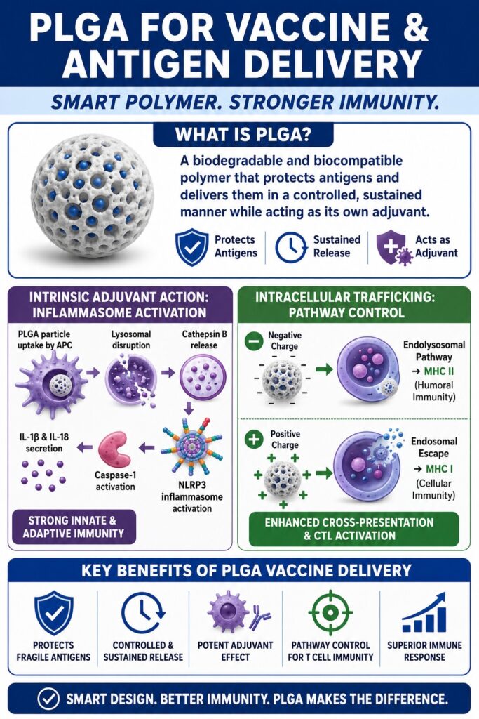
The design of modern subunit vaccines using PLGA for Vaccine and Antigen Delivery capitalizes on the particulate nature of the polymer to imitate the physical characteristics of bacterial pathogens, allowing PLGA particles to function as intrinsic adjuvants. Both micro- and nanoparticles are efficiently internalized by antigen-presenting cells (APCs), initiating cellular activation through lysosomal disruption and subsequent inflammasome signaling. The magnitude of this immunostimulatory response depends largely on particle size, surface charge, and overall structural organization. Nanoparticles measuring less than 500 nm are predominantly taken up by dendritic cells through receptor-mediated endocytosis, whereas larger microparticles ranging from 1 μm to 10 μm are more readily engulfed by macrophages or remain at the administration site as localized antigen depots that provide sustained release.
After cellular internalization, PLGA nanoparticles stimulate activation of the intracellular NLRP3 (nucleotide-binding oligomerization domain-like receptor pyrin domain-containing 3) inflammasome pathway. Uptake of these rigid polymeric particles induces lysosomal swelling, mechanical disruption of the lysosomal membrane, and eventual lysosomal destabilization. Damage to the lysosome results in the release of acidic lysosomal enzymes, including cathepsin B, into the cytoplasm. This intracellular danger signal, together with the generation of reactive oxygen species (ROS) and potassium (K⁺) efflux, promotes recruitment and assembly of the NLRP3 inflammasome complex.
Following assembly, the inflammasome recruits the adaptor protein ASC together with pro-caspase-1, resulting in activation of caspase-1. Activated caspase-1 subsequently cleaves the inactive precursors of the pro-inflammatory cytokines interleukin-1 beta (IL-1β) and interleukin-18 (IL-18), converting them into their biologically active forms for secretion. The release of these cytokines establishes a highly immunostimulatory microenvironment that supports robust activation of innate and adaptive immune responses.
The intrinsic adjuvant properties of PLGA particles can be further enhanced through co-encapsulation or surface functionalization with Toll-like receptor (TLR) ligands such as lipopolysaccharides (LPS), CpG oligodeoxynucleotides, or monophosphoryl lipid A (MPL). As an example, surface modification of PLGA nanoparticles with LPS has been shown to improve antigen encapsulation efficiency by approximately 16%, increasing ovalbumin loading from 37 μg to 44 μg per milligram of particles. This enhancement is attributed to the amphipathic characteristics of LPS, which function as a surfactant and stabilize the primary emulsion droplets during particle fabrication.
Simultaneous delivery of both the antigen and the TLR agonist to the same dendritic cell generates a synergistic immunological response. This coordinated activation promotes enhanced maturation of antigen-presenting cells, increased expression of costimulatory molecules, and stronger downstream cellular and humoral immune responses, ultimately improving the overall efficacy of PLGA-based vaccine formulations.
Advanced Encapsulation Strategies for Fragile Antigens
Maintaining the structural integrity and native conformation of sensitive antigens during microparticle fabrication requires manufacturing techniques that minimize both mechanical stress and exposure to water-organic solvent interfaces. Although the conventional double emulsion process continues to be widely employed, newer encapsulation technologies—including microfluidic fabrication and active self-healing approaches—provide significant improvements in encapsulation efficiency while better preserving the biological activity of fragile biomolecules. Traditional water-in-oil-in-water (W₁/O/W₂) double emulsion solvent evaporation methods expose delicate protein antigens to organic solvents such as dichloromethane or ethyl acetate while simultaneously subjecting them to intense homogenization or probe sonication. These processing conditions frequently promote protein unfolding, aggregation, and chemical instability at the water-oil interface, ultimately reducing antigen integrity and diminishing vaccine immunogenicity.
Address systemic formulation bottlenecks early by referencing our study on Challenges in PLGA Microsphere Development.
To overcome these challenges, several advanced encapsulation strategies have been introduced that provide improved protection for sensitive biological payloads while enhancing manufacturing performance.
Nanoprecipitation (Interfacial Deposition):
Nanoprecipitation is a single-step fabrication technique in which both the polymer and a moderately hydrophobic therapeutic compound are dissolved in a water-miscible organic solvent, such as acetone or dimethyl sulfoxide (DMSO). The resulting solution is then added gradually into an aqueous phase, where spontaneous solvent displacement induces rapid phase separation and nanoparticle formation. Because this process eliminates the need for high-shear homogenization, it significantly reduces mechanical stress and helps preserve the structural integrity of delicate macromolecules, including plasmid DNA. However, despite these advantages, nanoprecipitation generally exhibits relatively low encapsulation efficiencies when highly hydrophilic proteins are incorporated.
Microfluidics-Assisted Synthesis:
Microfluidic fabrication employs microscale channels that direct continuous liquid streams under carefully controlled flow conditions. Through coaxial flow focusing and rapid microscale mixing, these systems produce highly monodisperse nanoparticles with exceptionally uniform particle sizes and narrow polydispersity indices. Compared with conventional batch manufacturing methods, microfluidic platforms offer greater reproducibility, higher encapsulation efficiencies, and substantially lower shear stress, thereby preserving the biological function of sensitive therapeutic molecules throughout the fabrication process.
Active Self-Healing Microencapsulation:
Active self-healing microencapsulation completely eliminates the direct exposure of proteins to organic solvents and high-shear processing conditions. Initially, highly porous blank PLGA microspheres are fabricated by incorporating a hydrophilic porogen such as trehalose into the internal aqueous phase. These porous microspheres are subsequently incubated with a dilute aqueous antigen solution under mild temperature conditions ranging from 10 °C to 38 °C. To facilitate efficient antigen loading, a protein-binding agent such as aluminum hydroxide (Al(OH)₃) or calcium phosphate is preloaded within the porous core of each microsphere. These basic inorganic materials adsorb protein molecules as they diffuse into the pores, allowing encapsulation efficiencies approaching 97% to 100%. Following antigen loading, the pores are sealed either by introducing a suitable polymer plasticizer or by gently heating the formulation slightly above the hydrated polymer’s glass transition temperature. Under these conditions, polymer chains undergo relaxation and flow, effectively sealing the pores while preserving antigen stability.
Evaluate payload specificities with our comprehensive look into Encapsulating Hydrophilic vs Hydrophobic APIs in PLGA.
| Encapsulation Technology | Physical Processing Parameters | Key Advantages for Biomolecules | Manufacturing Limitations | Typical Antigen Match |
|---|---|---|---|---|
| Traditional Double Emulsion (W₁/O/W₂) | High-shear homogenization, probe sonication, and organic-aqueous interfaces | High payload loading capacity with straightforward batch-scale production | Protein denaturation caused by mechanical shear and solvent interfaces | Robust peptides; inactivated toxins |
| Nanoprecipitation | Spontaneous solvent displacement through gradual addition of organic solution into water | Low mechanical stress that preserves genomic and macromolecular integrity | Limited encapsulation efficiency for highly hydrophilic antigens | Hydrophobic peptides; small plasmid DNA |
| Microfluidics-Assisted | Controlled microscale mixing with coaxial flow focusing | Highly uniform particle size distribution and reproducible release kinetics | Specialized instrumentation and scale-up through chip parallelization | Recombinant proteins; therapeutic mRNA |
| Active Self-Healing | Ambient-temperature adsorption followed by thermal pore sealing | No solvent or shear exposure with encapsulation efficiencies approaching 100% | Requires incorporation of protein-trapping agents such as Al(OH)₃ | Highly labile enzymes; subunit proteins |
Mitigating Acidic Microenvironments to Ensure Protein Stability
The formation of localized acidic microenvironments within degrading PLGA matrices represents a major challenge for maintaining the structural stability and biological activity of encapsulated proteins. This problem can be substantially reduced by incorporating alkaline ceramic buffering agents that neutralize acidic degradation products throughout polymer erosion. Because PLGA degrades through a bulk erosion mechanism, water penetrates the entire polymer matrix much faster than the resulting acidic degradation products can diffuse outward. Consequently, lactic acid and glycolic acid accumulate within the particle core, creating highly concentrated acidic microdomains where the internal pH may decrease to approximately 3.0. Such acidic conditions accelerate peptide bond hydrolysis, promote both covalent and non-covalent protein aggregation, and frequently lead to incomplete release of the encapsulated antigen.
To counteract this localized acidification, formulation scientists commonly incorporate alkaline ceramic excipients such as magnesium hydroxide (Mg(OH)₂) or calcium carbonate directly into the PLGA matrix. As acidic degradation products accumulate, these basic compounds dissolve and consume excess protons, thereby buffering the internal microenvironment and maintaining a pH closer to physiological conditions.
Despite its buffering capability, unmodified Mg(OH)₂ possesses high hydrophilicity and readily partitions into the external aqueous phase during emulsification, resulting in poor retention within PLGA particles. Surface modification of Mg(OH)₂ using hydrophobic lipids such as ricinoleic acid, producing ricinoleic acid-modified magnesium hydroxide (RA-MH), markedly improves its dispersion within the organic polymer phase. This hydrophobic surface treatment increases antacid encapsulation approximately four-fold, provides prolonged pH buffering during polymer degradation, and contributes additional anti-inflammatory benefits within surrounding tissues.
An alternative strategy involves loading mesoporous nano-hexagonal Mg(OH)₂ nanostructures into anhydrous reverse micelles (MNS-RM), thereby minimizing burst release while enhancing stabilization of sensitive proteins such as vascular endothelial growth factor (VEGF). Additionally, PLGA composites containing approximately 5–10 wt% elemental magnesium can be engineered to provide intrinsic self-neutralization throughout degradation while simultaneously improving the overall biocompatibility of the delivery system.
Understand the mechanisms governing protein stabilization through our resource on the Characterization of Long-Acting Biologics.
Intracellular Trafficking and Immune Pathway Directing
The intracellular fate of encapsulated antigens, including their presentation through either the major histocompatibility complex (MHC) Class I or MHC Class II pathway, is strongly influenced by the surface chemistry and electrostatic properties of PLGA carriers. Carefully engineered surface modifications enable nanoparticles to bypass conventional endolysosomal degradation, thereby promoting efficient cytoplasmic delivery and enhanced antigen cross-presentation.
Native PLGA nanoparticles generally possess a negative surface charge because of terminal carboxyl functional groups. Following internalization by antigen-presenting cells, these negatively charged particles are transported through the classical endolysosomal pathway before ultimately fusing with lysosomes. Within the acidic lysosomal environment, typically maintained at a pH between 4.5 and 5.0, encapsulated antigens undergo enzymatic degradation into peptide fragments. These peptides are subsequently loaded onto MHC Class II molecules and transported to the cell surface, where they stimulate CD4⁺ T-helper cells and initiate humoral immune responses characterized by antibody production.
Generation of robust cellular immunity requires cytosolic delivery of antigens for presentation through MHC Class I molecules, leading to activation of cytotoxic CD8⁺ T lymphocytes (CTLs). Achieving this outcome depends on successful escape of nanoparticles from the endosomal compartment. One effective strategy involves reversing the surface charge of PLGA nanoparticles through coating with cationic polymers such as polyethylenimine (PEI). The resulting positive surface charge greatly enhances electrostatic interactions with negatively charged cellular membranes, promoting more efficient cellular uptake.
Following endocytosis, the amine-rich PEI coating induces the well-characterized “proton-sponge effect” within the acidic endosomal compartment. Progressive proton accumulation is accompanied by chloride ion influx and osmotic water uptake, causing swelling of the endosome and eventual rupture of the surrounding membrane. This process releases the encapsulated antigen directly into the cytoplasm, where it undergoes proteasomal degradation before transport into the endoplasmic reticulum for loading onto MHC Class I molecules and subsequent activation of cytotoxic T-cell responses.
An alternative strategy utilizes polymer blend nanoparticles composed of PLGA combined with the pH-responsive terpolymer poly(DMAEMA-co-PAA-co-BMA) in a 1:1:2 molar ratio. These hybrid delivery systems promote simultaneous MHC Class I and Class II antigen presentation by integrating pH-sensitive membrane-disruptive activity within early endosomal compartments, thereby supporting both cellular and humoral immunity.
Targeted delivery can be further enhanced by functionalizing nanoparticle surfaces with peptide ligands that selectively bind the dendritic cell receptor DEC-205. Upon receptor-mediated uptake, these engineered nanoparticles rapidly escape from early endocytic vesicles while avoiding colocalization with LAMP-1 (lysosome-associated membrane protein 1) and EEA-1 (early endosome antigen 1). This trafficking pathway bypasses conventional endolysosomal processing and delivers encapsulated antigens directly into the cytosol, allowing immediate access to the intracellular MHC Class I antigen presentation machinery and significantly improving cross-presentation efficiency.
Clinical and Preclinical Advancements in PLGA Vaccine Formulations
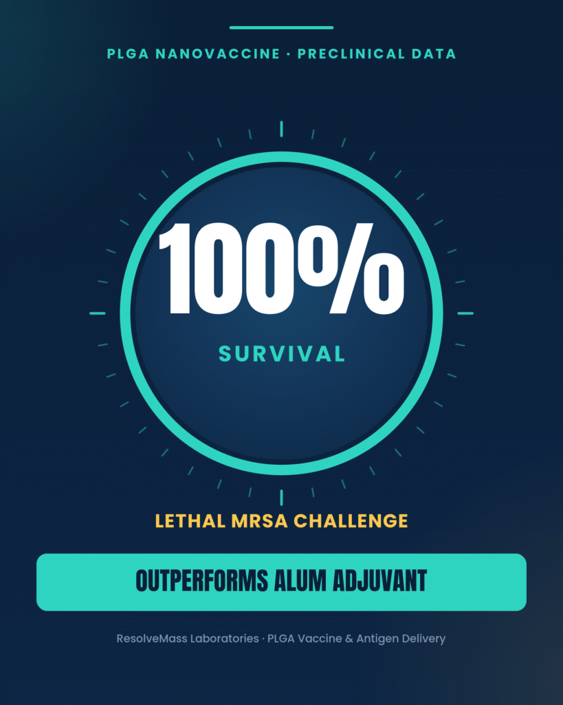
The translation of PLGA-based antigen delivery systems from laboratory research into clinical development has demonstrated substantial improvements in protective immunity and survival across a wide range of infectious disease and oncology models when compared with conventional vaccine adjuvants. Numerous preclinical investigations have shown that carefully controlling antigen presentation kinetics through rational carrier engineering can significantly strengthen both the magnitude and duration of immune responses. In one notable study, non-toxic variants of the Staphylococcus aureus exotoxin Hla (rHlaH35L) and the cell wall-associated protein SpA (rSpAm) were covalently conjugated onto PLGA-PEG 25% nanoparticles to create an advanced nanovaccine platform. These engineered nanoparticles generated markedly stronger cellular and humoral immune responses than traditional aluminum-based (alum) adjuvants.
When experimental animals were challenged with a lethal dose of methicillin-resistant Staphylococcus aureus (MRSA), vaccination with the PLGA-based nanovaccine achieved complete (100%) survival, demonstrating the superior protective efficacy of this particulate delivery system compared with conventional alum formulations.
PLGA-based vaccine carriers have also expanded opportunities for non-invasive immunization strategies that eliminate the need for conventional needle injections. In one approach, inactivated influenza A (H1N1 and H3N2) viral antigens were encapsulated within PLGA microparticles and administered to mice using transdermal dissolving microneedle arrays. This innovative delivery platform successfully induced high systemic concentrations of IgG, IgG1, and IgG2a antibodies while simultaneously stimulating strong mucosal IgA responses within the lungs, providing immune protection directly at the primary site of viral infection.
To facilitate successful clinical translation of these sophisticated vaccine platforms, ResolveMass Laboratories Inc. provides comprehensive analytical services that include advanced polymer characterization, peptide-oligonucleotide conjugate (POC) sameness assessments, and high-resolution mass spectrometry (HRMS). These analytical capabilities support regulatory compliance while ensuring batch-to-batch consistency throughout the development of complex investigational formulations.
Review testing protocols required for regulatory compliance via our detailed report on Q1/Q2 Polymer Equivalence Assessment.
Analytical Characterization and Quality Control of Vaccine Carriers
Achieving regulatory approval and ensuring the clinical safety of particulate vaccine formulations require an extensive range of advanced physicochemical characterization techniques. Comprehensive assessment of polymer molecular weight, compositional purity, thermal behavior, and processing-induced structural changes forms a critical component of regulatory submissions. To satisfy the stringent quality expectations established by both Health Canada and the U.S. Food and Drug Administration (FDA), vaccine developers must implement validated analytical methodologies capable of monitoring the physicochemical integrity of polymer carriers throughout formulation development, manufacturing scale-up, and commercial production.
Thermal Analysis (DSC & TGA):
Differential Scanning Calorimetry (DSC) is employed to characterize the polymer’s glass transition temperature (Tg), melting behavior, and degree of crystallinity. These measurements provide valuable insight into polymer stability, processing behavior, and drug release characteristics. Thermogravimetric Analysis (TGA) complements DSC by evaluating thermal degradation profiles, decomposition onset temperatures, and residual moisture content, all of which are essential parameters for predicting long-term formulation stability, storage performance, and shelf-life.
Structural and Compositional Verification (NMR & FTIR):
Quantitative Nuclear Magnetic Resonance (qNMR) spectroscopy enables precise determination of the lactide-to-glycolide (L:G) ratio while also confirming monomer sequence distribution throughout the copolymer backbone. Fourier Transform Infrared (FTIR) spectroscopy provides complementary structural information by verifying successful polymer surface modification, peptide conjugation, functional group integrity, and degradation-related chemical changes that occur during formulation development.
Absolute Molecular Weight Determination (GPC-MALS):
Conventional Gel Permeation Chromatography (GPC) methods that rely on single-angle refractive index detection together with polystyrene calibration standards frequently produce molecular weight inaccuracies of up to 70% when analyzing PLGA polymers. Coupling GPC with Multi-Angle Light Scattering (MALS) enables direct determination of absolute molecular weight and polydispersity without dependence on polymer conformation or external calibration standards, providing substantially greater analytical accuracy for polymer characterization.
Discover the technical impacts of molecular weight distributions in our analysis of PLGA PDI in Pharmaceutical Formulations.
Impurity Profiling and Safety Testing (HRMS):
High-Resolution Mass Spectrometry (HRMS), particularly when performed using Orbitrap-based platforms, enables highly sensitive identification and quantification of trace-level impurities. These analyses include detection of potentially genotoxic nitrosamines, polymer degradation products, and extractables and leachables (E&L) originating from manufacturing equipment, packaging materials, or container-closure systems. Such comprehensive impurity profiling plays an essential role in demonstrating product safety and supporting regulatory approval.
Secure a clear regulatory trajectory with our overview of PLGA Polymer Sameness for ANDA Submission.
ResolveMass Laboratories Inc. is a highly specialized pharmaceutical Contract Research Organization (CRO) and Contract Development and Manufacturing Organization (CDMO) located in Canada. The laboratory operates under a certified ISO 9001:2015 Quality Management System, maintains a Health Canada Drug Establishment Licence (3-002945-A) for Good Manufacturing Practice (GMP) compliance, and is registered with the U.S. Food and Drug Administration under Establishment Identifier 3042696771. By integrating advanced polymer synthesis, High-Resolution Mass Spectrometry, Gel Permeation Chromatography, Differential Scanning Calorimetry, and quantitative Nuclear Magnetic Resonance within a single facility, ResolveMass provides the analytical validation, quality documentation, and regulatory support required to advance complex vaccine and drug delivery products from early-stage research through clinical development.
Conclusion
The successful clinical and commercial advancement of PLGA for Vaccine and Antigen Delivery depends on a comprehensive understanding of polymer chemistry, sophisticated formulation technologies, and precise control of the local microenvironment surrounding encapsulated biomolecules. Although conventional manufacturing approaches continue to face challenges such as protein denaturation caused by high-shear processing and instability resulting from acidic microenvironments generated during polymer degradation, modern formulation strategies have substantially improved vaccine performance. Technologies including microfluidics-assisted synthesis, active self-healing encapsulation, and lipid-modified buffering systems preserve both the structural integrity and biological activity of highly sensitive therapeutic biomolecules throughout manufacturing and controlled release.
Furthermore, careful engineering of particle characteristics—including size, morphology, and surface charge—enables formulation scientists to regulate cellular uptake mechanisms, intracellular trafficking, and antigen presentation pathways with greater precision. These design strategies ultimately promote stronger cellular and humoral immune responses while improving the overall effectiveness of advanced vaccine formulations. Organizations requiring custom polymer synthesis, comprehensive physicochemical characterization, or GMP-compliant formulation development can obtain specialized technical guidance and analytical support from the scientific team at ResolveMass Laboratories Inc. through the Contact Us page.
Frequently Asked Questions
PLGA undergoes bulk erosion because water rapidly diffuses throughout the entire polymer matrix before extensive polymer chain cleavage occurs. This causes degradation to take place simultaneously within both the interior and exterior of the particle instead of only at its surface. During vaccine delivery, this degradation pattern generally produces an initial burst release, followed by a controlled diffusion phase and a final accelerated release as the polymer structure gradually disintegrates.
The glass transition temperature (Tg) represents the point at which PLGA changes from a rigid glassy material to a flexible rubbery state. During storage, maintaining formulations below the dry Tg helps preserve particle stability and minimizes premature antigen leakage. After administration, water entering the polymer acts as a plasticizer, lowering the Tg below physiological temperature. This increase in polymer chain mobility promotes controlled diffusion and sustained release of the encapsulated antigen.
Polyethylenimine (PEI) provides PLGA nanoparticles with a positive surface charge, which enhances their interaction with the negatively charged membranes of antigen-presenting cells. This electrostatic attraction improves cellular uptake through endocytosis. Inside acidic endosomes, PEI induces the proton-sponge effect, causing osmotic swelling and disruption of the endosomal membrane. This process enables antigen release into the cytoplasm, facilitating MHC Class I presentation and activation of cytotoxic CD8⁺ T cells.
Active self-healing microencapsulation is an advanced loading technique designed to protect fragile proteins during PLGA formulation. Porous PLGA microspheres are first prepared and then incubated with an aqueous antigen solution under mild conditions to avoid solvent exposure and mechanical stress. Protein-trapping agents such as aluminum hydroxide (Al(OH)₃) efficiently capture antigens within the pores. Gentle heating above the hydrated glass transition temperature subsequently seals the pores, producing highly efficient antigen encapsulation while preserving protein stability.
As PLGA gradually degrades, it produces lactic acid and glycolic acid, creating an acidic internal environment that can destabilize sensitive proteins. Magnesium hydroxide (Mg(OH)₂) functions as an internal buffering agent by neutralizing these acidic degradation products. Maintaining a more physiological pH helps prevent protein denaturation, aggregation, and loss of biological activity. This buffering strategy ultimately improves antigen stability and supports more reliable release throughout the degradation process.
Following internalization by antigen-presenting cells, PLGA nanoparticles can disrupt lysosomal integrity due to their particulate nature. Lysosomal destabilization releases enzymes such as cathepsin B into the cytoplasm while also promoting reactive oxygen species (ROS) generation and potassium ion efflux. These intracellular stress signals stimulate assembly of the NLRP3 inflammasome complex. Subsequent activation of caspase-1 leads to the maturation and secretion of inflammatory cytokines, including IL-1β and IL-18, thereby strengthening immune activation.
Microfluidics-assisted synthesis offers greater control over particle formation than conventional double emulsion techniques. Continuous flow through microscale channels minimizes exposure to high shear forces and reduces contact with damaging solvent interfaces, helping preserve delicate biomolecules. This approach consistently produces nanoparticles with uniform size distributions and improved reproducibility. Enhanced encapsulation efficiency and superior batch-to-batch consistency make microfluidics particularly valuable for advanced vaccine development.
Multi-angle static light scattering (MALS) determines the absolute molecular weight of PLGA without relying on calibration standards. In contrast, conventional GPC measurements often depend on polystyrene standards, which can produce significant inaccuracies because PLGA exhibits different physicochemical behavior in solution. MALS directly analyzes light scattering at multiple angles, providing highly accurate molecular weight and polydispersity measurements. This improved analytical precision supports reliable polymer characterization and regulatory quality assessments.
Reference:
- Silva, A. L., Soema, P. C., Slütter, B., Ossendorp, F., & Jiskoot, W. (2016). PLGA particulate delivery systems for subunit vaccines: Linking particle properties to immunogenicity. Human Vaccines & Immunotherapeutics, 12(4), 1056–1069. https://doi.org/10.1080/21645515.2015.1117714
- Allahyari, M., & Mohit, E. (2016). Peptide/protein vaccine delivery system based on PLGA particles. Human Vaccines & Immunotherapeutics, 12(3), 806–828. https://doi.org/10.1080/21645515.2015.1102804
- Morker, K., Purohit, D., & Patel, R. (2025). PLGA based nanomaterials in drug delivery for cancer treatment: Innovations and emerging applications. International Journal of Pharmaceutical Sciences, 3(3), 2068–2080. https://doi.org/10.5281/zenodo.15064506
- Lee, B. Y., & Schwendeman, S. P. (2014). Active self-healing encapsulation of vaccine antigens in PLGA microspheres. Advanced Drug Delivery Reviews, 74, 68–78. https://doi.org/10.1016/j.addr.2013.09.005
- Wang, Y., Li, X., Zhang, Y., Liu, J., & Wang, C. (2024). Recent advances in PLGA-based drug delivery systems for enhancing therapeutic efficacy. Pharmaceutics, 16(9), 1188. https://doi.org/10.3390/pharmaceutics16091188
- Demento, S. L., Eisenbarth, S. C., Foellmer, H. G., Platt, C., Caplan, M. J., Mark Saltzman, W., Mellman, I., Ledizet, M., Fikrig, E., Flavell, R. A., & Fahmy, T. M. (2009). Inflammasome-activating nanoparticles as modular systems for optimizing vaccine efficacy. Vaccine, 27(23), 3013–3021. https://doi.org/10.1016/j.vaccine.2009.03.034
- Saluja, S. S., Hanlon, D. J., Sharp, F. A., Hong, E., Khalil, D., Robinson, E., Tigelaar, R., Fahmy, T. M., & Edelson, R. L. (2014). Targeting human dendritic cells via DEC-205 using PLGA nanoparticles leads to enhanced cross-presentation of a melanoma-associated antigen. International Journal of Nanomedicine, 9, 5231–5246. https://doi.org/10.2147/IJN.S66639
Get In Touch With Us
About The Author


