

Selectivity and Specificity in Bioanalytical Method Validation: What They Mean and How to Demonstrate Them
 By Anusha Sinha
•
August 5, 2026
By Anusha Sinha
•
August 5, 2026
Introduction
Selectivity and Specificity in Bioanalytical Method Validation are fundamental parameters that determine the scientific reliability of pharmacokinetic and pharmacodynamic data by ensuring that the measured analytical response originates solely from the target analyte and remains unaffected by endogenous biological constituents or structurally similar compounds. Within contemporary drug development programs, advancing a pharmaceutical candidate from early discovery through clinical evaluation requires convincing regulatory agencies—including the FDA, EMA, and Health Canada—that quantitative analytical methods can consistently perform despite the inherent complexity and variability of biological systems. The harmonized ICH M10 guideline has significantly increased expectations surrounding these validation parameters, compelling bioanalytical laboratories to utilize sophisticated liquid chromatography-tandem mass spectrometry (LC-MS/MS) platforms and carefully optimized ligand-binding assays (LBA) capable of accurately detecting target analytes within highly complex biological matrices. ResolveMass Laboratories Inc., an ISO 9001:2015 certified and Health Canada GMP-compliant Contract Research Organization (CRO), demonstrates the level of scientific precision and regulatory compliance necessary to meet these evolving industry requirements. This detailed report explores the advanced biochemical principles, regulatory expectations, and laboratory methodologies required to conclusively establish selectivity and specificity in modern bioanalytical applications.

Looking for a trusted CRO partner to accelerate your program? Learn more about our comprehensive Bioanalytical Strategy for Drug Development.
Need Help Demonstrating Selectivity and Specificity in Your Bioanalytical Method Validation?
Whether you need support with selectivity assessments, specificity investigations, matrix effect studies, or full bioanalytical method validation under ICH M10, our experts can help. Contact us
Article Summary Key Takeaways
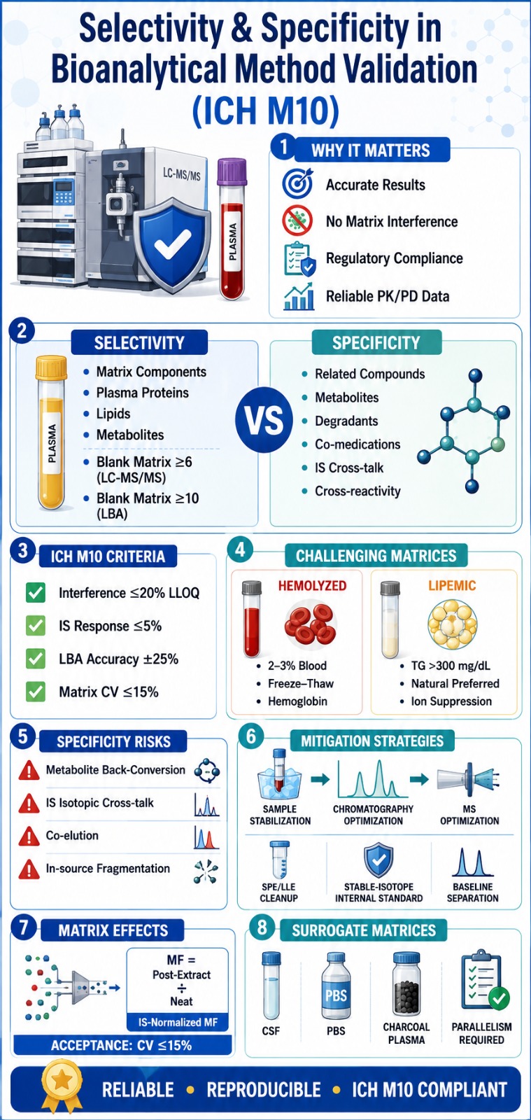
- Selectivity ensures the analyte is accurately measured without interference from endogenous biological matrix components, while specificity confirms it can be distinguished from structurally similar compounds, metabolites, degradants, and co-administered drugs.
- ICH M10 requires independent validation of selectivity and specificity, including testing multiple blank matrix sources, evaluating hemolyzed and lipemic samples, and meeting strict acceptance criteria for interference, accuracy, and matrix variability.
- Selectivity studies challenge analytical methods using complex biological matrices such as hemolyzed and lipemic plasma to verify that matrix constituents do not compromise analyte quantification.
- Specificity assessments evaluate potential interference from metabolites, isotopically labeled internal standards, degradation products, and concomitant medications while ensuring minimal cross-talk and accurate analyte identification.
- Matrix effects are measured using the IS-normalized Matrix Factor across multiple biological lots to identify ion suppression or enhancement, with a coefficient of variation (CV) of ≤15% required for regulatory compliance.
- Advanced mitigation strategies, including optimized chromatography, stable-isotope internal standards, sample stabilization, SPE/LLE cleanup, and MS parameter optimization, help prevent metabolite back-conversion and improve assay reliability.
- Surrogate matrices and parallelism testing support validation when authentic biological matrices are limited, ensuring bioanalytical methods remain accurate, reproducible, and fully compliant with global regulatory expectations.
Regulatory Framework for Selectivity and Specificity in Bioanalytical Method Validation
The regulatory framework governing bioanalytical method validation, particularly through the ICH M10 guideline, requires bioanalytical methods to provide quantitative evidence that matrix-derived background interference and structurally related compounds do not compromise analyte measurement. These requirements are essential for achieving regulatory approval and ensuring the reliability of bioanalytical data. Before the implementation of ICH M10 in 2022, differences between FDA and EMA validation expectations often resulted in duplicate validation efforts and additional cross-validation studies. The introduction of the harmonized guideline has standardized global requirements and clearly distinguishes selectivity and specificity as separate validation characteristics that must each undergo independent evaluation.
Navigating global regulatory hurdles? Read our deep dive on EMA vs. FDA Bioanalytical Method Validation Differences to align your filing strategy.
Selectivity focuses on managing the biological complexity of the sample matrix. It verifies that the analytical procedure can accurately quantify the analyte in the presence of naturally occurring matrix constituents, including plasma proteins, phospholipids, electrolytes, metabolites, and variations in lipid composition that may differ substantially among individuals.
Specificity addresses the challenge of distinguishing the analyte from structurally similar substances. It demonstrates that the analytical method is unaffected by compounds that may possess comparable chemical structures, isotopic characteristics, or fragmentation patterns. Within chromatographic assays, this includes endogenous analogs, Phase I and Phase II metabolites, degradation products, isotopically labeled internal standards, internal standard cross-talk, and concomitantly administered medications. In ligand-binding assays, specificity focuses on evaluating potential cross-reactivity of the binding reagent with related molecular species, isoforms, or co-administered therapeutic agents.
| Validation Parameter | ICH M10 Definition | Chromatography Acceptance Criteria | LBA Acceptance Criteria |
|---|---|---|---|
| Selectivity | Ability to differentiate the analyte from endogenous matrix components. | Blank matrix from ≥ 6 sources. Interference ≤ 20% of LLOQ and ≤ 5% of IS response. | Blank matrix from ≥ 10 sources. Interference below LLOQ in at least 80% of sources. |
| Specificity | Ability to differentiate the analyte from structurally related compounds. | Matrix spiked with analogs and metabolites. Interference ≤ 20% of LLOQ and ≤ 5% of IS response. | Matrix spiked with potentially cross-reactive compounds. Accuracy must remain within ± 25% at LLOQ and ULOQ. |
| Matrix Effect | Influence of matrix constituents on analyte response, including ion suppression or enhancement. | IS-normalized Matrix Factor CV ≤ 15% across ≥ 6 lots, including lipemic and hemolyzed matrices. | Evaluated together with selectivity, emphasizing recovery and parallelism assessments. |
Swipe horizontally to view the full table on smaller screens.
Need help implementing the latest standards? Discover how our team ensures strict adherence to ICH M10 Bioanalytical Method Validation Guidelines.
Demonstrating Selectivity: Overcoming Complex Biological Matrices
Establishing selectivity requires the analysis of blank biological matrices obtained from at least six independent sources for chromatographic methods and ten independent sources for ligand-binding assays. This evaluation must conclusively demonstrate that endogenous matrix components do not generate interfering responses at the retention times corresponding to the analyte and internal standard. Given the significant biological variability present within clinical populations, demonstrating acceptable performance in a single, idealized matrix lot is inadequate for regulatory acceptance.
Selectivity assessments must also extend beyond healthy donor samples and include biologically altered matrices that represent realistic clinical conditions. These matrices are commonly encountered during clinical studies, therapeutic monitoring programs, and forensic or postmortem investigations. Consequently, current ICH M10 validation practices require dedicated evaluation of both hemolyzed and lipemic samples.
Hemolyzed and Lipemic Matrix Protocols
Hemolysis, resulting from the rupture of erythrocytes, and lipemia, characterized by elevated concentrations of circulating lipids and triglycerides, can introduce substantial analytical complications. These effects may include analyte degradation, altered extraction efficiency, ion suppression, signal distortion, and compromised assay performance.
To adequately challenge the analytical method during validation, laboratories must generate representative worst-case matrices that mimic these physiological conditions.
Hemolyzed Matrix Preparation
A controlled hemolyzed matrix is typically prepared by adding 2% to 3% whole human blood to a standard blank biological matrix such as plasma. The mixture is then subjected to at least one freeze-thaw cycle to promote complete erythrocyte disruption. This process releases substantial quantities of hemoglobin, intracellular proteins, enzymes, iron-containing species, and other cellular constituents into the matrix, thereby creating a rigorous test environment for evaluating method selectivity.
Lipemic Matrix Preparation
Lipemic matrices may be obtained from naturally hyperlipidemic plasma samples containing triglyceride concentrations greater than 300 mg/dL. Alternatively, laboratories may simulate lipemia by adding lipid emulsions such as Intralipid to blank plasma. Although artificial lipid supplementation is widely used, numerous comparative investigations have demonstrated that synthetic emulsions do not fully replicate the complexity of naturally occurring hyperlipidemic plasma. As a result, naturally lipemic samples are generally regarded as the preferred option for thoroughly assessing extraction efficiency, recovery performance, and ionization suppression effects.
| Matrix Challenge | Primary Source of Analytical Interference | Validated Preparation Protocol | Consequence of Selectivity Failure |
|---|---|---|---|
| Hemolysis | Hemoglobin, intracellular enzymes, iron-containing species. | Spike blank matrix with 2–3% whole blood followed by freeze-thaw lysis. | Analyte degradation, altered protein binding, and optical interference in LBA measurements. |
| Lipemia | Elevated triglycerides, cholesterol, and phospholipids. | Utilize plasma containing >300 mg/dL triglycerides or supplement with Intralipid. | Significant ESI ion suppression, reduced extraction recovery, and chromatographic column contamination. |
Swipe horizontally to view the full table on smaller screens.
Demonstrating Specificity: Mitigating Cross-Reactivity and Structural Interferences
Demonstrating specificity requires the intentional addition of known structurally related compounds to blank biological matrices. These compounds may include metabolites, degradation products, internal standards, isotopic variants, and co-administered medications. The purpose of this evaluation is to experimentally verify that such substances do not co-elute, cross-react, or otherwise contribute to the measured analyte signal. When interference is observed, method optimization becomes necessary through adjustments to chromatographic separation conditions, mass spectrometric acquisition parameters, or multiple reaction monitoring (MRM) transitions to ensure complete analytical discrimination.
In therapeutic fields where polypharmacy is common, including oncology, infectious disease, and transplant medicine, specificity assessments become particularly critical. The analytical method must demonstrate that concomitant medications and their circulating metabolites do not generate overlapping precursor-to-product ion transitions or produce responses at the same chromatographic retention time as the target analyte.
For ligand-binding assays, specificity challenges are primarily associated with the selectivity of the antibody or binding reagent. Validation studies require spiking blank matrices with structurally related compounds, endogenous isoforms, or concomitant medications at concentrations representing the highest expected clinical exposure. Under these conditions, measured analyte concentrations at both the lower limit of quantification (LLOQ) and upper limit of quantification (ULOQ) must remain within ± 25% of their nominal values. Successful performance demonstrates that the binding reagent maintains exclusive recognition of the intended target molecule despite the presence of potentially interfering substances.
Internal Standard Isotopic Purity and Cross-Talk
The use of a stable-isotope-labeled internal standard (SIL-IS) remains one of the most powerful approaches for achieving superior selectivity and specificity in LC-MS/MS assays because it closely mirrors the extraction, chromatographic, and ionization behavior of the target analyte. This similarity allows the internal standard to compensate for matrix effects and analytical variability while maintaining accurate quantification.
Despite these advantages, producing a SIL-IS with complete isotopic purity presents significant scientific and manufacturing challenges. Trace quantities of unlabeled analyte present within the internal standard material, or overlapping isotopic distributions between the analyte and internal standard, can generate substantial isotopic cross-talk. Such interference may artificially increase analyte responses near the lower limit of quantification and compromise assay accuracy.
Consequently, specificity evaluations must clearly demonstrate that signal contribution from the internal standard to the analyte transition does not exceed 20% of the LLOQ response. Similarly, the analyte must not contribute more than 5% of the internal standard response. Meeting these requirements confirms that isotopic interference is adequately controlled and that the analytical method can reliably distinguish between the analyte and its isotopically labeled counterpart.
The Critical Challenge of Metabolite Back-Conversion in Specificity
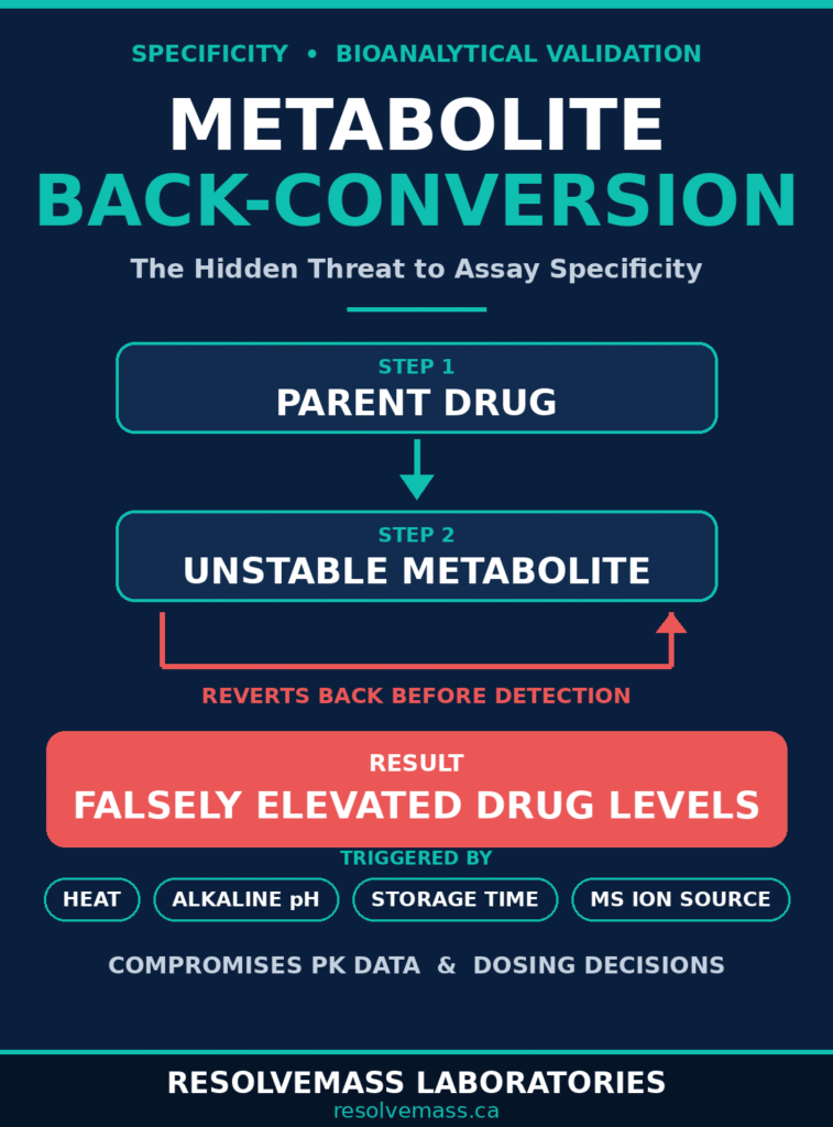
Metabolite back-conversion represents one of the most significant threats to specificity in bioanalytical method validation. This phenomenon occurs when unstable Phase II metabolites, including acyl glucuronides and N-oxide derivatives, revert to the parent compound during sample preparation, storage, extraction procedures, or within the mass spectrometer itself. As a result, the measured concentration of the parent drug can be artificially elevated, leading to inaccurate pharmacokinetic interpretations, erroneous exposure calculations, and potentially serious dosing decisions. Therefore, robust stabilization strategies and effective chromatographic separation are essential components of any bioanalytical method designed to accurately assess drug concentrations.
Among unstable metabolites, acyl glucuronides are particularly challenging because of their inherent chemical reactivity. These electrophilic ester conjugates can readily undergo hydrolysis under relatively mild conditions, regenerating the parent aglycone. Factors such as alkaline pH, elevated extraction temperatures, prolonged sample storage, and extended bench-top exposure can accelerate this conversion process. In addition to chemical instability during sample handling, acyl glucuronides may also undergo fragmentation within the mass spectrometer. Even when a glucuronide metabolite remains intact throughout sample preparation, the high thermal energy and elevated declustering potentials present in electrospray ionization (ESI) sources can trigger collision-induced dissociation (CID). This process removes the glucuronic acid moiety and generates precursor and product ions that are indistinguishable from those of the parent drug.
To adequately demonstrate specificity in the presence of potentially unstable metabolites, the analytical method must provide complete baseline chromatographic separation between the parent compound and its metabolite. Since glucuronide conjugates are substantially more polar than their parent molecules, optimized chromatographic gradients typically allow these metabolites to elute significantly earlier than the parent drug, thereby minimizing the risk of analytical overlap.
Facing unexpected validation hurdles or assay instability? See how our experts solve complex issues through our Bioanalytical Method Validation Failure Solutions.
Advanced Mitigation Strategies for Unstable Metabolites
Pre-Analytical Stabilization
Immediate stabilization following sample collection is often the most effective strategy for preventing metabolite back-conversion. Clinical protocols frequently require sample handling under tightly controlled conditions, including the use of ice-cold processing environments, protection from light exposure for photosensitive compounds such as fenofibric acid, specialized anticoagulants such as oxalate fluoride, and immediate pH adjustment using acidification buffers. These measures help suppress enzymatic activity and reduce the likelihood of chemical hydrolysis occurring before analysis.
Chromatographic Resolution
Achieving complete chromatographic separation between unstable metabolites and parent compounds is essential for maintaining specificity. This may involve selecting specialized stationary phases, such as phenyl-hexyl columns, or optimizing mobile phase composition and pH through additives such as 0.1% formic acid. Such adjustments enhance the separation of highly polar metabolites from more lipophilic parent molecules, reducing the possibility of co-elution.
MS Source Optimization
Mass spectrometric parameters must be carefully optimized to prevent in-source fragmentation. Lowering cone voltage, reducing capillary temperature, and minimizing declustering potential can significantly decrease the amount of internal energy transferred to analyte molecules. These adjustments help preserve metabolite integrity and reduce the risk of collision-induced cleavage occurring within the ion source.
Matrix Effects: Quantifying Ion Suppression and Enhancement
Matrix effects are assessed through the calculation of the internal standard (IS)-normalized matrix factor, which compares the response of analytes in post-extraction spiked samples with the response observed in neat solutions. This evaluation is critical because selectivity and specificity cannot be fully established without understanding how residual matrix components influence analyte ionization within the mass spectrometer.
Following sample preparation, residual phospholipids, salts, proteins, and other endogenous substances may still be present in the extracted sample. When these compounds enter the ion source simultaneously with the analyte, they compete for charge during the electrospray ionization process. This competition can substantially alter ionization efficiency.
When matrix constituents dominate the available charge on the surface of evaporating droplets, the analyte may fail to ionize efficiently, resulting in ion suppression and reduced signal intensity. Conversely, certain matrix components may facilitate analyte transfer into the gas phase by altering droplet surface properties, leading to ion enhancement and artificially elevated signal responses.
To quantitatively assess the impact of these effects on assay performance, bioanalytical laboratories commonly employ the Matuszewski post-extraction addition approach to calculate both the absolute Matrix Factor (MF) and the IS-normalized Matrix Factor.
The procedure requires the preparation of quality control samples at both low and high concentration levels using at least six independent matrix lots.
Sample Sets Used for Matrix Effect Evaluation
- Set A (Neat Solution)
The analyte and internal standard are prepared directly in the mobile phase without any biological matrix components. - Set B (Post-Extraction Spiked Samples)
A blank biological matrix undergoes extraction, after which the analyte and internal standard are added to the processed extract.
The absolute Matrix Factor is calculated as:
The IS-normalized Matrix Factor is subsequently calculated as:
Under ICH M10 requirements, the coefficient of variation (CV) for the IS-normalized Matrix Factor across the six independent matrix lots, including both hemolyzed and lipemic samples, must not exceed 15%. Compliance with this criterion demonstrates that the stable-isotope-labeled internal standard effectively compensates for matrix-induced ionization variability.
Experienced CROs such as ResolveMass Laboratories Inc. recognize that failure to meet this acceptance criterion often indicates inadequate sample cleanup. In such cases, laboratories may need to replace simple protein precipitation (PPT) techniques with more selective sample preparation approaches, including Solid Phase Extraction (SPE) or Liquid-Liquid Extraction (LLE), to remove problematic matrix constituents before LC-MS/MS analysis.
Ion suppression or phospholipid interference causing variable results? Read our overview on handling Matrix Effects in LC-MS/MS Bioanalysis.
Managing Rare and Surrogate Matrices in Method Validation
The validation of bioanalytical methods for uncommon biological matrices, including cerebrospinal fluid (CSF), vitreous humor, ocular tissues, and other difficult-to-obtain specimens, presents unique challenges. Obtaining multiple independent blank lots from these matrices may be impractical, ethically restricted, or physically impossible. Under such circumstances, surrogate matrices are employed as alternatives. Their use requires rigorous scientific justification and comprehensive parallelism assessments to demonstrate that they accurately replicate the behavior of authentic biological samples.
This strategy is particularly important for endogenous biomarker quantification and supports the pharmaceutical industry’s commitment to the 3Rs principles (Reduce, Refine, Replace) by minimizing the use of animal-derived materials during preclinical research.
A surrogate matrix may consist of artificial cerebrospinal fluid, phosphate-buffered saline (PBS), or specially processed biological matrices such as charcoal-stripped plasma from which endogenous analytes have been removed. The absence of endogenous target compounds allows investigators to establish calibration curves without interference from naturally occurring analyte concentrations.
Need expert bioanalytical support for central nervous system or tissue-specific targets? Explore our Tissue and CSF Bioanalytical Services.
However, demonstrating acceptable performance in a surrogate matrix alone is insufficient for regulatory acceptance. To confirm that selectivity and specificity remain valid in clinical applications, a comprehensive parallelism assessment must be performed.
Parallelism testing evaluates whether the analytical response generated in the surrogate matrix accurately reflects the response observed in authentic biological samples. The process typically involves selecting a study sample containing a high concentration of the analyte, performing a series of serial dilutions using the surrogate matrix, and comparing the resulting response profile with expected values.
Successful parallelism is demonstrated when the diluted samples produce concentration-response relationships that remain linear, proportional, and consistent with the original sample. If significant divergence in slope or response is observed, this indicates that the surrogate matrix does not adequately reproduce the extraction recovery, matrix effects, or analytical behavior of the authentic biological fluid. Under such circumstances, the surrogate matrix cannot be considered suitable for method validation or regulatory submission.
Moving into clinical testing? Discover how our Bioanalytical CRO Partnership supports rapid progression into First-in-Human Studies.
Conclusion
Demonstrating Selectivity and Specificity in Bioanalytical Method Validation remains one of the most important scientific requirements for ensuring that pharmacokinetic assessments, biomarker measurements, bioequivalence studies, and exposure-response analyses accurately reflect true biological conditions rather than analytical artifacts or unintended interferences. With regulatory agencies worldwide adopting the harmonized expectations outlined in the ICH M10 guideline, traditional validation strategies based on limited matrix evaluations are no longer sufficient.
Modern bioanalytical validation requires a comprehensive understanding of the biochemical environment surrounding the assay. Laboratories must rigorously evaluate challenging matrices such as hemolyzed and lipemic samples, establish effective chromatographic separation of unstable metabolites such as acyl glucuronides, control metabolite back-conversion, and quantify matrix effects through detailed IS-normalized matrix factor assessments across diverse biological sources. These activities collectively ensure that analytical results remain accurate, reproducible, and scientifically defensible.
Successfully executing these complex validation activities requires a combination of advanced instrumentation, extensive scientific expertise, and a thorough understanding of evolving global regulatory expectations. Through the strategic use of surrogate matrices, careful management of isotopic internal standards, optimized sample preparation techniques, and robust chromatographic methodologies, laboratories can transform analytical challenges into high-quality, submission-ready datasets that support regulatory success.
Collaborating with a specialized Contract Research Organization that possesses demonstrated experience in FDA, EMA, and Health Canada-compliant bioanalysis can significantly reduce development risk and accelerate regulatory acceptance. Organizations such as ResolveMass Laboratories Inc. provide the technical capabilities and regulatory insight necessary to support complex bioanalytical programs throughout the drug development lifecycle.
For expert consultation, method development support, and regulatory-compliant execution of Selectivity and Specificity in Bioanalytical Method Validation, contact the scientific team at ResolveMass Laboratories Inc. through their official website:
Frequently Asked Questions
What are the specific acceptance criteria for selectivity in LC-MS/MS bioanalysis?
For LC-MS/MS methods, selectivity is demonstrated by evaluating blank matrices obtained from multiple independent sources. Any signal detected at the analyte retention time must not exceed 20% of the response observed at the lower limit of quantification (LLOQ), while interference at the internal standard retention time must remain below 5% of the internal standard response. These limits help ensure accurate quantification in diverse biological samples.
Why must hemolyzed and lipemic matrices be explicitly tested during method validation?
Hemolyzed and lipemic samples represent challenging real-world clinical conditions that can significantly influence analytical performance. Hemolysis introduces intracellular constituents such as hemoglobin and enzymes that may alter analyte stability, while lipemia increases lipid content that can affect extraction efficiency and ionization. Evaluating these matrices confirms that the method remains reliable even when analyzing compromised clinical specimens.
How is a hemolyzed matrix prepared for validation purposes?
A hemolyzed matrix is generally prepared by adding approximately 2% to 3% whole human blood to a blank biological matrix, such as plasma. The mixture is then subjected to at least one freeze-thaw cycle to ensure complete rupture of red blood cells. This process releases intracellular components into the matrix, creating conditions that closely resemble hemolyzed clinical samples encountered during routine bioanalysis.
What is metabolite back-conversion, and why does it pose a major specificity risk?
Metabolite back-conversion occurs when unstable metabolites, particularly certain Phase II conjugates such as acyl glucuronides, transform back into the parent drug during sample handling, extraction, storage, or mass spectrometric analysis. This process can artificially increase the measured concentration of the parent compound and lead to inaccurate pharmacokinetic data. As a result, controlling back-conversion is essential for maintaining assay specificity and data integrity.
How do bioanalysts prevent the in-source fragmentation of glucuronide metabolites?
Preventing in-source fragmentation requires careful optimization of mass spectrometric operating conditions. Analysts typically reduce parameters such as cone voltage, source temperature, and declustering potential to minimize excessive molecular activation. In addition, chromatographic conditions are optimized to achieve complete separation between glucuronide metabolites and the parent compound before they enter the ion source, further reducing the risk of analytical interference.
What is the Matuszewski approach to evaluating matrix effects?
The Matuszewski approach is a widely accepted strategy for assessing matrix effects in LC-MS/MS bioanalysis. It involves comparing the analytical response of an analyte added to an extracted blank matrix with the response of the same analyte prepared in a matrix-free solution. This comparison allows scientists to quantify the extent of ion suppression or ion enhancement caused by residual biological matrix components.
What does the IS-normalized matrix factor prove about a bioanalytical assay?
The IS-normalized matrix factor demonstrates how effectively an internal standard compensates for matrix-related variations in analyte ionization. By normalizing analyte response to the internal standard response, analysts can evaluate whether matrix effects are being adequately controlled. According to ICH M10 recommendations, the coefficient of variation (CV) of the IS-normalized matrix factor across multiple matrix lots should not exceed 15%, indicating acceptable method robustness.
When is the use of a surrogate matrix permitted under ICH M10 guidelines?
Surrogate matrices may be used when obtaining sufficient quantities of authentic blank biological matrix is impractical or impossible. This situation commonly arises during the analysis of endogenous biomarkers or when working with rare matrices such as cerebrospinal fluid, ocular fluids, or specialized tissue homogenates. The surrogate matrix must be scientifically justified and shown to provide analytical performance comparable to the authentic matrix.
How is parallelism used to mathematically validate a surrogate matrix?
Parallelism is used to confirm that a surrogate matrix accurately reproduces the analytical behavior of the authentic biological matrix. The assessment involves serial dilution of real study samples followed by comparison of the resulting concentration-response relationship with the calibration curve prepared in the surrogate matrix. When both responses exhibit comparable slopes and proportionality, it demonstrates that the surrogate matrix does not introduce significant analytical bias and can be considered suitable for quantitative analysis.
Reference:
- U.S. Food and Drug Administration. (2018, May). Bioanalytical method validation: Guidance for industry. U.S. Department of Health and Human Services, Food and Drug Administration, Center for Drug Evaluation and Research, and Center for Veterinary Medicine. https://www.fda.gov/media/128343/download
- International Council for Harmonisation of Technical Requirements for Pharmaceuticals for Human Use. (2022). ICH harmonised guideline M10: Bioanalytical method validation and study sample analysis (Step 4, adopted 24 May 2022). https://database.ich.org/sites/default/files/M10_Guideline_Step4_2022_0524.pdf
- Prime Minister’s Office of Lebanon. (2023). Guidelines for good clinical practice (GCP) (Decision No. 1348/2023). Government of Lebanon. PCM Lebanon PDF
- Bonfiglio, R., King, R. C., Olah, T. V., & Merkle, K. (1999). The effects of sample preparation methods on the variability of the electrospray ionization response for model drug compounds. Rapid Communications in Mass Spectrometry, 13(12), 1175–1185. https://doi.org/10.1002/(SICI)1097-0231(19990630)13:12%3C1175::AID-RCM639%3E3.0.CO;2-0
- International Council for Harmonisation of Technical Requirements for Pharmaceuticals for Human Use. (2019, March 20). ICH M10: Bioanalytical method validation—Expert Working Group (EWG) Step 2 presentation. https://database.ich.org/sites/default/files/M10_EWG_Step2_Presentation.pdf
- U.S. Food and Drug Administration. (2022). M10 bioanalytical method validation and study sample analysis: Guidance for industry. U.S. Department of Health and Human Services, Food and Drug Administration, Center for Drug Evaluation and Research (CDER). https://www.fda.gov/media/162903/download
- European Medicines Agency. (2011). Guideline on bioanalytical method validation (EMEA/CHMP/EWP/192217/2009 Rev. 1 Corr. 2). European Medicines Agency. https://www.ema.europa.eu/en/documents/scientific-guideline/guideline-bioanalytical-method-validation_en.pdf
Need Help Demonstrating Selectivity and Specificity in Your Bioanalytical Method Validation?
Whether you need support with selectivity assessments, specificity investigations, matrix effect studies, or full bioanalytical method validation under ICH M10, our experts can help.
Contact usAbout The Author
Anusha Sinha
Anusha Sinha, B.Pharm, is an experienced pharma professional with a strong background in Analytical Chemistry and Polymer Chemistry. With a passion for translating complex scientific data into clear, accessible content, she plays a vital role in communicating ResolveMass Laboratories Inc.’s advanced testing capabilities. In addition to her scientific expertise, Anusha leads Business Development initiatives, helping clients across pharmaceutical, biotechnology, and materials science sectors find tailored analytical solutions. Her combined experience in science and strategy positions her at the forefront of client engagement and technical communication.

