

Introduction
Non-enzymatic Deamidation and Aspartate Isomerization in Therapeutic Peptides are significant chemical degradation pathways that can modify peptide backbone structure, surface charge, biological activity, and pharmacokinetic behavior throughout biopharmaceutical development and storage. Governed by the primary amino acid sequence and surrounding environmental conditions, these spontaneous chemical modifications transform uncharged asparagine or standard aspartic acid residues into complex mixtures containing aspartyl, isoaspartyl, and cyclic succinimide species.
Therapeutic peptides occupy a distinctive structural position between small-molecule pharmaceuticals and large, folded protein therapeutics. Because they lack the rigid tertiary structure that provides shielding in globular proteins, synthetic and recombinant peptides often expose their backbone amide nitrogens and side-chain functional groups directly to the surrounding aqueous solvent. This high degree of solvent exposure increases their vulnerability to intramolecular nucleophilic attacks. When a neutral asparagine residue is converted into a negatively charged aspartic acid residue, molecular hydrophilicity increases and a net negative charge (Δq = -1) is introduced at physiological pH. At the same time, aspartate isomerization inserts a methylene group (-CH2-) directly into the peptide backbone, extending the main chain by 1.4 Å. Such structural changes may modify receptor binding affinity, reduce metabolic half-life, and promote non-native self-assembly or immunogenic aggregation.
Effective control of these critical quality attributes (CQAs) depends on robust analytical platforms that can detect, localize, and quantify subtle chemical modifications that are frequently isobaric. The integration of ultra-high-performance liquid chromatography (UHPLC), high-resolution tandem mass spectrometry (MS/MS), radical fragmentation approaches, and gas-phase ion mobility spectrometry provides the analytical resolution needed to characterize complicated degradation profiles. In addition, combining these analytical technologies with physics-based computational developability screening supports rational sequence engineering and formulation optimization during the early stages of drug candidate selection.
Explore ResolveMass approaches for peptide physicochemical characterization and analytical characterization.
Explore Peptide Physicochemical Characterization Services
Share via:
Quick Summary:
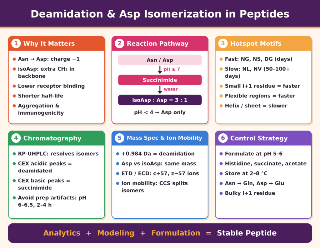
- What happens: Asparagine and aspartate residues in peptides degrade without any enzyme involved. Asn turns into Asp or isoAsp, which adds a negative charge, and isoAsp also lengthens the backbone by one CH₂ group. These changes can weaken receptor binding, shorten half-life, and trigger aggregation or immune reactions.
- Mechanism: At neutral to alkaline pH, the residue forms a cyclic succinimide intermediate. Water then opens the ring, giving roughly 3 parts isoAsp to 1 part Asp, with small amounts of D-forms from racemization. Below pH 4, Asn instead hydrolyzes directly and gives only Asp.
- Sequence hotspots: The residue that follows Asn or Asp sets the speed of degradation.
- Small or flexible neighbors such as Gly and Ser react fast, with half-lives of days.
- Bulky neighbors such as Leu and Val slow it to months.
- Rigid helices and sheets protect the site; flexible, solvent-exposed regions speed it up.
- Chromatography:
- RP-UHPLC with shallow gradients and TFA/DFA additives separates the Asp and isoAsp isomers.
- CEX shows deamidated forms as acidic peaks and succinimide intermediates as basic peaks.
- Low-pH, short, low-temperature sample prep is needed to avoid creating artificial deamidation.
- Mass spectrometry:
- Deamidation shows up as a +0.984 Da mass shift, but Asp and isoAsp have identical mass.
- ETD/ECD fragmentation produces diagnostic c+57 and z−57 ions that pinpoint isoAsp.
- UVPD and ion mobility (CCS measurements) separate the isomers quickly.
- Formulation control: The most stable range is pH 5–6. Non-catalytic buffers (histidine, succinate, acetate) are preferred, along with 2–8 °C storage and viscosity-raising excipients such as sucrose or trehalose. Phosphate and bicarbonate should be avoided because they speed up degradation.
- Sequence engineering and prediction: When the residue isn’t needed for activity, swapping Asn for Gln or Asp for Glu, or adding a bulky neighboring residue, removes the hotspot. QSAR and molecular dynamics modeling can flag risky sites before the peptide is synthesized.
Reaction Pathways and Kinetics of Deamidation and Aspartate Isomerization in Therapeutic Peptides
Non-enzymatic degradation involving asparagine and aspartic acid residues generally occurs through a cyclic five-membered aspartyl-succinimide intermediate (Asu), which subsequently undergoes rapid hydrolysis to produce a characteristic 3:1 kinetic ratio of L-isoaspartyl (L-isoAsp) to L-aspartyl (L-Asp) products. Under acidic conditions (pH < 4.0), however, asparagine deamidation proceeds through a different mechanism, bypassing the succinimide pathway and undergoing direct, water-assisted hydrolysis that produces exclusively L-aspartic acid.
Under neutral to alkaline pH conditions (pH ≥ 7.0), the succinimide pathway becomes predominant. The process begins when the deprotonated backbone amide nitrogen of the adjacent C-terminal residue (i+1) performs an intramolecular nucleophilic attack on the side-chain carbonyl carbon (Cγ) of the target asparagine (Asni) or aspartic acid (Aspi) residue. In the case of asparagine, nucleophilic ring closure results in the elimination of ammonia (NH3), producing the metastable cyclic succinimide intermediate. For aspartic acid, a comparable dehydration process takes place, with protonation of the side-chain carboxyl group occurring before imide formation. Following formation, the strained succinimide ring undergoes base-catalyzed nucleophilic attack by water at either of its two carbonyl carbons. Attack at the side-chain carbonyl restores the conventional α-peptide linkage (L-Asp), whereas attack at the main-chain carbonyl opens the ring and produces an L-isoAsp residue, thereby converting the peptide backbone into a β-peptide linkage. Since attack at the main-chain carbonyl is both thermodynamically and sterically favored, L-isoAsp generally accumulates as the major degradation product.
Beyond structural isomerization, the planar configuration of the cyclic succinimide intermediate lowers the energy barrier associated with α-carbon proton abstraction, thereby promoting enantiomeric racemization. Hydrolysis of these racemized imides produces smaller quantities of D-Asp and D-isoAsp enantiomers, which may accumulate under elevated temperatures or when general base catalysts such as bicarbonate are present. At low pH (pH < 4.0), the backbone nitrogen remains completely protonated, which prevents it from acting as a nucleophile. Instead, direct hydrolysis of the side-chain carboxamide group by the solvent occurs, generating pure L-Asp without formation of cyclic succinimides or isoaspartyl isomers.
The primary amino acid sequence has a major influence on intrinsic chemical degradation rates. Common deamidation sequence motifs include Asn-Gly (NG), Asn-Ser (NS), Asn-Ala (NA), Asn-Thr (NT), and Asn-His (NH). Common aspartate isomerization motifs include Asp-Gly (DG), Asp-Ser (DS), Asp-Asp (DD), and Asp-His (DH). The steric characteristics and electronic properties of the i+1 side chain are the principal sequence-level factors controlling reaction velocity:
- Unhindered i+1 residues such as glycine (Gly) provide very little steric resistance, allowing the peptide backbone to adopt the constrained dihedral angles (φ, ψ) necessary for nucleophilic ring closure.
- Hydrophilic i+1 residues such as serine (Ser) and threonine (Thr) can stabilize transition-state intermediates through local hydrogen bonding and proton transfer catalysis.
- Sterically hindered or hydrophobic i+1 residues such as leucine (Leu), isoleucine (Ile), and valine (Val) position bulky side chains near the reaction site, physically shielding the side-chain carbonyl carbon and extending deamidation half-lives from days to months.
Conformational flexibility and the local microenvironment also contribute substantially to kinetic susceptibility. Unstructured random coil regions generally maintain high solvent accessibility (SASA) and limited backbone rigidity, conditions that facilitate succinimide formation. In contrast, locating an Asn or Asp residue within a rigid α-helix or β-sheet restricts backbone movement, making it more difficult for the i+1 nitrogen to align with the Cγ carbonyl and thereby substantially reducing degradation rates.
| Sequence Motif | Degradation Pathway | Dominant End-Products | Half-Life Range (τ1/2) | Kinetic Drivers & Environmental Factors |
|---|---|---|---|---|
| Asn-Gly (NG) | Succinimide Ring Closure | L-isoAsp, L-Asp (~3:1 ratio) | 1 to 5 Days | High backbone flexibility; alkaline pH (≥ 7.5) |
| Asn-Ser (NS) | Succinimide Ring Closure | L-isoAsp, L-Asp | 7 to 14 Days | Hydrogen-bonding assistance from i+1 hydroxyl |
| Asn-Leu / Asn-Val | Succinimide Ring Closure | L-isoAsp, L-Asp | 50 to 100+ Days | Steric hindrance at i+1 position suppressing attack |
| Asp-Gly (DG) | Dehydration to Imide | L-isoAsp, L-Asp, Asu | 3 to 10 Days | Accelerated under slightly acidic to neutral pH |
| Asn-X (pH < 4) | Direct Amide Hydrolysis | Pure L-Asp exclusively | Temperature-Dependent | Acid catalysis; complete absence of isoaspartyl variants |
High-Performance Chromatographic Detection and Quantification of Deamidation and Aspartate Isomerization in Therapeutic Peptides
High-performance chromatographic approaches, including reversed-phase UHPLC (RP-UHPLC) and cation exchange chromatography (CEX), can separate deamidated and isomerized peptide variants by taking advantage of relatively subtle changes in hydrophobicity, local conformation, and surface charge.
Reversed-Phase Ultra-High-Performance Liquid Chromatography (RP-UHPLC) is widely used as a primary analytical approach for separating peptide degradation variants. Conversion of asparagine to aspartic acid introduces a hydrophilic carboxyl group, which reduces retention on C18 stationary phases and generally causes deamidated species to elute before the native peptide. In contrast, isomerization of aspartate to isoaspartate changes the local backbone connectivity while leaving the net formula weight unchanged. The additional backbone methylene group in isoAsp disrupts localized hydrophobic interactions and can consequently modify retention behavior compared with the native Asp peptide. Baseline chromatographic resolution of isobaric Asp and isoAsp species requires careful optimization of both column chemistry and mobile phase conditions:
- Sub-2 µm particle stationary phases incorporating polar-embedded or C18-amide chemistries can improve selectivity for structural isomers.
- Volatile ion-pairing additives, including 0.05–0.1% trifluoroacetic acid (TFA) or difluoroacetic acid (DFA), can suppress silanol interactions and improve peak shape.
- Ultra-shallow organic solvent gradients, such as 0.1–0.3% acetonitrile per minute, combined with optimized column temperatures of 30–40°C, are often required to resolve closely eluting isomeric peaks.
Learn more about analytical strategies used to evaluate peptide stability and degradation behavior.
Explore GLP-1 Peptide Stability and Analytical Methods
Ion Exchange Chromatography (IEX), particularly Cation Exchange Chromatography (CEX), can be used to quantify charge heterogeneity associated with deamidation and imide intermediates:
- Acidic Variant Formation: Deamidation changes an uncharged carboxamide (Asn) into a negatively charged carboxylate (Asp or isoAsp), decreasing the overall isoelectric point (pI). When CEX columns are operated under acidic pH conditions (pH 4.0–5.5), deamidated variants show reduced retention and elute as distinct acidic charge variants ahead of the main peak.
- Basic Variant Formation: Accumulation of unhydrolyzed cyclic succinimide intermediates (Asu) neutralizes a side-chain carboxyl group (Asp → Asu). The resulting reduction in negative charge increases the local pI and causes succinimide species to appear as basic charge variants after the main peak.
Hydrophobic Interaction Chromatography (HIC) and Hydrophilic Interaction Liquid Chromatography (HILIC) offer additional orthogonal selectivity. HIC can separate conformational changes associated with isoaspartyl backbone extension under non-denaturing salt gradients, while HILIC is particularly effective for separating highly hydrophilic, short peptide fragments.
One important challenge in chromatographic quantification is avoiding artifacts introduced by the analytical method itself. Standard sample handling and enzymatic digestion procedures performed under alkaline pH conditions (pH 8.0–8.5) or at elevated temperatures (55–60°C) can accelerate artificial, sample-preparation-induced deamidation. To maintain analytical accuracy, sample preparation should use optimized low-pH digestion protocols (pH 6.0–6.5) with histidine, succinate, or citrate buffers, together with shorter incubation periods of 2–4 hours and low processing temperatures.
See how peptide characterization programs can be structured around comprehensive analytical data and defined CRO deliverables.
View the Peptide Characterization CRO Deliverables Checklist
Mass Spectrometry and Ion Mobility Spectrometry for Structural Characterization
High-resolution tandem mass spectrometry (MS/MS) and gas-phase ion mobility spectrometry enable precise localization of deamidation sites through a +0.984 Da monoisotopic mass shift and support differentiation of isobaric Asp/isoAsp isomers through radical dissociation methods and collision cross section (CCS) measurements.
Full MS analysis can readily identify asparagine deamidation because replacement of the side-chain -NH2 group with -OH produces a monoisotopic mass increase of +0.984 Da (Asn → Asp/isoAsp). High-resolution FT-ICR and Orbitrap mass spectrometers operating at resolving powers above 60,000 (at m/z 200) can resolve this mass change from overlapping natural isotopic envelopes, including the second isotopic peak containing two 13C atoms (Δm = 1.0033 Da).
Nevertheless, mass measurement by itself cannot distinguish native aspartic acid (Asp) from its isomerized counterpart, isoaspartic acid (isoAsp), because the isomerization reaction is strictly isobaric (Δm = 0.000 Da). Conventional low-energy Collision-Induced Dissociation (CID) and Higher-Energy Collisional Dissociation (HCD) tandem mass spectrometry depend on vibrational excitation along the lowest-energy fragmentation pathways. These techniques cleave conventional peptide amide bonds and generate identical b- and y-type fragment ion series for both Asp and isoAsp isomers. As a result, distinguishing the two structures at the specific modification site can be challenging without prior chromatographic separation.
Explore advanced mass spectrometry approaches for intact and structural characterization of therapeutic peptides.
Learn About Native Mass Spectrometry for Therapeutic Peptide Characterization
Electron-based radical dissociation approaches, including Electron Transfer Dissociation (ETD) and Electron Capture Dissociation (ECD), address this analytical limitation. Rather than relying on vibrational heating, ETD transfers an electron to a multiply protonated peptide, promoting non-ergodic cleavage of backbone N-Cα bonds and producing c- and z-type fragment ions. When an isoAsp residue is present, radical cleavage of the modified β-peptide backbone produces characteristic diagnostic fragment ions:
- cn + 57 Diagnostic Ion: A c-type fragment ion containing an additional 57.021 Da mass increment associated with radical rearrangement of the isoaspartyl side-chain backbone linkage.
- zm – 57 Diagnostic Ion: A complementary z-type fragment ion exhibiting a corresponding loss of 57.021 Da.
Ultraviolet Photodissociation (UVPD) at 193 nm offers another fragmentation mechanism. Photonic excitation promotes high-energy backbone cleavage and produces a, b, c, x, y, and z fragment ions. Relative abundance ratios among specific b/y and a/x ions located adjacent to the modification site can support unambiguous structural assignment of Asp versus isoAsp residues.
Gas-Phase Ion Mobility Spectrometry (IMS), including Trapped Ion Mobility Spectrometry (TIMS) and High-Resolution Structures for Lossless Ion Manipulations (SLIM), adds a gas-phase conformational separation dimension before mass analysis. Ion mobility separates gaseous peptide ions according to their charge, mass, and three-dimensional structure, with this behavior quantified through their Rotationally Averaged Collision Cross Section (CCS, Å2). Because insertion of isoaspartyl residues extends the peptide backbone, isoAsp-containing peptides can adopt a more extended gas-phase conformation than the compact structure associated with native Asp peptides. HRIM platforms can resolve these small ΔCCS differences within milliseconds, supporting rapid, multi-attribute monitoring (MAM) without dependence on lengthy chromatographic run times.
Discover how Multi-Attribute Monitoring (MAM) can support comprehensive peptide characterization workflows.
Explore Multi-Attribute Monitoring (MAM) for Peptide Characterization
| Analytical Method | Target Modification | Separation / Identification Mechanism | Primary Analytical Readout | Capabilities & Technical Considerations |
|---|---|---|---|---|
| High-Res LC-MS/MS | Asn Deamidation | Exact mass defect (m/z) measurement | +0.984 Da monoisotopic mass shift | High sensitivity; requires narrow mass tolerances to exclude 13C2 overlaps. |
| RP-UHPLC (TFA/DFA) | Asp vs. isoAsp Isomers | Differential hydrophobic column retention | Resolved chromatographic retention times (RT) | Quantitative; requires shallow organic gradients and optimized stationary phases. |
| Cation Exchange (CEX) | Deamidation & Succinimide | Net surface charge and pI alterations | Acidic (Deamidated) & Basic (Asu) charge peaks | Excellent for intact/subunit charge variant profiling; native conditions. |
| ETD / ECD Mass Spec | Isoaspartate (isoAsp) | Radical-driven N-Cα backbone cleavage | Diagnostic cn+57 and zm-57 fragment ions | Unambiguous site localization; lower fragmentation efficiency on low charge states. |
| Ion Mobility (SLIM/TIMS) | Isobaric Isomers (Asp/isoAsp) | Gas-phase collision cross section (CCS) | Resolved gas-phase drift time peaks (ΔCCS) | Ultra-fast separation (milliseconds); enables high-throughput MAM workflows. |
Formulation Design and Sequence Engineering Controls
Controlling deamidation and aspartate isomerization requires placing therapeutic peptides within an appropriate stability range of pH 5.0–6.0, selecting buffer systems with minimal catalytic activity, and applying physics-based molecular modeling to support strategic sequence modifications.
Formulation pH is one of the most influential variables controlling non-enzymatic degradation kinetics. At pH > 6.5, base-catalyzed deprotonation of the adjacent backbone amide nitrogen promotes succinimide ring closure and can result in rapid accumulation of deamidated and isomerized products. In contrast, at pH < 4.0, acid-catalyzed direct solvent hydrolysis of asparagine side chains becomes the dominant degradation route. Between these two kinetic regimes, the optimal formulation stability window for many therapeutic peptides is generally pH 5.0 to pH 6.0, where the combined rates of imide formation and direct hydrolysis approach a minimum.
Explore analytical approaches for evaluating peptide stability during development and storage.
Read More About GLP-1 Peptide Stability and Analytical Methods
Buffer composition and ionic strength can also exert substantial effects on chemical stability:
- Buffer Species Effects: Anions such as phosphate, bicarbonate, and imidazole can function as general base catalysts, accelerating proton abstraction from the backbone amide nitrogen and facilitating succinimide formation and enantiomeric racemization. Non-catalytic or weakly catalytic buffer systems, including histidine, succinate, or acetate at low concentrations (10–20 mM), should therefore be considered.
- Viscosity and Temperature Controls: Storage of peptide formulations at 2–8°C or in frozen conditions (-80°C) decreases reaction rates. The addition of tonicity modifiers and excipients such as trehalose, sucrose, or mannitol can increase solution micro-viscosity, restrict backbone flexibility, and help stabilize the peptide against transition-state configurations.
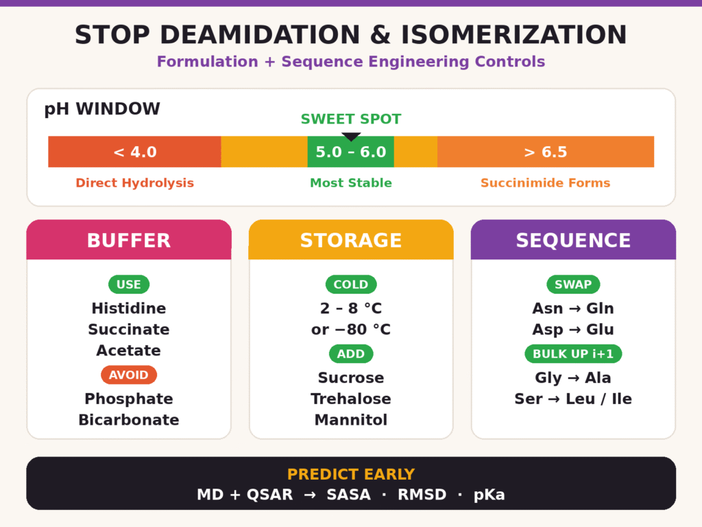
During early lead optimization, sequence engineering can provide a means of permanently removing chemical degradation hotspots. When an Asn or Asp residue does not directly participate in receptor binding or biological function, conservative amino acid substitutions may be introduced:
- Asn Substitutions: Substitution of Asn with glutamine (Gln) increases the cyclic intermediate ring size from a five-membered succinimide to a six-membered glutarimide, thereby slowing degradation kinetics by several orders of magnitude.
- Asp Substitutions: Replacing Asp with glutamic acid (Glu) can likewise suppress cyclic imide formation because six-membered ring closure requires a higher activation energy.
- Neighboring (i+1) Substitutions: Replacing an unhindered i+1 glycine (Gly) or serine (Ser) with a sterically bulky residue such as alanine (Ala), leucine (Leu), or isoleucine (Ile) creates additional steric shielding and reduces nucleophilic attack by the backbone amide nitrogen.
Learn how sequence confirmation can support the characterization and development of synthetic therapeutic peptides.
Explore Sequence Confirmation Services for Synthetic Therapeutic Peptides
Predictive computational approaches can accelerate identification of degradation hotspots before synthesis. Quantitative Structure-Activity Relationship (QSAR) models used together with molecular dynamics (MD) simulations can assess structural descriptors such as side-chain Solvent Accessible Surface Area (SASA), backbone root-mean-square deviation (RMSD), local hydrogen-bonding networks, and backbone amide nitrogen acidity (pKa). Evaluating these parameters across simulated conformational ensembles allows computational biophysicists to rank candidate stability and proactively support sequence engineering strategies.
Use complementary structural characterization techniques to evaluate peptide conformation and higher-order structural behavior.
Explore 2D NMR for Peptide Characterization
Conclusion
Effective control of Deamidation and Aspartate Isomerization in Therapeutic Peptides depends on the integration of high-resolution analytical characterization, predictive structural modeling, and rational formulation design. Advanced chromatographic techniques, including RP-UHPLC and CEX, together with high-resolution mass spectrometry, radical fragmentation (ETD/ECD), and gas-phase ion mobility spectrometry, enable detailed identification and quantification of isobaric degradation products. Integrating these analytical approaches with optimized formulation conditions (pH 5.0–6.0, non-catalytic buffers) and targeted sequence engineering can support long-term product stability, helping preserve therapeutic efficacy and maintain safety profiles throughout the drug product lifecycle.
Explore comprehensive analytical characterization services for therapeutic peptide development.
Explore Peptide Physicochemical Characterization Services
For expert analytical characterization, custom peptide mapping, and regulatory-compliant biopharmaceutical stability testing, visit ResolveMass Laboratories Inc. at:
Frequently Asked Questions
Electron Transfer Dissociation (ETD) can generate characteristic cₙ + 57 and zₘ − 57 diagnostic ions associated with isoaspartyl residues. These ions result from radical-driven rearrangement involving the modified β-peptide backbone during N–Cα bond cleavage. Their detection provides a useful approach for distinguishing isoAsp from conventional α-linked Asp and determining the location of the modification.
Local secondary structure influences deamidation by controlling peptide backbone flexibility and orientation. Flexible random coil regions allow the i+1 backbone amide nitrogen to approach the side-chain carbonyl carbon more easily, promoting succinimide formation. In contrast, rigid α-helices and β-sheets restrict the necessary backbone geometry and can significantly reduce the rate of deamidation.
Sample digestion near pH 6.0 helps minimize artificial deamidation and isomerization during peptide mapping. Conventional digestion under alkaline conditions such as pH 8.0–8.5 can promote base-catalyzed succinimide formation and introduce preparation-related degradation. Maintaining a mildly acidic pH of 6.0–6.5 helps preserve the original degradation profile and provides more reliable analytical results.
Hydrolysis of the cyclic aspartyl-succinimide intermediate generally produces L-isoAsp and L-Asp in an approximate 3:1 ratio. Preferential hydrolysis at the main-chain carbonyl favors formation of the isoaspartyl product. Smaller quantities of racemized D-isoAsp and D-Asp can also be generated when reaction conditions promote racemization of the intermediate.
Deamidation converts the neutral carboxamide group of Asn into a negatively charged carboxylate group, resulting in a net charge change of Δq = -1 and lowering the peptide pI. Deamidated species therefore generally appear as acidic charge variants during cation exchange chromatography (CEX). In contrast, formation of unhydrolyzed Asu removes a negative charge, increases the local pI, and produces a basic charge variant.
The identity of the i+1 residue strongly influences the accessibility and reactivity of the backbone amide nitrogen. Small residues such as glycine (Gly) and serine (Ser) provide less steric resistance and can facilitate succinimide formation. Bulkier residues such as leucine (Leu), valine (Val), and isoleucine (Ile) provide steric shielding that can restrict nucleophilic attack and improve peptide chemical stability.
High-Resolution Ion Mobility Spectrometry (HRIM), including Trapped Ion Mobility Spectrometry (TIMS) and Structures for Lossless Ion Manipulations (SLIM), can distinguish isobaric Asp and isoAsp species without conventional liquid chromatography. This separation is based on differences in gas-phase molecular conformation and Rotationally Averaged Collision Cross Section (CCS). The additional backbone methylene group associated with isoAsp can produce a different gas-phase structure from native Asp.
At neutral to alkaline pH, particularly at pH ≥ 6.5, deamidation mainly occurs through base-catalyzed intramolecular attack followed by cyclic succinimide formation. Hydrolysis of this intermediate can produce both Asp and isoAsp. At pH < 4.0, protonation of the backbone nitrogen suppresses succinimide formation, favoring direct acid-catalyzed hydrolysis that produces L-Asp.
Physics-based molecular dynamics (MD) and QSAR models can evaluate several structural characteristics to identify potential degradation hotspots. Solvent Accessible Surface Area (SASA) indicates exposure of relevant side chains, while root-mean-square deviation (RMSD) provides information about conformational flexibility. Backbone amide nitrogen acidity (pKa) can further indicate the likelihood of deprotonation and subsequent involvement in succinimide formation.
Reference:
- Lu, X., Nobrega, R. P., Lynaugh, H., Jain, T., Barlow, K., Boland, T., Sivasubramanian, A., Vásquez, M., & Xu, Y. (2019). Deamidation and isomerization liability analysis of 131 clinical-stage antibodies. mAbs, 11(1), 45–57. https://doi.org/10.1080/19420862.2018.1548233
- Adav, S. S. (2025). Advances in the study of protein deamidation: Unveiling its influence on aging, disease progression, forensics and therapeutic efficacy. Proteomes, 13(2), 24. https://doi.org/10.3390/proteomes13020024
- Yu, X., Sargaeva, N. P., Thompson, C. J., Costello, C. E., & Lin, C. (2015). In-source decay characterization of isoaspartate and β-peptides. International Journal of Mass Spectrometry, 390, 101–109. https://doi.org/10.1016/j.ijms.2015.07.013
- Erckes, V., Chamera Rendueles, L., Misiek, A., & Steuer, C. (2025). Revealing deamidation and isoaspartate formation during peptide analysis, purification and storage by tandem mass spectrometry. RSC Medicinal Chemistry, 17(2), 1144–1154. https://doi.org/10.1039/D5MD01025J
- De Sciscio, M. L., Nardi, A. N., Centola, F., Rossi, M., & Guarnera, E. (2023). Molecular modeling of the deamidation reaction in solution: A theoretical-computational study. The Journal of Physical Chemistry B, 127(44), 9550–9559. https://doi.org/10.1021/acs.jpcb.3c04662
- Hoffmann, D., Bauer, J., Kossner, M., Henry, A., Karow-Zwick, A. R., & Licari, G. (2024). Predicting deamidation and isomerization sites in therapeutic antibodies using structure-based in silico approaches. mAbs, 16(1), 2333436. https://doi.org/10.1080/19420862.2024.2333436
Get In Touch With Us
About The Author


