


Introduction
PEGylated Peptide Characterization represents a comprehensive analytical framework designed to assess batch-to-batch consistency, chemical integrity, structural conformation, and purity of peptide-polyethylene glycol bioconjugates. This analytical approach provides precise control over polymer polydispersity, modification site selectivity, and degradation profiles, supporting compliance with global regulatory requirements for biopharmaceutical development. The covalent conjugation of monomethoxy polyethylene glycol (mPEG) to therapeutic peptides can substantially increase hydrodynamic volume, extend circulating plasma half-life, mask immunogenic epitopes, and reduce renal clearance. Nevertheless, the attachment of a polydisperse synthetic polymer to a monodisperse biological peptide creates considerable analytical challenges. In contrast to un-modified synthetic peptides, PEGylated peptides demonstrate micro-heterogeneity resulting from polymer chain-length distributions, differences in site occupancy, and structural positional isomers. Therefore, analytical protocol development must address these complexities through high-resolution mass spectrometry, multi-dimensional chromatography, and universal aerosol detection to support the Investigational New Drug (IND) guidelines established by regulatory agencies, including the FDA and EMA.
Explore advanced peptide characterization capabilities:
LC-MS Characterization of GLP-1 Peptides
Share via:
Quick Summary:
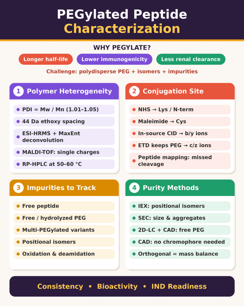
- Why PEGylate: Attaching PEG to a therapeutic peptide extends its half-life, masks immunogenic sites, and reduces renal clearance. But the PEG chains vary in length and can attach at different sites, which makes the product much harder to characterize.
- Polymer heterogeneity: PEG chains differ by 44 Da ethoxy units, so the polymer is described by Mn, Mw and the polydispersity index. Pharma-grade mPEG usually has a PDI of 1.01–1.05.
- Mass spectrometry for intact mass: High-resolution ESI-MS (Q-ToF or Orbitrap) with MaxEnt deconvolution resolves the overlapping charge states. MALDI-TOF complements it by giving simpler, singly charged spectra.
- Conjugation site mapping: In-source fragmentation trims the bulky PEG before CID, giving b/y ions. ETD keeps the PEG linkage intact and gives c/z ions. Peptide mapping finds the modified residue through missed cleavages.
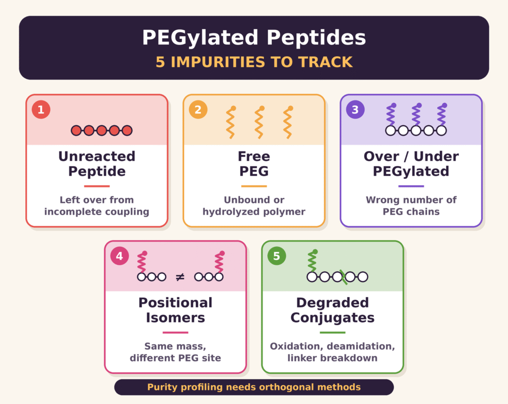
- Impurity profiling: The impurities to track are unreacted peptide, free or hydrolyzed PEG, multi-PEGylated variants, positional isomers, and degradants such as oxidized or deamidated species.
- Orthogonal separation methods: IEX resolves positional isomers through charge changes. SEC separates species by hydrodynamic size and detects aggregates. 2D-LC with CAD measures free PEG, which UV can’t detect because PEG has no chromophore.
- Conclusion: No single technique is enough. An integrated MS and chromatography strategy is needed to show batch consistency, preserve bioactivity, and meet FDA/EMA expectations for IND filings.
Resolving Polymer Heterogeneity in PEGylated Peptide Characterization
Resolving polymer heterogeneity in PEGylated Peptide Characterization requires the application of high-resolution mass spectrometry together with specialized chromatographic separation techniques to determine polymer dispersity and deconvolute complicated oligomeric mass spectra. Synthetic polyethylene glycol reagents inherently contain a distribution of chain lengths that differ according to their repeating ethoxy units, making accurate determination of molecular weight averages essential.
Learn more about advanced peptide physicochemical characterization:
Peptide Physicochemical Characterization Services
Synthetic polyethylene glycol reagents are produced through anionic ring-opening polymerization of ethylene oxide. This manufacturing process generates a Poisson distribution of polymer chains that differ by individual ethoxy monomer units (-CH₂CH₂O-, Δm = 44.05 Da). This intrinsic polydispersity influences the physical and spectroscopic properties of the resulting bioconjugate. The polymer distribution is described using the number-average molecular weight (Mn) and weight-average molecular weight (Mw), with their ratio defining the Polydispersity Index (PDI):
Mn = ∑ NiMi / ∑ Ni
Mw = ∑ NiMi2 / ∑ NiMi
PDI = Mw / Mn
A completely monodisperse material has a PDI of 1.00, whereas commercially available pharmaceutical-grade mPEGs generally demonstrate PDI values between 1.01 and 1.05.
Molecular Weight Distribution and Charge-State Deconvolution
In Electrospray Ionization Mass Spectrometry (ESI-MS), the inherent polydispersity of the PEG moiety is combined with the multi-charging behavior of the peptide backbone. Individual PEG oligomers within the molecular weight distribution can accept multiple protons or cations during electrospray ionization, resulting in hundreds of overlapping mass-to-charge (m/z) signals. High molecular weight PEGs, including 10 kDa, 20 kDa, and 40 kDa branched architectures, generate highly dense m/z spectra in which adjacent charge states from different oligomers can overlap directly. Consequently, specialized Maximum Entropy (MaxEnt) approaches or automated high-resolution deconvolution algorithms are necessary to resolve individual mass components, calculate Mn and Mw, and establish the precise PDI. High-resolution accurate mass (HRAM) instruments, including Quadrupole Time-of-Flight (Q-ToF) and Orbitrap mass spectrometers, offer the resolving power required to differentiate isobaric overlaps and distinguish minor chemical modifications, including oxidation or deamidation, from the 44.05 Da shift associated with the polymer repeating unit.
Chromatographic Dispersity and Retentivity Mechanisms
In Reversed-Phase High-Performance Liquid Chromatography (RP-HPLC), the retention characteristics of PEGylated peptides are governed by both the hydrophobicity of the peptide core and the chain-length dispersity of the attached PEG polymer. As PEG chain length increases, the polymer increasingly shields hydrophobic regions of the peptide core, resulting in reduced retention on conventional C8 or C18 stationary phases. Polymer dispersity can also produce considerable peak broadening because multiple oligomeric chain lengths may co-elute. Addressing this behavior requires optimized organic modifier gradients, commonly based on acetonitrile with trifluoroacetic acid (TFA), along with elevated column temperatures of approximately 50°C to 60°C to improve mass transfer kinetics.
Explore peptide characterization approaches for complex analytical workflows:
Peptide Characterization CRO Deliverables Checklist
MALDI-TOF MS Matrix and Cationization Optimization
Matrix-Assisted Laser Desorption/Ionization Time-of-Flight Mass Spectrometry (MALDI-TOF MS) provides a robust complementary approach to ESI-MS by predominantly generating singly charged [M+H]+ species and thereby reducing the complexity associated with overlapping multi-charge envelopes. The choice of matrix has a major influence on desorption efficiency and helps minimize undesirable in-source laser degradation. Commonly used matrices include 2,5-dihydroxybenzoic acid (DHB), trans-2-[3-(4-tert-butylphenyl)-2-methyl-2-propenylidene]malononitrile (DCTB), and α-cyano-4-hydroxycinnamic acid (CHCA). In addition, alkali salt dopants such as silver trifluoroacetate, sodium iodide, or sodium trifluoroacetate can stabilize ionization pathways by facilitating the formation of [M+Na]+ or [M+Ag]+ adducts. This approach helps suppress irregular protonation behavior along the ether oxygen backbone.
Elucidating Conjugation Sites: Advanced Tandem Mass Spectrometry Strategies
Determination of conjugation sites in PEGylated peptides depends on tandem mass spectrometry (MS/MS) approaches, including in-source fragmentation combined with collision-induced dissociation (CID), electron-transfer dissociation (ETD), and bottom-up proteomic mapping, to identify the exact modified amino acid residue. Accurate attachment-site mapping is critical because non-specific coupling can generate positional isomers that exhibit different biological activities and pharmacokinetic properties.
Discover specialized peptide sequencing workflows for detailed structural characterization:
GLP-1 Peptide Sequencing CRO Services
Chemical Conjugation Pathways and Isomeric Heterogeneity
PEGylation strategies employ functionalized PEG linkers designed to react with specific amino acid side chains:
- Amine-reactive coupling uses N-hydroxysuccinimide (NHS) esters or propionaldehydes to target lysine ε-amines and the peptide N-terminus. When several free lysine residues are accessible, non-specific coupling can generate complex mixtures of positional isomers.
- Thiol-reactive coupling uses maleimide-functionalized mPEGs to target cysteine thiols. Thiol modification generally provides greater site selectivity because free cysteine residues occur at relatively low natural abundance.
- Histidine modification uses specialized haloacetyl or carbamate linkers and requires comprehensive site mapping to verify the selectivity of the intended modification.
Review a dedicated GLP-1 analog sequencing workflow:
GLP-1 Analog Peptide Sequencing Workflow
In-Source Fragmentation Combined with CID-MS/MS
Direct collision-induced dissociation (CID) of intact, high-molecular-weight PEGylated peptides can be inefficient because the flexible PEG polymer backbone functions as an energy sink. It absorbs a substantial portion of the collision energy, thereby reducing effective cleavage of the peptide backbone. An in-source fragmentation strategy can be used before tandem mass analysis to overcome this energy-dissipation effect.
Increasing atmospheric pressure ionization (API) source voltages or cone voltages promotes thermal and electrical fragmentation of labile PEG ether bonds located near the linkage site. This in-source activation removes much of the high-molecular-weight polymer and leaves a lower-molecular-weight truncated ethylene glycol tag attached to the modified residue. The resulting trimmed precursor ion is then isolated within the quadrupole and subjected to conventional CID MS/MS. Further fragmentation produces complete b- and y-type fragment ion series, enabling unequivocal localization of the conjugation site through the resulting localized mass shifts.
Radical-Driven Electron-Transfer Dissociation (ETD)
For bioconjugate linkages that are particularly labile, Electron-Transfer Dissociation (ETD) or Electron-Capture Dissociation (ECD) offers an alternative fragmentation strategy. Radical-driven ETD cleaves peptide backbone N-Cα bonds to produce c- and z-type fragment ions while maintaining fragile PEG polymer linkages. By preserving the polymer modification on the amino acid from which it originated, this approach minimizes the generation of confounding internal polymer fragments and supports more reliable conjugation-site identification.
Explore multi-attribute analytical approaches for peptide characterization:
Multi-Attribute Monitoring (MAM) for Peptide Characterization
Enzymatic Bottom-Up Mapping and Missed Cleavage Profiles
Enzymatic digestion with proteases such as trypsin, Lys-C, Glu-C, or chymotrypsin breaks bioconjugates into smaller surrogate peptides that are appropriate for LC-MS/MS analysis. Bulky PEG modifications can sterically restrict protease access to nearby cleavage sites. For instance, PEGylation of a lysine residue can prevent tryptic cleavage at that particular location, producing a predictable missed cleavage product. Comparison of LC-MS/MS profiles from native and conjugated peptide digests allows analysts to identify the disappearance of native fragments and the formation of modified surrogate peptide species.
Purity Determination and Impurity Profiling in PEGylated Peptide Characterization
Purity determination in PEGylated Peptide Characterization requires orthogonal chromatographic workflows capable of separating and quantifying unreacted reagents, free polymer contaminants, multi-PEGylated species, and positional isomers. Establishing comprehensive purity profiles is essential for maintaining therapeutic consistency and reducing potential immunogenic risks associated with free PEG or degraded bioconjugate variants.
Learn more about advanced peptide impurity characterization:
GLP-1 Peptide Impurity Characterization
Profiling the Spectrum of Bioconjugate Impurities
Analytical purity assessments must characterize a range of product-related impurities:
- Unreacted Native Peptide: Residual un-modified precursor peptide that remains because of incomplete coupling reactions.
- Free Unreacted or Hydrolyzed Polymer: Non-conjugated mPEG-OH or hydrolyzed reagent species that remain within the analytical matrix.
- Over- and Under-PEGylated Variants: Unintended di-, tri-, or multi-PEGylated species generated as a consequence of sub-optimal reaction stoichiometry.
- Positional Isomers: Structural variants that possess identical molecular masses but differ in the locations at which the polymer is attached along the peptide backbone.
- Degraded Bioconjugates: Oxidized methionine or cysteine residues, deamidated species, and hydrolyzed linker conjugates that may develop during downstream purification or storage.
Ion-Exchange Chromatography for Positional Isomer Resolution
Ion-Exchange Chromatography (Cation Exchange, CEX; Anion Exchange, AEX) is an important separation mode for resolving positional isomers and charge variants. Covalent modification of a lysine ε-amine neutralizes a positive charge at physiological pH and consequently produces a substantial change in the overall surface charge and pI of the peptide. Site-specific charge shielding can modify retention behavior on CEX columns, enabling separation of distinct mono-PEGylated positional isomers that may remain unresolved through size-exclusion or conventional reversed-phase mechanisms.
Size-Exclusion Chromatography and Hydrodynamic Volume Inflation
Size-Exclusion Chromatography (SEC) or Gel Permeation Chromatography (GPC) separates bioconjugates primarily according to their hydrodynamic radius (Rh). PEG is highly hydrated in aqueous environments and coordinates a substantial shell of water molecules. As a result, the conjugated polymer can exhibit an unusually large Stokes radius compared with its actual formula mass. For example, a 20 kDa PEG chain attached to a 3 kDa peptide may elute at a retention volume corresponding to a globular protein standard in the 60–100 kDa range. SEC can therefore readily distinguish unreacted single peptide molecules from mono-PEGylated and di-PEGylated species because of the significant differences in hydrodynamic volume.
Explore analytical approaches for peptide aggregation and higher-order characterization:
Peptide Aggregation Analysis
Multi-Dimensional Liquid Chromatography with Aerosol Detection
Accurate quantification of free PEG and unreacted peptide species can be challenging with UV/Vis detection because PEG lacks chromophores, while peptide absorbance at 214 nm or 280 nm can vary considerably according to aromatic amino acid composition. To address this detector-response limitation, comprehensive multi-dimensional liquid chromatography (2D-LC) coupled with a Charged Aerosol Detector (CAD) or Evaporative Light Scattering Detector (ELSD) can be employed. Charged Aerosol Detection involves nebulizing the column eluate into aerosol droplets, drying these droplets to form particle residues, charging the resulting particles through a corona discharge, and measuring the total charge transferred to an electrometer. CAD provides a relatively uniform, mass-proportional response that is less dependent on chemical structure or the presence of optical chromophores. This enables accurate mass-balance determination of unreacted PEG, free peptide, and bioconjugate components within a single analytical workflow.
Explore advanced 2D analytical characterization strategies:
2D NMR for Peptide Characterization
Comprehensive Analytical Methodologies for PEGylated Peptide Characterization
The characterization of PEGylated bioconjugates requires analytical techniques to be matched with specific molecular critical quality attributes (CQAs). The table below summarizes the analytical performance characteristics, primary mechanisms, and technical limitations associated with key characterization platforms.
| Analytical Method | Primary Characterization Target | Separation / Ionization Mechanism | Mass & Resolution Capabilities | Key Advantages | Limitations & Technical Challenges |
|---|---|---|---|---|---|
| ESI-QTOF / Orbitrap LC-MS | Intact mass, oligomer distributions, and charge state profiles | Electrospray ionization; high-resolution mass analysis | Resolves m/z up to 8,000+ with <5 ppm mass accuracy | High mass accuracy; resolves multicharged species and adducts | Overlapping spectra require specialized software deconvolution |
| LC-MS/MS (In-Source CID / ETD) | Attachment site mapping and sequence verification | API in-source fragmentation combined with CID or ETD | Fragment-level resolution (b/y and c/z ion series) | Pinpoints exact modified amino acid residues; resolves positional isomers | Requires specialized source tuning and optimization of breakdown curves |
| MALDI-TOF MS | Average molecular weight (Mn, Mw) and polymer dispersity | Matrix-assisted laser desorption/ionization; time-of-flight | Single charge detection beyond 100 kDa | Produces singly charged ions ([M+H]+); simple spectral interpretation | Potential matrix adduct formation and laser-induced degradation |
| Ion-Exchange Chromatography (IEX-HPLC) | Positional isomers and surface charge variants | Charge-based retention on CEX/AEX stationary phases | High resolution for charge-altering modifications | Resolves positional isomers with identical intact masses | Non-volatile buffer salts require offline desalting prior to MS characterization |
| Size-Exclusion Chromatography (SEC/GPC) | Hydrodynamic volume (Rh), stoichiometry, and aggregation | Size-based exclusion through porous stationary phase | Molecular weight range dictated by column pore size | Assesses hydrodynamic radius; identifies high-MW aggregates | Low chromatographic resolution; inflates apparent molecular mass |
| 2D-LC coupled with CAD / MS | Purity profiling and absolute quantitation of free PEG and peptide | Orthogonal separations (IEX × RP) with aerosol detection | Broad analyte gradient compatibility | Structure-independent mass response for accurate impurity quantitation | Complex system setup; mobile phase non-volatility constraints require solvent splitting |
Conclusion
In conclusion, rigorous PEGylated Peptide Characterization requires an integrated analytical strategy capable of addressing polymer heterogeneity, conjugation-site localization, and bioconjugate purity. Understanding the complex interaction among PEG polymer heterogeneity, positional isomerism, and trace impurity profiles is critical for demonstrating manufacturing consistency, preserving product bioactivity, and fulfilling stringent global regulatory expectations. By combining targeted fragmentation approaches, including in-source CID and ETD, with advanced separation technologies such as IEX and 2D-LC-CAD, analytical scientists can comprehensively deconvolute complex bioconjugate mixtures and facilitate the progression of therapeutic peptide pipelines from development through clinical approval.
For comprehensive peptide characterization and specialized analytical support, connect with ResolveMass Laboratories:
ResolveMass Laboratories – Regulatory Requirements for GLP-1 Peptide Characterization
For expert analytical services, high-resolution LC-MS mapping, and specialized bioconjugate testing, visit the ResolveMass Laboratories Contact Page.
Frequently Asked Questions
PEGylation can significantly modify chromatographic behavior because the attached polymer shields hydrophobic regions of the peptide surface. This generally decreases retention on conventional $C_{18}$ columns, while differences in PEG chain length can broaden chromatographic peaks. The combined effects of steric shielding and polymer dispersity therefore complicate separation.
High molecular weight PEG chains can absorb collision energy during conventional CID MS/MS, limiting fragmentation of the peptide backbone. In-source fragmentation partially removes the PEG chain near the conjugation point before precursor isolation. The resulting smaller tagged species can then undergo CID more efficiently, generating $b$- and $y$-type ions for reliable site localization.
Ion-Exchange Chromatography (IEX) distinguishes positional isomers through differences in their surface charge characteristics. PEG attachment at different sites, such as individual lysine residues or the N-terminus, can produce different local charge environments and pI values. These variations result in distinct interactions with cation or anion exchange stationary phases and consequently different retention times.
PEG has a strong capacity to hydrate and retain surrounding water molecules, giving the conjugate a larger hydrodynamic or Stokes radius than its formula mass would suggest. During Size-Exclusion Chromatography (SEC), this expanded hydrodynamic size causes earlier elution. Consequently, PEGylated peptides can appear substantially heavier when compared with globular protein calibration standards.
Free PEG is difficult to measure by conventional UV detection because PEG does not contain strong UV chromophores. Multi-dimensional liquid chromatography or reversed-phase chromatography coupled with a Charged Aerosol Detector (CAD) or Evaporative Light Scattering Detector (ELSD) can overcome this limitation. CAD provides a broadly mass-responsive signal, supporting more reliable quantification of free PEG.
MALDI-TOF MS analysis of PEG conjugates commonly uses matrices such as 2,5-dihydroxybenzoic acid (DHB) and DCTB to promote efficient analyte desorption and ionization. Cationization agents including silver trifluoroacetate and sodium iodide can further promote formation of stable metal-adducted ions. Appropriate matrix and dopant selection can improve spectral consistency and reduce unwanted adduct complexity.
Enzymatic digestion breaks PEGylated bioconjugates into smaller peptide fragments that can be effectively analyzed using LC-MS/MS. The bulky PEG modification may hinder protease access to nearby cleavage sites, resulting in characteristic missed cleavage products. These modified fragments provide useful sequence information and help establish the location of the PEG conjugation site.
Reference:
- Shimizu, T., Nakanishi, M., & Tani, M. (2012). Aspartic acid isomerization in proteins and peptides: A review. Journal of Pharmaceutical Sciences, 101(10), 3567–3579. https://doi.org/10.1002/jps.23257
- Wang, Z., Zhang, Q., Shen, H., Yang, P., & Zhou, X. (2021). Optimized MALDI-TOF MS strategy for characterizing polymers. Frontiers in Chemistry, 9, 698297. https://doi.org/10.3389/fchem.2021.698297
- Lu, X., Gough, P. C., DeFelippis, M. R., & Huang, L. (2010). Elucidation of PEGylation site with a combined approach of in-source fragmentation and CID MS/MS. Journal of the American Society for Mass Spectrometry, 21(5), 810–818. https://doi.org/10.1016/j.jasms.2010.01.011
- Belén, L. H., Rangel-Yagui, C. de O., Lissabet, J. F. B., Effer, B., Lee-Estevez, M., Pessoa, A., Castillo, R. L., & Farías, J. G. (2019). From synthesis to characterization of site-selective PEGylated proteins. Frontiers in Pharmacology, 10, 1450. https://doi.org/10.3389/fphar.2019.01450
- Hyldbakk, A., Hansen, T., Hak, S., & Borgos, S. E. F. (2024). Polyethylene glycol (PEG) as a broad applicability marker for LC-MS/MS-based biodistribution analysis of nanomedicines. Journal of Controlled Release, 366, 611–620. https://doi.org/10.1016/j.jconrel.2024.01.016
Get In Touch With Us
About The Author


