


Introduction:
Bioanalytical Method Development for Oligonucleotide Therapeutics is the process of building and validating a quantitative assay that measures an oligonucleotide drug, and often its metabolites, in biological matrices such as plasma and tissue. It generates the reliable PK and tissue distribution data that nucleic acid medicines need during development. Our team has outlined the broader framework in our guide to bioanalytical method development and validation, and this case study applies it to one of the harder analyte classes.
Oligonucleotide therapeutics modulate gene expression through sequence-specific mechanisms. ASOs bind target RNA and can alter its processing or promote its degradation, while siRNAs guide RNA-induced silencing complexes to complementary RNA sequences. Their activity depends on sequence, chemical modification, cellular uptake, tissue distribution and intracellular availability. Those same features make them difficult to measure. Plasma contains proteins, salts, enzymes and endogenous nucleic acids that interfere with analysis, and tissue adds differences in cellular composition, lipid content, nuclease activity and drug accumulation across organs.
ResolveMass Laboratories Inc. is a Canadian analytical CRO/CDMO with working experience in mass spectrometry, nucleic acid therapeutics and regulated bioanalysis. The same principles apply across modalities, as shown in our work on bioanalytical method development for mRNA therapeutics and peptide-oligonucleotide conjugate (POC) therapeutics.
About this case study: it is a representative development scenario that illustrates how a method can be designed and evaluated for an oligonucleotide in plasma and tissue. It is technically illustrative and is not a report of a specific completed client project. No client-specific experimental results are implied.
Summary:
- Bioanalytical Method Development for Oligonucleotide Therapeutics requires specialized strategies to quantify antisense oligonucleotides (ASOs) and small interfering RNAs (siRNAs) accurately in plasma and tissue, because these molecules are large, polyanionic, nuclease-sensitive, protein-bound and chemically heterogeneous.
- Plasma and tissue need different sample preparation strategies. Protein binding, endogenous nucleases, tissue composition and extraction efficiency all affect analytical performance.
- Hybridization-based ligand-binding assays and liquid chromatography–mass spectrometry (LC-MS) are complementary approaches, and neither is universally superior.
- This representative case study uses solid-phase extraction (SPE) with ion-pair reversed-phase LC-MS/MS and a structurally matched internal standard.
- Validation should address selectivity, sensitivity, accuracy, precision, recovery, matrix effects, stability and dilution integrity, in line with ICH M10 and FDA expectations.
- Tissue results need careful interpretation: total tissue concentration is not the same as pharmacologically available drug.
- A well-designed workflow supports pharmacokinetic (PK) evaluation, tissue distribution studies, dose selection, preclinical development and regulatory submissions.
1: What Was the Study Objective?
The objective was to develop a fit-for-purpose method that quantifies a chemically modified oligonucleotide in plasma and selected tissues, with reliable measurements across the expected range while managing matrix interference, analyte degradation and differences in sample composition.
The program focused on five goals:
- Establish a selective analytical approach for the target oligonucleotide.
- Develop effective extraction procedures for plasma and tissue homogenates.
- Achieve sensitivity suitable for anticipated PK and tissue distribution studies.
- Evaluate analytical performance across matrices and concentration levels.
- Define a validation strategy aligned with the intended use of the data.
| Parameter | Illustrative Target |
|---|---|
| Analyte | 20-mer phosphorothioate gapmer ASO (2′-MOE wings) |
| Matrices | Rat plasma (K2EDTA), liver, kidney |
| Technique | SPE + ion-pair RP-LC-MS/MS |
| Plasma range | 1–1,000 ng/mL |
| Tissue range | 50–50,000 ng/g |
| Guidance | ICH M10, FDA bioanalytical guidance |
Method selection depends on sequence, backbone chemistry, sugar modifications, conjugation status, expected concentration range, and whether the study needs intact parent drug, total oligonucleotide-related material or a specific metabolite. These distinctions matter because different methods measure different analyte populations. A method that detects a sequence-related fragment may not selectively measure intact parent drug.
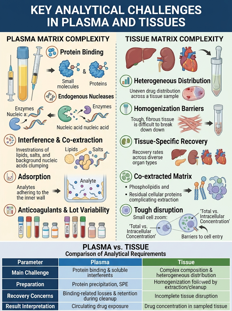
2: What Are the Key Analytical Challenges in Plasma and Tissue?
The main challenge is obtaining a representative measurement without losses, degradation or interference during sample preparation and detection. Each matrix creates its own problems.
Plasma Matrix Complexity
Plasma contains proteins, salts, lipids, endogenous nucleic acids and enzymes that affect sample preparation and signal. Common challenges include:
- Protein binding that complicates extraction
- Endogenous nucleases that degrade susceptible structures
- Co-extracted components that suppress or enhance MS signal
- Background interference from endogenous nucleic acids
- Variability between plasma lots and anticoagulant types
- Analyte loss through adsorption to containers and processing materials
Modifications such as phosphorothioate linkages and modified sugars improve nuclease resistance, but their effect on protein binding and analytical behavior must still be evaluated.
Tissue Matrix Complexity
Drug distribution is not uniform within an organ, so homogenization, cell disruption, analyte binding and extraction conditions all influence the result. Key considerations include:
- Differences in tissue composition and endogenous interference
- Incomplete homogenization or inconsistent tissue-to-buffer ratios
- Variable extraction recovery across organs
- Residual proteins, phospholipids and other co-extractives
- Potential degradation during collection and processing
- Differences between extracellular, intracellular and tissue-associated drug pools
A high concentration in a tissue homogenate does not automatically show that the drug has reached its intracellular site of action. Total tissue concentration is an analytical measurement, while pharmacologically available concentration depends on additional biological factors.
Plasma Versus Tissue: Different Requirements
| Parameter | Plasma | Tissue Homogenate |
|---|---|---|
| Main matrix challenge | Protein binding and soluble interferents | Complex composition and heterogeneous distribution |
| Sample preparation | Protein precipitation, SPE or other extraction | Homogenization followed by extraction or cleanup |
| Key recovery concern | Binding-related losses and retention during cleanup | Incomplete tissue disruption and tissue-specific efficiency |
| Matrix effect assessment | Multiple independent plasma lots | Relevant tissue lots, ideally each intended organ |
| Stability considerations | Collection, processing, storage, freeze–thaw | Collection, homogenization, storage, freeze–thaw |
| Result interpretation | Circulating drug exposure | Drug-associated concentration in sampled tissue |
The best method may share one detection platform but use different sample preparation for plasma and for each tissue.
3: How Do You Select the Analytical Platform?
The platform should match the required specificity, sensitivity, throughput and intended interpretation of results. Hybridization-based assays and LC-MS are the two main options, and the right choice depends on the program.
Hybridization-Based Ligand-Binding Assays
These assays use sequence complementarity to recognize the target. A capture probe and a detection probe bind the analyte, and the signal is related to concentration. Potential advantages include high sensitivity for suitable designs, high throughput and sequence-directed recognition. However, performance depends on probe design, hybridization conditions, target accessibility and detection specificity. Shortened metabolites that retain enough complementary sequence may cross-react, so the ability to distinguish intact parent drug must be demonstrated, not assumed.
Liquid Chromatography–Mass Spectrometry
LC-MS combines chromatographic separation with mass-based detection, giving molecular-mass selectivity and the ability to investigate intact oligonucleotides and selected metabolites. Challenges include complex charge-state distributions, adsorption and recovery issues, and ion suppression from matrix. Sensitivity depends on the analyte, ionization behavior, sample preparation, instrumentation and the required lower limit of quantification (LLOQ). Our guidance on bioanalytical method development covers how these trade-offs are weighed for different molecule types.
Comparing the Two Approaches
| Feature | Hybridization-Based Assay | LC-MS |
|---|---|---|
| Detection principle | Sequence-specific molecular recognition | Chromatographic separation and mass-based detection |
| Key strength | Potentially high sensitivity and throughput | Molecular-mass selectivity and characterization potential |
| Main limitation | Cross-reactivity with related sequences or metabolites | Ion suppression, complex ionization, extraction losses |
| Intact parent specificity | Depends on probe design and configuration | Depends on chromatographic and MS selectivity |
| Development priority | Probe specificity and hybridization performance | Recovery, chromatographic selectivity, ionization |
For complex programs, an integrated strategy can work well: a hybridization assay supports routine quantification while LC-MS provides complementary evidence on identity and related species. In this scenario, LC-MS/MS was selected for its selectivity against metabolites.
4: How Did We Approach Method Development?
We worked in stages: define the analytical target profile, screen extraction and chromatography options, optimize, then validate. This order reduces rework and exposes matrix risks early.
- Define the analytical target profile. Set LLOQ, dynamic range, metabolite selectivity, sample volume and throughput before lab work begins.
- Tune the mass spectrometer. Use negative ESI, compare charge states, select the most intense and stable one for quantification, and keep a second transition for confirmation.
- Optimize chromatography. Ion-pair reversed-phase chromatography with a volatile amine ion-pairing agent and a fluorinated alcohol modifier gives good peak shape and MS sensitivity. Screen columns, temperature and gradient to resolve the parent from n-1 shortmers.
5: Which Sample Extraction Strategy Worked Best?
Mixed-mode weak anion exchange (WAX) SPE gave the best balance of recovery, cleanliness and reproducibility in both plasma and tissue. Simple protein precipitation was fast but left too much matrix and produced inconsistent ionization.
| Approach | Recovery | Matrix Effect | Verdict |
|---|---|---|---|
| Protein precipitation | Moderate | High suppression | Not suitable alone |
| Liquid-liquid extraction (phenol-based) | Good | Moderate | Labor-intensive, low throughput |
| Mixed-mode SPE (WAX) | Consistent | Low | Selected |
| Hybridization capture | High specificity | Low | Option for lower LLOQ needs |
6: How Should Tissue Samples Be Handled?
Homogenize tissue in a buffer containing a lysis agent, digest with proteinase K to release protein-bound oligonucleotide, then extract by SPE. These conditions should be optimized and fixed in the method, because tissue recovery depends heavily on them.
- Keep a consistent tissue-to-buffer ratio so calibrators match study samples.
- Use surrogate matrix or matrix-matched calibration, justified by parallelism testing.
- Test dilution integrity, since liver and kidney concentrations can exceed the upper limit.
- Verify nuclease inhibition during homogenization to prevent ex vivo degradation.
- Assess recovery and matrix effects in each intended organ, not one tissue alone.
How Is Selectivity Against Metabolites Demonstrated?
Analyze blank matrix from multiple individual sources, spike known n-1 and n-2 metabolites at relevant levels, and confirm chromatographic resolution or unique mass transitions. Shortmers can otherwise cause overestimation of the parent drug. Document whether the method reports parent only or total oligonucleotide so the data are interpreted correctly.
7: What Does Validation Include?
Validation covers selectivity, calibration, accuracy and precision, carryover, matrix effect, recovery, dilution integrity and stability, judged against ICH M10 criteria. For a deeper look at the process, see our overview of bioanalytical method development and validation at a CDMO.
| Parameter | Typical Acceptance Criterion (ICH M10) |
|---|---|
| Accuracy | Within ±15% of nominal (±20% at LLOQ) |
| Precision (CV) | ≤15% (≤20% at LLOQ) |
| Selectivity | Interference ≤20% of LLOQ response |
| Carryover | ≤20% of LLOQ after ULOQ |
| Stability | Bench-top, freeze-thaw, long-term, post-preparative, stock |
| Incurred sample reanalysis | ≥67% of repeats within ±20% of original |
Stability needs special attention because oligonucleotides can be sensitive to nucleases, freeze-thaw cycles and storage conditions. Test each condition in the same matrix and container type used for the study.
8: What Are the Key Lessons?
- Optimize extraction first. Chromatography cannot fix poor recovery or heavy matrix effects.
- Control adsorption. Low-bind plastics and well-chosen diluents protect low-concentration samples.
- Use a matched internal standard. A structurally similar oligonucleotide improves correction for recovery and ionization.
- Treat metabolites as a design input, not an afterthought.
- Interpret tissue data carefully. Total tissue concentration does not equal intracellular availability.
- Plan incurred sample reanalysis early. It exposes problems that spiked QCs can hide.
Regulatory Expectations
- ICH M10 on bioanalytical method validation and study sample analysis
- FDA guidance on bioanalytical method validation and on clinical pharmacology for oligonucleotide therapeutics
- GLP (21 CFR Part 58) for nonclinical safety studies
- ALCOA+ data integrity principles across the data lifecycle
If your program involves a related modality, our work on mRNA therapeutics bioanalysis shows how these expectations carry across nucleic acid platforms.
Conclusion:
Bioanalytical Method Development for Oligonucleotide Therapeutics succeeds when the method is designed around the molecule’s chemistry, the matrix and the regulatory goal from the start. Matrix-specific extraction, metabolite-aware selectivity, the right platform choice and rigorous validation turn a difficult analyte into a defensible dataset.
Frequently Asked Questions:
Different tissues contain varying amounts of proteins, lipids, salts, enzymes, and endogenous nucleic acids that can influence extraction and detection. A method optimized for plasma may not perform adequately in liver, kidney, muscle, or brain homogenates. Tissue-specific evaluation helps establish suitable homogenization, extraction recovery, matrix-effect control, and sensitivity. This improves the reliability of tissue distribution measurements.
Yes, appropriate methods can distinguish intact oligonucleotides from selected metabolites, but the ability depends on assay design and analytical selectivity. LC-MS may differentiate species based on mass and chromatographic behavior, while hybridization assays may also detect shortened sequences that retain the recognized binding region. The method must demonstrate that it measures the intended analyte population rather than assuming that all detected signal represents intact parent drug.
Bioanalysis provides concentration measurements across biological matrices and sampling time points. Plasma measurements support the assessment of systemic exposure, while tissue measurements help characterize organ distribution and persistence. These data can contribute to pharmacokinetic analysis, dose selection, and comparisons between candidate formulations. However, total tissue concentration alone does not establish intracellular delivery, target engagement, or pharmacological activity.
Matrix effects occur when co-extracted biological components alter the ionization or detection response of the target analyte. In LC-MS, this commonly appears as ion suppression or enhancement. Proteins, salts, phospholipids, and other endogenous components may contribute to these effects. Appropriate extraction, chromatographic separation, matrix-matched calibration, and suitable internal standards can help manage matrix-related variability.
Chemical modifications, including phosphorothioate linkages and modified sugar groups, can influence nuclease resistance, protein binding, chromatographic retention, and ionization behavior. These properties may affect extraction efficiency and analytical response. Therefore, the bioanalytical method should be developed for the specific oligonucleotide structure rather than transferred directly from an unmodified sequence without adequate evaluation.
Oligonucleotides may adsorb to sample containers, pipette tips, filters, and other laboratory materials under certain conditions. Such losses can reduce measured concentrations, particularly at low analyte levels. Evaluating suitable materials, sample handling procedures, and compatible processing conditions can help minimize adsorption. Recovery experiments should confirm that the selected workflow performs consistently across the intended concentration range.
Solid-phase extraction (SPE) can remove interfering biological components and, where suitable, concentrate an oligonucleotide before analysis. Sorbent selection, loading conditions, washing steps, and elution conditions must be optimized for the target molecule. SPE performance should be evaluated for recovery, selectivity, reproducibility, and matrix effects to ensure that the cleanup process does not introduce unacceptable analyte losses.
Reference
- Li P, Gong Y, Kim J, Liu X, Gilbert J, Kerns HM, Groth R, Rooney M. Hybridization liquid chromatography–tandem mass spectrometry: an alternative bioanalytical method for antisense oligonucleotide quantitation in plasma and tissue samples. Analytical chemistry. 2020 Aug 4;92(15):10548-59.https://pubs.acs.org/ancham/article/92/15/10548/828205
- Tremblay GA, Oldfield PR. Bioanalysis of siRNA and oligonucleotide therapeutics in biological fluids and tissues. Bioanalysis. 2009 Jun 1;1(3):595-609.https://www.tandfonline.com/doi/abs/10.4155/bio.09.66
- Ewles M, Ledvina AR, Powers B, Thomas CE. Observations from a decade of oligonucleotide bioanalysis by LC-MS. Bioanalysis. 2024 Jun 17;16(12):615-29.https://www.tandfonline.com/doi/abs/10.4155/bio-2024-0007
- Cen Y, Li X, Liu D, Pan F, Cai Y, Li B, Peng W, Wu C, Jiang W, Zhou H. Development and validation of LC–MS/MS method for the detection and quantification of CpG oligonucleotides 107 (CpG ODN107) and its metabolites in mice plasma. Journal of pharmaceutical and biomedical analysis. 2012 Nov 1;70:447-55.https://www.sciencedirect.com/science/article/pii/S0731708512003548
- Sanford EJ, Chen J, Tran J, Korboukh I, Zhang G. Development of an LC-MS/MS assay to analyze a lipid-conjugated siRNA by solid phase extraction (SPE) in mouse plasma and tissue using a stable isotope labeled internal standard (SILIS). Bioanalysis. 2025 Jul 18;17(14):901-11.https://www.tandfonline.com/doi/abs/10.1080/17576180.2025.2535953
About the Author


