
Introduction
The chemical synthesis of glycoproteins enables the precise, cell-free assembly of full-length, functional proteins containing uniform oligosaccharide chains. This approach addresses the structural microheterogeneity commonly associated with biological cell-expression systems. By combining solid-phase peptide synthesis (SPPS), native chemical ligation (NCL), and chemoselective desulfurization in a multistep process, researchers have demonstrated the total chemical synthesis of human glycosyl-interferon β-1a containing a complex biantennary N-glycan without relying on living cellular machinery.
For related analytical approaches, explore peptide physicochemical characterization services to understand how complementary testing can support the evaluation of complex therapeutic molecules.
Human interferon β-1a (IFN-β-1a) is a 166-amino-acid cytokine with a glycosylated molecular mass of approximately 22.5 kDa. Its polypeptide backbone has the empirical formula C908H1408N246O252S7, with a reported backbone molecular mass of approximately 20,027 Da. As a disease-modifying therapy for relapsing forms of multiple sclerosis (MS), IFN-β-1a binds to the heterodimeric type I interferon receptor complex, consisting of IFNAR1 and IFNAR2. This interaction activates the receptor-associated Janus kinases JAK1 and TYK2, which promote the phosphorylation of signal transducers and activators of transcription (STAT1 and STAT2). These signaling events regulate downstream genes involved in antiviral defense, antiproliferative activity, and immunomodulation.
Traditionally, therapeutic glycoproteins have been produced using recombinant mammalian host cells, including Chinese hamster ovary (CHO) cells. However, enzymatic glycosylation within cellular organelles is not directly dictated by a genetic template, resulting in heterogeneous glycoform populations with variations in sialylation, core fucosylation, and antennary branching. This microheterogeneity can contribute to analytical variability, batch-to-batch differences, and changes in pharmacokinetic behavior. Total chemical synthesis offers an alternative by enabling controlled assembly of the peptide backbone and the desired glycan structure without requiring a living production host.
Share via:
Quick Summary:
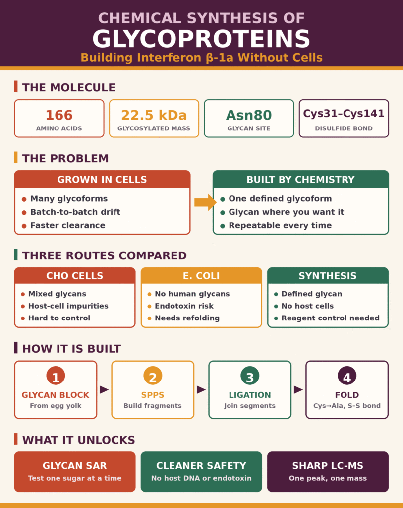
- What was done: Full-length human interferon β-1a — a 166-amino-acid, ~22.5 kDa cytokine — was assembled entirely by chemistry, carrying a single defined complex biantennary N-glycan, with no living cells involved.
- Why it matters: Recombinant systems (CHO cells) glycosylate enzymatically rather than from a genetic template, so batches contain mixed glycoforms; asialo species with exposed galactose get cleared faster through hepatic asialoglycoprotein receptors. E. coli-expressed interferon β-1b carries no glycan at all.
- The target’s architecture: Five amphipathic α-helices stabilized by a Cys31–Cys141 disulfide, with Cys17 free, and the complex-type sialylated glycan sitting on Asn80, where it can shield hydrophobic surface, shape the local loop near the IFNAR1/IFNAR2 interface, and add terminal sialic acid.
- Shortcut for the sugar: Rather than building an ~11-sugar oligosaccharide from monosaccharides, the sialylglycopeptide is extracted from hen egg yolk, trimmed to SGP-Asn, and Fmoc-protected into a ready-made glyco-amino acid that drops straight into standard SPPS.
- Backbone assembly: The 166-residue sequence is split into fragments joined by native chemical ligation — a C-terminal thioester reacts with an N-terminal cysteine (6 M Gn·HCl, phosphate, MPAA, TCEP, pH ~7.0–7.5), then an intramolecular S-to-N acyl shift converts the thioester intermediate into a native amide bond.
- Finishing the protein: Temporary cysteines placed at alanine positions to enable ligation are stripped back to alanine by radical desulfurization (TCEP/VA-044) while native cysteines sit protected as Acm; Acm removal and oxidative folding in a glutathione or Cys/CySS redox buffer then set the Cys31–Cys141 bond.
- What it buys you: Defined glycoform panels for genuine structure–activity work, freedom from host-cell protein, DNA, viral and endotoxin burdens (though reagents, solvents and by-products still need control), no risk of Neu5Gc or α-Gal, and sharper LC-MS, peptide-mapping and glycopeptide-mapping signatures — with feasibility still gated by sequence length, yield, purification and cost.
Comparative Analysis: Chemical Synthesis of Glycoproteins vs. Recombinant Expression
Recombinant cell-expression systems can generate mixtures of structurally diverse glycoprotein isoforms because glycosylation involves multiple enzymatic reactions within the endoplasmic reticulum and Golgi apparatus. In contrast, the chemical synthesis of glycoproteins can produce a defined glycoform with controlled glycan composition and attachment-site occupancy. This precision facilitates the investigation of glycan-dependent biological properties, although improvements in pharmacokinetics, stability, and biological activity must be established experimentally for each molecule.
For additional context on analytical expectations for therapeutic peptides, review the regulatory requirements for GLP-1 peptide characterization and the role of comprehensive characterization in supporting pharmaceutical quality assessments.
In recombinant expression systems, the polypeptide chain is synthesized according to the messenger RNA (mRNA) sequence, whereas glycan attachment and processing depend on the availability of glycosyltransferases, glycosidases, and activated sugar-nucleotide donors. Consequently, individual production batches may contain multiple glycoforms. One important example is the presence of asialoglycoforms, which lack terminal N-acetylneuraminic acid (sialic acid) residues. Exposed terminal galactose residues can bind to hepatic asialoglycoprotein receptors, promoting receptor-mediated uptake and clearance from the bloodstream.
In contrast, non-glycosylated variants produced in prokaryotic hosts, such as interferon β-1b expressed in Escherichia coli, lack carbohydrate chains. The absence of glycans can influence protein folding, solubility, stability, and susceptibility to aggregation. However, these effects depend on the protein’s sequence, formulation, and structural characteristics. Chemical synthesis provides a route to incorporating a defined glycan at the intended position, enabling systematic assessment of its effects on protein stability, receptor interactions, and circulation time.
| Parameter | Recombinant CHO Cell Expression | Prokaryotic Expression (E. coli) | Chemical Synthesis of Glycoproteins |
|---|---|---|---|
| Product homogeneity | May contain multiple glycoforms with variations in glycan structure and occupancy. | Typically produces an unglycosylated protein backbone. | Can produce a defined glycoform when assembly and purification are adequately controlled. |
| Glycan attachment site | Site occupancy and glycan processing depend on the expression system and protein sequence. | Conventional bacterial expression does not provide human-like N-linked glycosylation. | Allows site-specific incorporation of a preassembled glycan building block. |
| Biological safety profile | Requires appropriate control of host-cell proteins, residual DNA, and other process-related impurities. | Requires control of bacterial host-cell proteins, DNA, and endotoxins. | Avoids host-cell-derived contaminants associated with biological expression, but chemical reagents, process impurities, and other contaminants must still be controlled. |
| Sialylation control | Depends on host-cell glycosylation pathways and process conditions. | Conventional expression produces no human-like sialylated N-glycans. | Allows defined terminal sialylation when the selected glycan is incorporated successfully. |
| Physical stability | Depends on protein structure, glycan distribution, formulation, and processing. | Depends on folding, aggregation behavior, and formulation; some proteins require refolding. | Can support systematic optimization of glycan-dependent stability, but superior stability is not guaranteed. |
Structural Architecture and Glycan Topography of Interferon β-1a
Human interferon β-1a consists of a 166-residue polypeptide chain that adopts a five-helix bundle fold. Its structure is stabilized by a disulfide bond between Cys31 and Cys141. The protein also contains an N-linked complex biantennary oligosaccharide attached to Asn80. This glycan can influence protein folding, aqueous solubility, conformational stability, and interactions with biological receptors.
The tertiary structure of IFN-β-1a contains five amphipathic α-helices, designated A through E, connected by loop regions. The native protein contains three cysteine residues: Cys31 and Cys141 form the intramolecular disulfide bond, while Cys17 remains as a free thiol under the conventional native structural assignment.
The protein’s N-glycosylation site is located at asparagine 80 (Asn80). The attached carbohydrate is a complex-type biantennary oligosaccharide built on a core containing three mannose and two N-acetylglucosamine residues, represented as Man3GlcNAc2. Additional branches contain N-acetylglucosamine, galactose, and terminal N-acetylneuraminic acid (sialic acid) residues.
Steric Masking of Hydrophobic Surfaces
The bulky biantennary carbohydrate chain can influence the accessibility of nearby protein surfaces, including regions containing hydrophobic residues. Depending on its orientation and local protein structure, glycan-mediated shielding may reduce nonspecific intermolecular interactions and help limit self-association or aggregation.
To understand analytical approaches for evaluating aggregation-related behavior, explore peptide aggregation analysis and its relevance to assessing intermolecular interactions and product quality.
Receptor Affinity Modulation
The spatial arrangement of the Asn80 glycan can influence the conformational properties of the surrounding loop and the accessibility of nearby protein surfaces. These effects may alter receptor interactions with IFNAR1 and IFNAR2. The magnitude and direction of any change in receptor affinity depend on the specific glycan structure and the molecular context.
Metabolic Protection
Terminal sialic acid residues contribute negative charge and influence interactions between glycoproteins and circulating biological components. Sialylation can also reduce recognition by hepatic asialoglycoprotein receptors by masking terminal galactose residues. Its effects on circulation time and susceptibility to proteolytic degradation depend on the glycoprotein’s structure and physiological environment.
For further insight into how analytical methods assess the stability of therapeutic peptides, read about GLP-1 peptide stability and analytical methods and the role of complementary testing in evaluating molecular integrity.
Chemical Synthesis of Glycoproteins: Synthetic Assembly of Homogeneous Interferon β-1a
The total chemical synthesis of glycoproteins such as interferon β-1a uses a convergent segment-assembly strategy. Peptide fragments and glycopeptide segments prepared through solid-phase peptide synthesis are joined using native chemical ligation (NCL). Following ligation, chemoselective desulfurization can convert auxiliary cysteine residues into alanine residues, allowing the intended native amino acid sequence to be restored before protein folding.
Sialylglycopeptide Extraction and Glyco-Building Block Preparation
Homogeneous biantennary sialyloligosaccharides can be obtained from natural sources, including hen egg yolk, in the form of sialylglycopeptides (SGPs). Using an isolated glycan-containing precursor can reduce the number of individual chemical glycosylation steps required to construct a complex oligosaccharide from simple monosaccharides.
De novo construction of a complex, approximately 11-sugar biantennary sialyloligosaccharide can require numerous synthetic steps, including protecting-group installation, selective glycosylation, and deprotection. To simplify the process, researchers can isolate sialylglycopeptides from hen egg yolk powder and process them to obtain a purified sialyloligosaccharide attached to a single asparagine residue, designated SGP-Asn.
The isolated SGP-Asn is then chemically modified at its amino group using a 9-fluorenylmethoxycarbonyl (Fmoc) protecting group to produce an Fmoc-protected glyco-amino acid building block, represented as Fmoc-Asn(sialylglycan)-OH. This preassembled glyco-amino acid can be incorporated into a growing peptide chain during conventional Fmoc-based solid-phase peptide synthesis. Appropriate reaction conditions and protecting-group strategies are necessary to preserve sensitive glycosidic linkages, including α(2→6) sialic acid linkages where present in the selected glycan structure.
Native Chemical Ligation and Fragment Coupling
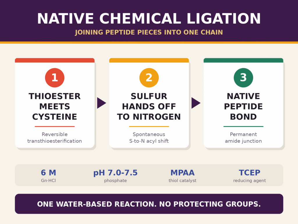
Native chemical ligation joins peptide segments through a chemoselective reaction in an aqueous buffer. The reaction typically involves a peptide bearing a C-terminal thioester and another peptide bearing an N-terminal cysteine residue. Following an initial sulfur-mediated coupling step, an intramolecular rearrangement produces a native amide bond at the ligation junction.
To construct the 166-amino-acid backbone of IFN-β-1a, the target sequence can be divided into shorter synthetic segments. Fragment lengths and ligation junctions are selected according to peptide synthesis performance, solubility, sequence compatibility, and the availability of native or temporary cysteine residues at suitable positions.
For an example of how sequence identity is evaluated in therapeutic peptide analysis, see the GLP-1 peptide sequencing workflow, which illustrates the importance of sequence-level characterization in confirming molecular identity.
Reversible Transthioesterification
The C-terminal thioester of an upstream peptide fragment reacts with the thiol group of an N-terminal cysteine residue on the adjacent downstream fragment. This reaction can be performed in an aqueous buffer containing guanidine hydrochloride (Gn·HCl), sodium phosphate, and a suitable thiol catalyst, such as 4-mercaptophenylacetic acid (MPAA). Tris(2-carboxyethyl)phosphine (TCEP) may be included where appropriate to maintain reducing conditions.
Representative reaction conditions may include 6 M Gn·HCl, 200 mM Na2HPO4, and a pH of approximately 7.0–7.5. The exact formulation depends on the peptide sequences, thioester chemistry, solubility, and compatibility of the glycan-containing fragment.
Spontaneous S-to-N Acyl Shift
The initially formed thioester intermediate undergoes an intramolecular S-to-N acyl shift. This rearrangement transfers the acyl group from sulfur to nitrogen, generating a stable native peptide bond at the ligation junction.
Native chemical ligation reaction
Peptide1–C(=O)–SR + H2N–Cys–Peptide2
⇌ Peptide1–C(=O)–S–Cys–Peptide2
→ Peptide1–C(=O)–NH–Cys–Peptide2
The final step represents the intramolecular S-to-N acyl shift that forms the native peptide bond.
Sequential N-to-C Assembly
The peptide and glycopeptide segments are coupled sequentially to generate the complete 166-residue glycosylated polypeptide backbone. After each ligation, the reaction mixture may require purification, buffer exchange, or other processing before the next segment is introduced. The resulting full-length intermediate must then undergo sequence restoration, disulfide-bond formation, and final purification.
For broader information about analytical approaches used to investigate peptide structure and molecular conformation, review 2D NMR for peptide characterization and its applications in structural analysis.
Chemoselective Desulfurization and Redox Protein Folding
Radical-mediated desulfurization removes the sulfur functionality from auxiliary cysteine residues and converts them into alanine residues. The resulting polypeptide can then undergo oxidative folding under controlled redox conditions to promote formation of the native Cys31–Cys141 disulfide bond.
Because human IFN-β-1a contains three native cysteine residues—Cys17, Cys31, and Cys141—temporary cysteine residues may be introduced at suitable alanine positions to facilitate ligation. These auxiliary cysteines must subsequently be converted back into alanine to restore the intended native sequence. The choice of ligation sites and protecting groups must preserve the native cysteines required for the final protein structure.
In a suitable strategy, the native cysteine residues are protected using acetamidomethyl (Acm) groups during auxiliary-cysteine desulfurization. The assembled glycopeptide is treated with a reducing agent such as TCEP, a suitable radical initiator such as 2,2′-azobis[2-(2-imidazolin-2-yl)propane] dihydrochloride (VA-044), and an appropriate thiol-containing reagent system. Under compatible reaction conditions, the carbon–sulfur bond of the auxiliary cysteine is replaced by a carbon–hydrogen bond, converting the residue to alanine.
Simplified transformation: Auxiliary cysteine → alanine, with the side-chain transformation represented as –CH2SH → –CH3.
After desulfurization, the Acm protecting groups can be removed using an appropriate deprotection procedure, which may involve iodine-mediated oxidation. The deprotected polypeptide is then transferred into an oxidative folding buffer containing a reduced and oxidized glutathione system or a cysteine/cystine (Cys/CySS) redox system. A suitable pH and redox potential help promote formation of the native Cys31–Cys141 disulfide bond while limiting unwanted oxidation and intermolecular disulfide formation.
Folding conditions must be optimized for the specific glycoprotein and intermediate. Circular dichroism spectroscopy can provide complementary information about protein secondary structure and conformational changes during development. For additional background, see CD spectroscopy for peptide secondary structure characterization.
Folding conditions must be optimized for the specific glycoprotein and intermediate. After folding, high-performance liquid chromatography (HPLC) and complementary analytical techniques can be used to isolate the target product and evaluate its purity, structural integrity, and glycan composition.
| Synthetic Phase | Target Reaction or Process | Key Reagents or Materials | Functional Outcome |
|---|---|---|---|
| Phase I: Glyco-building block preparation | SGP extraction and Fmoc functionalization | Hen egg yolk-derived SGP, Fmoc-based protection chemistry, and suitable organic solvents such as DMF | Preparation of the Fmoc-protected glyco-amino acid building block |
| Phase II: Fragment assembly | Automated Fmoc-based SPPS and peptide fragment preparation | Resin support, protected amino acids, coupling reagents such as HATU, and a base such as DIPEA | Preparation of peptide and glycopeptide segments suitable for ligation |
| Phase III: Ligation | Sequential transthioesterification and S-to-N acyl shift | Guanidine hydrochloride, phosphate buffer, MPAA, and TCEP where appropriate | Assembly of the full-length 166-amino-acid glycopeptide |
| Phase IV: Sequence restoration and folding | Radical desulfurization, deprotection, oxidative folding, and purification | TCEP, VA-044, suitable thiol reagents, and a Cys/CySS or glutathione redox system | Isolation of the correctly assembled and folded glycosylated IFN-β-1a product |
Strategic Impact on Biopharmaceuticals and Glycoengineering
Total chemical protein synthesis provides a platform for investigating glycan-specific biological functions, evaluating pharmacokinetic properties, and preparing defined glycoforms for research and development. By avoiding living-cell expression, this approach can reduce dependence on biological production systems and their associated host-derived impurities. Nevertheless, the practical suitability of chemical synthesis depends on molecular size, sequence complexity, synthetic yield, purification requirements, and manufacturing cost.
The chemical synthesis of human glycosyl-interferon β-1a demonstrates the feasibility of assembling a structurally complex cytokine containing a complex-type sialylated N-glycan through a combination of peptide chemistry, glycopeptide building blocks, ligation, and controlled folding.
Structure–Activity Relationship (SAR) Resolution
Preparing specific glycoform variants, including mono-sialylated, di-sialylated, asialo, and truncated-core structures, can help researchers systematically investigate how individual carbohydrate branches influence receptor binding, biological activity, tissue distribution, and clearance. Comparisons between defined glycoforms support the identification of relationships between glycan architecture and protein function. These findings can inform the rational design and optimization of therapeutic glycoproteins.
Safety and Contaminant Control
Because chemical synthesis uses defined chemical building blocks rather than living-cell expression, it avoids host-cell proteins, residual host-cell DNA, and viruses associated specifically with biological production systems. It also avoids bacterial endotoxin introduced through bacterial expression. However, synthetic processes still require controls for residual reagents, protecting groups, catalysts, solvents, by-products, and other process-related impurities.
Defined chemical glycosylation also allows the glycan composition to be selected deliberately, reducing the risk of incorporating unwanted non-human glycans that may be associated with particular biological expression systems. Examples include N-glycolylneuraminic acid (Neu5Gc) and galactose-α-1,3-galactose (α-Gal), where relevant to the selected production platform and product.
Quality Assurance and Mass Spectrometry Verification
Single-glycoform therapeutics can produce more clearly defined mass-spectrometric signals and chromatographic profiles than heterogeneous glycoprotein mixtures. However, the extent of this analytical advantage depends on product purity, glycan composition, ionization behavior, and the analytical method used.
Liquid chromatography–mass spectrometry (LC-MS) can support molecular-mass confirmation, impurity profiling, and assessment of product heterogeneity. Peptide mapping can help verify the amino acid sequence and identify modifications, while glycopeptide mapping can establish glycan attachment sites and characterize glycan composition. Additional methods, including HPLC, nuclear magnetic resonance (NMR), and appropriate biological assays, may be required to evaluate purity, structural integrity, folding, and biological activity.
For complementary information about mass spectrometric evaluation of molecular structure and noncovalent interactions, review native mass spectrometry for therapeutic peptide characterization.
Conclusion
The total chemical synthesis of glycoproteins represents an important advancement in chemical biology and glycoengineering. It demonstrates how complex biopharmaceutical molecules, including glycosylated cytokines such as interferon β-1a, can be assembled with controlled protein sequences and defined carbohydrate structures without relying on living host cells.
By combining solid-phase peptide synthesis, isolated sialylglycopeptide building blocks, native chemical ligation, chemoselective desulfurization, and oxidative folding, researchers can control both the polypeptide backbone and the selected glycan structure. This approach offers an alternative to recombinant expression for producing defined glycoforms and investigating how glycosylation influences molecular stability, biological activity, and pharmacokinetics.
As biopharmaceutical development increasingly emphasizes structural characterization and product consistency, robust analytical verification remains essential. Mass spectrometry, peptide mapping, glycopeptide analysis, chromatographic purity assessment, and functional testing can collectively support the characterization of complex glycoproteins throughout research and development.
How can advanced analytical testing support the characterization of complex glycoproteins? Contact the technical team at ResolveMass Laboratories Inc. to discuss mass spectrometry analysis, custom protein characterization, and analytical quality-control solutions for complex biologics.
Contact ResolveMass Laboratories Inc.
Frequently Asked Questions
Native chemical ligation (NCL) joins peptide segments by reacting a C-terminal peptide thioester with an N-terminal cysteine-containing peptide in an aqueous buffer. The reaction proceeds through transthioesterification, followed by a spontaneous \(S\rightarrow N\) acyl shift that forms a native amide bond at the ligation junction.
N-glycosylation at Asparagine-80 contributes to the structural stability and aqueous solubility of Interferon β-1a. The attached glycan helps shield exposed hydrophobic regions, reduces the tendency toward protein aggregation, and influences the cytokine’s pharmacokinetic behavior and interactions with its receptors.
Complex-type glycans can be obtained by isolating sialylglycopeptides (SGP) from natural sources, including hen egg yolk. The isolated material is processed into an Fmoc-protected glyco-amino acid building block, which can then be incorporated at the designated position during solid-phase peptide synthesis.
Post-ligation chemoselective desulfurization converts auxiliary cysteine residues introduced for peptide coupling into alanine residues, restoring the intended protein sequence. Radical-mediated chemistry using reagents such as TCEP and VA-044 replaces the thiol functionality with hydrogen, enabling the synthesis of proteins with native amino acid sequences.
Interferon β-1a is a glycosylated, 166-amino acid protein with an amino acid sequence identical to that of natural human interferon β. Interferon β-1b is an unglycosylated, 165-amino acid variant produced in E. coli that contains a Cys17Ser substitution and differs in its structural characteristics and specific biological activity.
High-resolution electrospray ionization mass spectrometry (HR-ESI-MS) and reversed-phase ultra-performance liquid chromatography (RP-UPLC) help assess molecular mass, product purity, and the presence of impurities. Appropriate peptide mapping, glycan characterization, and antiviral or cytostatic bioassays can further evaluate structural identity, glycan occupancy, and biological potency.
Correct disulfide bond formation is promoted by incubating the assembled polypeptide in an oxidative folding buffer containing a cysteine/cystine redox couple under controlled pH conditions. This environment supports the development of the native tertiary structure and facilitates formation of the Cys31-Cys141 disulfide bond in Interferon β-1a.
Chemical glycoprotein synthesis avoids the use of living expression systems, substantially reducing the risks associated with host-cell proteins, adventitious viruses, genomic material, and cell-derived contaminants. However, the final product must still undergo appropriate purification and quality control to assess chemical impurities, residual reagents, and other process-related contaminants.
Yes, chemical synthesis and ligation-based approaches can be used to construct other complex glycoproteins by assembling defined peptide segments and incorporating specific glycan structures. Erythropoietin (EPO), which contains three N-linked glycans and one O-linked glycan, is an example of a complex glycoprotein that has been investigated using advanced synthetic strategies.
Reference:
- Wang, L.-X., & Amin, M. N. (2014). Chemical and chemoenzymatic synthesis of glycoproteins for deciphering functions. Cell Chemical Biology, 21(1), 51–66. https://doi.org/10.1016/j.chembiol.2014.01.001
- Alam, J., Rogge, M., & Goelz, S. (1999). Recombinant human interferon beta-1a (IFN-beta-1a) formulation (WO Patent No. WO1999062542A1). World Intellectual Property Organization. https://patents.google.com/patent/WO1999062542A1/en
- Shirakawa, A., Manabe, Y., & Fukase, K. (2021). Recent advances in the chemical biology of N-glycans. Molecules, 26(4), Article 1040. https://doi.org/10.3390/molecules26041040
- European Medicines Agency. (2005). Rebif: EPAR—Scientific discussion. https://www.ema.europa.eu/en/documents/scientific-discussion/rebif-epar-scientific-discussion_en.pdf
- Tian, J., Li, Y., Ma, B., Tan, Z., & Shang, S. (2022). Automated peptide synthesizers and glycoprotein synthesis. Frontiers in Chemistry, 10, Article 896098. https://doi.org/10.3389/fchem.2022.896098
Get In Touch With Us
About The Author