


Introduction
Intact Mass Analysis for Biosimilar Characterization is a fundamental bioanalytical technique used to determine the total mass of a biotherapeutic molecule in its unfragmented state. It helps establish sequence fidelity, assess macro-purity, and verify subunit assembly. By measuring the mass of the complete molecule before enzymatic digestion, bioanalytical scientists can obtain an unbiased profile of its proteoforms, providing a baseline for biosimilar comparability testing.
Biopharmaceuticals, particularly recombinant monoclonal antibodies (mAbs) and fusion proteins, are complex macromolecules produced in biological host cell systems. Unlike small-molecule generic drugs, which have well-defined chemical structures, biotherapeutics exhibit inherent structural micro-heterogeneity resulting from post-translational modifications (PTMs), enzymatic processing, and manufacturing conditions. Regulatory authorities, including the U.S. Food and Drug Administration (FDA) and the European Medicines Agency (EMA), require comprehensive comparability assessments in accordance with International Council for Harmonisation (ICH) Q6B and Q5E guidelines. Within these regulatory frameworks, intact mass determination serves as an essential first-line screening technique. It provides an overall structural profile of the biotherapeutic, enabling scientists to verify correct primary assembly and identify major batch-to-batch variations before proceeding with destructive downstream characterization workflows.
Explore ResolveMass biosimilar characterization services for comprehensive analytical support throughout biosimilar development.
Share via:
Quick Summary:
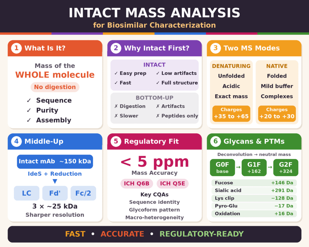
- What it is: Intact mass analysis measures the total mass of a biotherapeutic (such as a mAb) in its whole, undigested state. This confirms sequence fidelity, macro-purity and correct subunit assembly, and gives a baseline for comparing a biosimilar to the reference product.
- Why use it early: It needs very little sample preparation, usually just online desalting. That avoids the artifacts common in bottom-up peptide mapping, such as deamidation, oxidation and disulfide scrambling. It is also fast and keeps whole-molecule information like paired heavy-chain glycoforms.
- Two MS approaches: Denaturing LC-MS unfolds the protein using acidic, organic conditions and gives accurate covalent mass (charges around +35 to +65). Native MS uses neutral ammonium acetate buffers to keep the protein folded. This makes it useful for non-covalent complexes, aggregates and ADCs (charges around +20 to +30).
- Middle-up strategy: IdeS digestion combined with disulfide reduction splits the ~150 kDa antibody into three ~25 kDa subunits (LC, Fd’ and Fc/2). This sharpens resolution and helps detect low-abundance variants such as C-terminal lysine and glycoform differences.
- Regulatory relevance: The technique supports ICH Q6B and Q5E comparability requirements under a Quality by Design framework. High-resolution instruments such as Orbitraps achieve mass accuracy below 5 ppm. This enables checks on critical quality attributes like sequence identity, glycoform consistency and macro-heterogeneity.
- Deconvolution and PTM profiling: Software converts charge-state envelopes into neutral mass spectra. This reveals glycoforms (G0F, G1F +162 Da, G2F +324 Da) and modifications such as fucosylation, sialylation, lysine clipping, pyroglutamate formation and oxidation. Their relative peak areas show batch consistency and biosimilar comparability.
- Bottom line: Intact mass analysis is a rapid, accurate, first-line screening tool. It underpins biosimilar regulatory approval and stays essential as molecules grow more complex, including bispecifics, ADCs and fusion proteins.
Strategic Value of Intact Mass Analysis for Biosimilar Characterization in Early-Stage Development
Implementing Intact Mass Analysis for Biosimilar Characterization during the initial stages of development can minimize downstream analytical delays by providing a rapid, non-destructive assessment of primary sequence identity and overall proteoform distribution. This high-level screening helps establish early alignment with the reference medicinal product’s Quality Target Product Profile (QTPP) before resources are committed to complex and destructive analytical procedures.
Conventional “bottom-up” proteomic characterization depends on site-specific enzymatic digestion, such as trypsinization, to break down complex proteins into smaller peptides before Liquid Chromatography-Mass Spectrometry (LC-MS) analysis. Although bottom-up peptide mapping is indispensable for identifying localized single-amino acid modifications, the technique presents several operational challenges. Prolonged digestion at elevated temperatures (37°C) and slightly alkaline pH levels can introduce sample preparation artifacts, including artificial asparagine deamidation, methionine oxidation, and disulfide bond scrambling. Conversely, intact mass spectrometry requires very little sample manipulation, generally involving online desalting or buffer exchange. This limited preparation helps preserve the biotherapeutic’s original modification state without introducing unnecessary analytical artifacts.
Additionally, determining the mass of the complete molecule preserves essential structural relationships between different protein domains. When an intact IgG mAb (approximately 150 kDa) is digested into numerous individual peptides during bottom-up sample preparation, the stoichiometric relationships between modifications on separate chains, such as paired heavy-chain N-glycosylation states, are no longer retained. Intact mass spectrometry preserves this structural information, making it possible to identify the precise combinations of co-existing proteoforms present across the fully assembled molecule.
For a more detailed understanding of peptide-level characterization and protein sequence analysis, explore the proteomics approach for biosimilars.
| Analytical Parameter | Intact Mass Analysis | Bottom-Up Peptide Mapping |
|---|---|---|
| Primary Scope | Global mass determination, macro-purity assessment, and whole-molecule proteoform distribution | Site-specific amino acid sequencing, localized PTM mapping, and point mutation detection |
| Sample Preparation | Minimal preparation, typically involving rapid online desalting or buffer exchange | Complex, multi-step preparation involving denaturation, reduction, alkylation, and enzymatic digestion |
| Artifact Risk | Low; preserves the original molecular state with minimal risk of artificial modifications | Higher; susceptible to non-native deamidation, oxidation, and disulfide bond scrambling |
| Throughput & Speed | High throughput, with relatively short analysis times per sample | Moderate to slow, owing to extended incubation periods and longer chromatographic gradients |
| Structural Context | Maintains whole-molecule stoichiometry and chain-pairing information | Separates localized peptide modifications from their whole-molecule proteoform context |
| Mass Accuracy / Resolution | High-resolution deconvoluted mass profiles with mass accuracy below 5 ppm | Provides sequence coverage and localized characterization at the individual peptide level |
Core Analytical Methodologies for Intact Mass Analysis for Biosimilar Characterization
The analytical framework for Intact Mass Analysis for Biosimilar Characterization integrates High-Resolution Mass Spectrometry (HRMS) with denaturing, native, and subunit-level liquid chromatography techniques. These complementary analytical approaches help resolve charge-state distributions, characterize structural heterogeneity, and investigate non-covalent associations across different biotherapeutic modalities.
Denaturing LC-MS vs. Native Mass Spectrometry in Intact Mass Analysis for Biosimilar Characterization
Denaturing LC-MS employs organic solvents and acidic conditions to unfold protein structures, facilitate higher-charge ionization, and determine covalent molecular mass accurately. In contrast, native mass spectrometry uses neutral aqueous buffers to preserve the protein’s higher-order conformation and non-covalent molecular assemblies. Both approaches provide essential structural information for comprehensive biosimilar comparability assessments.
Denaturing intact mass analysis combines reversed-phase liquid chromatography (RP-HPLC or UHPLC) with Electrospray Ionization Mass Spectrometry (ESI-MS). Conventional denaturing separations use wide-pore stationary phases, such as C4 or C8 columns with pore sizes ranging from 300 Å to 1000 Å, operated at elevated temperatures of 60°C–70°C to reduce secondary interactions. The mobile phases generally consist of water and acetonitrile gradients acidified with 0.1% formic acid or 0.02–0.05% trifluoroacetic acid (TFA). Under these denaturing conditions, the biotherapeutic unfolds completely, exposing basic amino acid side chains and producing broad charge-state envelopes. Intact IgGs typically exhibit charge states ranging from +35 to +65 within an m/z transmission window of 1500–4500.
Native Mass Spectrometry (nMS) maintains the native folding state and non-covalent interactions of biotherapeutics by avoiding organic denaturants and non-volatile salts. This technique uses volatile, neutral physiological buffers, such as 50–200 mM ammonium acetate, in combination with online Size-Exclusion Chromatography (SEC) or Hydrophobic Interaction Chromatography (HIC). Because the protein remains tightly folded, fewer protonation sites are accessible to the solvent during electrospray ionization. Consequently, the resulting charge-state envelopes shift toward lower charge states, typically +20 to +30, and higher m/z ranges of 2500–8000 m/z. Native MS is particularly valuable for investigating non-covalent protein complexes, higher-order aggregates, and labile biotherapeutics, including non-covalently linked antibody-drug conjugates (ADCs).
Learn how native MS helps preserve and investigate non-covalent protein assemblies in native mass spectrometry for biosimilars.
Middle-Up Subunit Strategies for Enhanced Mass Resolution
Middle-up subunit strategies involve targeted proteolysis to divide complex intact antibodies into domain fragments measuring approximately 25–50 kDa before mass spectrometric analysis. This strategy substantially reduces spectral complexity and charge-state overlap, facilitating more precise characterization of localized post-translational modifications.
Although intact antibody mass analysis provides an essential whole-molecule profile at approximately 150 kDa, the accumulation of multiple micro-heterogeneous features can produce overlapping spectral signals. This overlap makes it more difficult to identify low-abundance variants present at levels below 5%. To address these resolution limitations, targeted enzymatic digestion with IdeS (Immunoglobulin-decomposing enzyme of Streptococcus pyogenes) selectively cleaves human IgG below the hinge region.
When IdeS digestion is combined with chemical reduction of inter-chain disulfide bonds using tris(2-carboxyethyl)phosphine (TCEP) or dithiothreitol (DTT), it generates three distinct fragments of approximately 25 kDa:
- Light Chain (LC)
- Heavy Chain Fd’ subunit (variable domain plus CH1)
- Fc/2 subunit (single-chain CH2-CH3 region containing the N-glycosylation site)
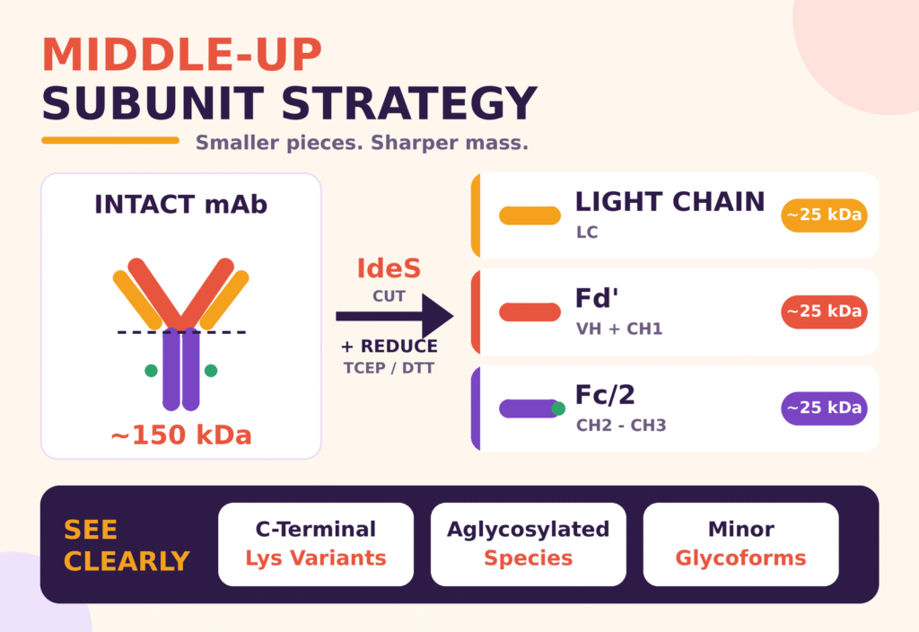
Reducing the molecular mass of these subunits shifts their isotopic envelopes into regimes that offer greater effective resolution. This facilitates the baseline separation of C-terminal lysine variants, aglycosylated species, and minor glycoform structures while preserving domain-level spatial context.
Regulatory Standards and Mass Accuracy in Intact Mass Analysis for Biosimilar Characterization
Regulatory guidelines established by the International Council for Harmonisation, particularly ICH Q6B and Q5E, require biosimilar developers to demonstrate structural identity and comparability against the reference drug product using high-precision analytical methods. Intact Mass Analysis for Biosimilar Characterization supports these regulatory objectives by providing mass measurements with accuracy below 5 ppm, consistent with the analytical rigor expected under Quality by Design (QbD) principles.
Within Quality by Design (QbD) regulatory frameworks, biosimilar applicants must demonstrate that Critical Quality Attributes (CQAs) remain within defined target ranges established through characterization of the reference product. Intact mass spectrometry directly evaluates several important CQAs:
- Primary Sequence Identity: Confirms the measured molecular mass against the theoretical mass calculated from the intended primary sequence.
- Glycoform Pattern Consistency: Assesses Fc glycosylation profiles, including G0F, G1F, and G2F, which can influence antibody effector functions.
- Macro-Heterogeneity Limits: Identifies abnormal expression products, including uncleaved signal peptides and major truncation products.
Modern High-Resolution Mass Spectrometers, particularly Orbitrap instruments capable of achieving resolving power of up to 240,000 (FWHM at m/z 200), routinely deliver experimental mass measurements with errors below 5 ppm. Mass measurement error, expressed in parts per million (ppm), is calculated using the following equation:
Mass Error (ppm) = [(Mexperimental − Mtheoretical) / Mtheoretical] × 106
Achieving mass accuracy below 5 ppm enables bioanalytical teams at ResolveMass Laboratories Inc. to verify target sequence expression and identify point mutations or amino acid misincorporations during clone selection and process scale-up.
To understand how quality attributes are identified, prioritized, and monitored during development, read about critical quality attributes (CQAs) in biosimilars.
Resolving Micro-Heterogeneity: Deconvolution and Glycoform Profiling
Automated charge-state deconvolution algorithms convert complex electrospray charge-state envelopes into neutral mass spectra, enabling the accurate identification and quantitative assessment of individual biotherapeutic proteoforms. This computational processing helps distinguish mass shifts associated with specific glycosylation patterns and post-translational modifications.
Electrospray ionization generates a complex distribution of multiply charged ions across the m/z spectrum. Advanced bioanalytical software applies deconvolution algorithms, including Maximum Entropy and Sliding Window processing, to combine individual charge states into a single neutral molecular weight spectrum representing the zero-charge state. The resulting deconvoluted mass spectrum displays distinct peaks corresponding to different structural variants of the biotherapeutic.
In therapeutic IgGs, N-linked glycosylation at Asn-297 within the Fc domain is a major contributor to structural micro-heterogeneity. Core oligosaccharide structures produce characteristic, additive mass increments relative to the aglycosylated protein backbone:
- G0F: A core biantennary heptasaccharide containing core fucose (C56H92N4O39), with an added mass of +1445.3 Da.
- G1F: Incorporation of one terminal galactose residue, resulting in a mass increase of +162.05 Da relative to G0F.
- G2F: Incorporation of two terminal galactose residues, resulting in a mass increase of +324.10 Da relative to G0F.
For a deeper look at glycan profiling methods and their importance in evaluating biosimilar similarity, explore glycosylation analysis of biosimilars.
| Post-Translational Modification / Variant | Delta Mass (Δm) | Structural Significance & Functional Impact |
|---|---|---|
| Galactosylation (+1 Hexose) | +162.05 Da | Common Fc glycan expansion (G0F → G1F → G2F); modulates complement-dependent cytotoxicity (CDC). |
| Fucosylation (+1 Fucose) | +146.06 Da | Core glycosylation feature; its absence can markedly enhance antibody-dependent cellular cytotoxicity (ADCC). |
| Sialylation (+1 Neu5Ac) | +291.09 Da | Terminal acidic sugar modification that influences anti-inflammatory activity and serum clearance rates. |
| C-Terminal Lysine Clipping | −128.09 Da per Lys | Enzymatic processing during cell culture; a major contributor to basic charge heterogeneity. |
| N-Terminal Pyroglutamate | −17.03 Da (−NH3) | Cyclization of N-terminal glutamine or glutamate; protects the biotherapeutic against aminopeptidase-mediated degradation. |
| Methionine Oxidation | +15.99 Da per oxygen | A chemical degradation product that can impair FcRn receptor binding and alter systemic half-life. |
| Non-Enzymatic Glycation | +162.05 Da per hexose | Uncontrolled attachment of reducing sugars during cell culture or storage, with potential implications for immunogenicity risk. |
By determining the relative integrated peak areas in deconvoluted mass spectra, bioanalytical laboratories can establish glycoform distribution ratios, assess batch-to-batch consistency, and demonstrate analytical comparability between the biosimilar candidate and the reference medicinal product.
Explore charge variant analysis in biosimilars using mass spectrometry to understand complementary approaches for investigating these variants.
Conclusion
Intact Mass Analysis for Biosimilar Characterization is an essential foundation of modern biopharmaceutical analytical programs. It provides rapid, unfragmented confirmation of molecular weight, sequence fidelity, and overall proteoform distribution. By assessing whole-molecule integrity while minimizing sample preparation artifacts, this top-down screening strategy supports regulatory alignment and helps guide subsequent characterization activities.
The commercial success and regulatory approval of biosimilar candidates depend on demonstrating structural comparability with their reference drug products. Intact mass spectrometry delivers an unbiased, highly accurate molecular profile that connects assessments of macro-purity with localized subunit characterization. As biotherapeutics continue to develop more complex architectures, including bispecific antibodies, antibody-drug conjugates, and multi-domain fusion proteins, high-resolution intact mass spectrometry remains a key analytical approach for achieving reliable structural characterization and measurement precision.
For additional support with analytical planning and regulatory documentation, explore the end-to-end biosimilar analytical package for a regulatory submission.
To discuss your biosimilar characterization, intact mass spectrometry, or regulatory comparability testing requirements with bioanalytical scientists, contact ResolveMass Laboratories Inc. directly through the ResolveMass Laboratories contact page.
Frequently Asked Questions
Intact mass analysis provides an early overview of the biotherapeutic’s molecular mass, assembly, and major structural variants. By identifying unexpected mass differences and potential manufacturing inconsistencies at an early stage, it helps scientists make informed decisions about further testing. This can reduce unnecessary analytical work and improve the efficiency of biosimilar development.
Intact mass analysis measures the molecular mass of the complete protein, preserving information about its overall structure and combinations of proteoforms. Bottom-up peptide mapping involves enzymatic digestion, followed by analysis of the resulting peptides to investigate amino acid sequences and localized post-translational modifications. Both methods provide complementary information for comprehensive biosimilar characterization.
High-Resolution Mass Spectrometry (HRMS) instruments, including hybrid Quadrupole-Orbitrap and high-resolution Time-of-Flight (Q-TOF) systems, are widely used for intact mass characterization. Their high resolving power, accurate mass measurements, and suitable mass-to-charge (m/z) ranges support the analysis of large biotherapeutic molecules. Instrument selection depends on the molecule’s properties, analytical objectives, and required sensitivity.
Native mass spectrometry examines biotherapeutics under non-denaturing conditions to preserve their higher-order structures and non-covalent interactions. It helps investigate protein complexes, oligomeric states, and molecular assemblies that may be disrupted by conventional denaturing techniques. This approach is particularly useful when structural integrity and non-covalent associations are important to the characterization strategy.
Electrospray ionization produces ions carrying multiple charges, resulting in several signals for the same protein across the mass-to-charge spectrum. Deconvolution software uses mathematical algorithms, such as Maximum Entropy, to interpret these signals and calculate the corresponding neutral molecular masses. The resulting spectrum helps distinguish individual proteoforms according to their molecular weights.
Intact mass spectrometry can assess major Fc N-glycoforms in monoclonal antibodies, including G0F, G1F, and G2F, by identifying their characteristic mass differences. The relative abundance of these glycoforms can be estimated by integrating the corresponding peaks in the deconvoluted mass spectrum. The reliability of quantitation depends on analytical conditions, signal quality, and the method’s ability to distinguish overlapping species.
Middle-up or subunit mass analysis involves selectively cleaving a large protein, such as a monoclonal antibody, into smaller structural units before mass spectrometric analysis. Enzymes such as IdeS can generate antibody fragments that are easier to resolve than the complete molecule. This strategy reduces spectral complexity and supports the characterization of glycosylation patterns, chain variants, and other structural differences.
ICH Q6B addresses specifications and analytical characterization of biotechnological and biological products, while ICH Q5E provides guidance on demonstrating comparability following manufacturing changes. These guidelines support the assessment of structural attributes and product quality when establishing similarity between a biosimilar and its reference product. Intact mass analysis contributes valuable molecular mass data alongside other orthogonal analytical techniques required for a comprehensive comparability assessment.
Intact mass spectrometry generally requires less sample preparation than bottom-up peptide mapping, reducing the risk of introducing artificial modifications during analysis. It can minimize preparation-related artifacts such as artificial deamidation, methionine oxidation, disulfide bond scrambling, and incomplete enzymatic digestion. However, it does not eliminate every potential artifact, as sample handling, storage conditions, and instrument settings can still influence analytical results.
Reference:
- Kristensen, D. B., Sloth, T. M., Ørgaard, M., & Jensen, P. F. (2021). Characterization of protein glycoforms at intact level by Orbitrap mass spectrometry. In Methods in Molecular Biology (Vol. 2271, pp. 23–45). Humana. https://doi.org/10.1007/978-1-0716-1241-5_2
- Sorensen, M., Harmes, D. C., Stoll, D. R., Staples, G. O., Fekete, S., Guillarme, D., & Beck, A. (2016). Comparison of originator and biosimilar therapeutic monoclonal antibodies using comprehensive two-dimensional liquid chromatography coupled with time-of-flight mass spectrometry. mAbs, 8(7), 1224–1234. https://doi.org/10.1080/19420862.2016.1203497
- Carillo, S., Pérez-Robles, R., Jakes, C., Ribeiro da Silva, M., Millán Martín, S., Farrell, A., Navas, N., & Bones, J. (2020). Comparing different domains of analysis for the characterisation of N-glycans on monoclonal antibodies. Journal of Pharmaceutical Analysis, 10(1), 23–34. https://doi.org/10.1016/j.jpha.2019.11.008
Get In Touch With Us
About The Author


