


Introduction
Functional assays for biosimilar characterization are essential for evaluating analytical bioequivalence, biological potency, and mechanism-driven functional comparability between proposed biotherapeutics and their originator reference products. These assays form a central component of the totality-of-evidence approach required by major global regulatory agencies to demonstrate that minor structural differences do not adversely affect clinical performance. Modern biopharmaceutical characterization has evolved considerably, moving away from heavy reliance on large-scale confirmatory clinical efficacy trials and placing greater emphasis on high-resolution analytical, biophysical, and functional bioequivalence datasets. By examining the complete biological mechanism of action (MoA), including primary target engagement, cell-mediated signaling, downstream biological cascades, and Fc-mediated effector pathways, investigators can establish whether minor variations in post-translational modifications, such as glycosylation or charge variants, influence clinical performance, safety, or immunogenicity.
Learn more about post-translational modifications in biosimilars and their importance in analytical characterization.
Share via:
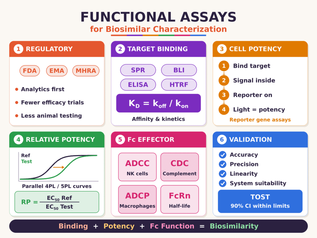
Quick Summary:
- Regulatory shift: The FDA, EMA, and MHRA now rely more on analytical and functional bioassay data than on large clinical trials. Strong analytical comparability, with PK and immunogenicity data, can make comparative efficacy trials and animal testing unnecessary.
- Target binding: Label-free biosensors such as SPR and BLI measure association (kon), dissociation (koff), and affinity (KD = koff/kon) for the biosimilar and the reference product. ELISA, HTRF, and SET confirm affinity under equilibrium conditions.
- Cell-based potency: Engineered reporter gene assays link target binding to a measurable luminescent or fluorescent signal through pathways such as NFAT, NF-κB, or STAT. They give better precision and less variability than primary cell assays.
- Relative potency: Dose-response curves are fitted with 4PL or 5PL models. Relative potency is calculated as the ratio of the reference EC50 to the test EC50, but only after the two curves are shown to be parallel, preferably by equivalence testing (TOST) rather than F-tests alone.
- Fc effector functions: Assays profile binding to the Fcγ receptors, FcRn, and C1q, and measure ADCC, CDC, and ADCP. These show how glycosylation differences, such as afucosylation or galactosylation, affect immune-mediated activity and half-life.
- Validation and equivalence: Bioassays are validated to USP requirements for accuracy, intermediate precision, linearity, and system suitability. Functional equivalence is shown when the 90% confidence interval falls within scientifically justified margins.
- Overall takeaway: Together, binding, potency, and Fc function assays form the core of the totality-of-evidence approach. They strengthen regulatory submissions and can shorten biosimilar development timelines.
Regulatory Frameworks Governing Functional Assays for Biosimilar Characterization
Regulatory frameworks governing functional assays for biosimilar characterization require comprehensive, orthogonal functional bioassays to establish biosimilarity and may reduce or eliminate the need for comparative clinical efficacy trials. Global regulatory agencies increasingly emphasize comparative analytical assessment datasets over large-scale clinical studies because appropriately designed bioassays can detect functional differences with greater analytical sensitivity.
Guidance and regulatory developments from the United States Food and Drug Administration (FDA), the European Medicines Agency (EMA), and the United Kingdom Medicines and Healthcare products Regulatory Agency (MHRA) emphasize the importance of comparative analytical assessments alongside a scientifically justified clinical development program. The FDA’s draft guidance describes an approach in which a proposed biosimilar demonstrating a high degree of similarity to its reference product through comparative analytical assessment may qualify for a streamlined development program. Such a program may rely on comparative pharmacokinetics (PK) and immunogenicity data, as appropriate, to support licensure under Section 351(k) of the Public Health Service Act. Comparative efficacy trials are not necessarily required in every development program because clinical endpoints may be less sensitive to subtle functional differences than carefully designed, multi-tiered bioassays conducted under controlled laboratory conditions.
Similarly, the EMA’s Committee for Medicinal Products for Human Use (CHMP) supports a scientifically tailored clinical development approach in which advanced analytical characterization, together with PK bioequivalence data and other relevant evidence, may support regulatory approval without additional efficacy trials when justified. The MHRA also supports evidence-based approaches to biosimilar development that reduce unnecessary animal testing and clinical studies when robust analytical and functional comparability data adequately address the relevant scientific questions. The precise requirements depend on the product, its mechanism of action, the available evidence, and the applicable regulatory guidance.
| Regulatory Agency | Primary Guidance Basis | Position on Comparative Clinical Efficacy Studies | Animal Testing Requirement | Primary Reliance for Biosimilarity |
|---|---|---|---|---|
| US FDA | Section 351(k) of the PHS Act; FDA draft guidance | May be omitted when the comparative analytical assessment and remaining evidence adequately establish biosimilarity | Generally minimized when scientifically justified; requirements depend on the development program | Orthogonal analytical, biophysical, and functional bioassays, supported by relevant PK/PD evidence |
| EMA | CHMP biosimilar guidelines and reflection paper | A tailored clinical pathway may permit omission when analytical and other evidence sufficiently establish comparability | Waived or limited when justified by the available evidence and applicable guidance | Detailed physicochemical, structural, and functional characterization of the mechanism of action |
| UK MHRA | National biosimilar guidance | May be omitted when the totality of evidence sufficiently supports biosimilarity | Minimized where scientifically justified and consistent with applicable guidance | Comparative analytical characterization, target-binding studies, and cell-based bioassay data |
Explore end-to-end biosimilar characterization services to support analytical comparability and regulatory development requirements.
Target Binding Kinetics and Affinity Profiling for Biosimilar Characterization
Target binding kinetics and affinity profiling determine the association rates (kon), dissociation rates (koff), and equilibrium dissociation constants (KD) that describe the interaction between a biotherapeutic and its therapeutic target. Within functional assays for biosimilar characterization, these biophysical measurements help assess higher-order structural integrity and primary molecular recognition mechanisms under cell-free conditions.
Surface Plasmon Resonance (SPR) and Bio-Layer Interferometry (BLI) are the principal label-free optical biosensor technologies used to investigate binding kinetics. SPR detects changes in the local refractive index near a sensor surface functionalized with immobilized target molecules or antibodies. By exposing the functionalized surface to a series of analyte concentrations, SPR captures real-time association and dissociation behavior. Bio-Layer Interferometry (BLI), in contrast, measures changes in white-light interference patterns reflected from the surface of a biosensor tip. When biomolecules bind to the tip, the optical thickness changes, producing a measurable spectral shift. Both SPR and BLI enable investigators to estimate kinetic and affinity parameters by fitting interaction curves to appropriate mathematical models, including the 1:1 Langmuir binding model:
A + B ⇌ AB
kon and koff govern the forward and reverse reactions, respectively.
The equilibrium dissociation constant (KD) is calculated from the kinetic rate constants as follows:
KD = koff / kon
A standardized target-binding characterization workflow typically involves the following operational steps:
- Biosensor Surface Functionalization: Immobilizing target proteins or capturing immunoglobulins on sensor surfaces using amine coupling, protein A, or anti-human Fc antibodies.
- Analyte Concentration Injections: Introducing serial dilutions of both the proposed biosimilar and the reference product through identical flow channels under comparable experimental conditions.
- Real-Time Kinetic Curve Capture: Monitoring association rates (kon) during sample injection and dissociation rates (koff) during subsequent buffer-washing steps.
- Mathematical Curve Fitting: Applying appropriate 1:1 Langmuir kinetic models to estimate kon, koff, and affinity KD values.
- Equivalence Testing: Statistically comparing kinetic rate constants and binding affinity values obtained for the biosimilar and reference product lots.
Alongside real-time, label-free biosensor techniques, equilibrium binding methods such as Enzyme-Linked Immunosorbent Assays (ELISA), Homogeneous Time-Resolved Fluorescence (HTRF), and Solution Equilibrium Titration (SET) are routinely used to assess and confirm target affinity under equilibrium conditions.
| Assay Platform | Detection Physics / Principle | Key Output Parameters | Advantages in Biosimilarity | Limitations |
|---|---|---|---|---|
| Surface Plasmon Resonance (SPR) | Refractive index changes near a gold sensor surface | kon, koff, KD | Real-time kinetic analysis, high sensitivity, and label-free detection | Susceptible to microfluidic mass transport limitations |
| Bio-Layer Interferometry (BLI) | Optical interference changes at a fiber-optic sensor tip | kon, koff, KD | No complex fluidics, higher throughput, and good tolerance to sample matrices | Generally less sensitive than high-end SPR instruments |
| Direct / Indirect ELISA | Colorimetric or chemiluminescent signal detection | EC50, equilibrium KD | Widely available, readily standardized, and suitable for routine release testing | Does not directly provide real-time kinetic parameters (kon, koff) |
| HTRF / FRET | Time-resolved fluorescence resonance energy transfer | EC50, relative binding | Solution-based measurement, minimal washing, and high throughput | Requires fluorophore labeling, which may alter native conformation |
Discover how native mass spectrometry for biosimilars can complement binding studies by providing insights into intact molecular structures and noncovalent interactions.
Cell-Based Potency Bioassays for Biosimilar Characterization
Cell-based potency bioassays quantify the biological responses produced by a biotherapeutic in intact cellular systems, providing insight into its integrated mechanism of action. In functional assays for biosimilar characterization, these methods capture physiological receptor signaling, gene activation, and downstream biological outcomes that cannot be fully assessed through cell-free binding assays alone.
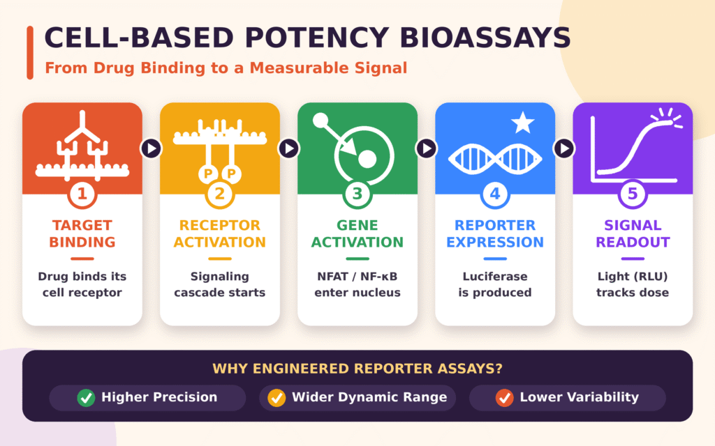
Modern biosimilar characterization increasingly incorporates engineered reporter gene assays instead of relying exclusively on traditional primary cell proliferation, apoptosis, or cytokine release assays. Reporter gene assays employ immortalized cell lines that have been genetically engineered to express target receptors alongside a responsive reporter gene, such as firefly luciferase, green fluorescent protein, or alkaline phosphatase. Reporter expression is regulated by a specific transcription factor promoter, such as NFAT, NF-κB, or STAT. Following ligand binding and pathway activation, transcription of the reporter gene produces a luminescent or fluorescent signal that varies with the drug concentration.
Engineered bioassays generally provide improved analytical precision, broader dynamic ranges, and lower operator-dependent variability than many primary cell culture methods. Although primary cell bioassays can offer greater physiological relevance, they may be affected by donor-to-donor variability, low signal-to-noise ratios, and demanding cell culture requirements. Using ready-to-use, cryopreserved, division-arrested cells can reduce assay variability and improve the reproducibility of analytical comparability studies.
The cell-mediated mechanism of action assessed through engineered bioassays generally follows a defined sequence:
- Target Binding: The therapeutic antibody or fusion protein selectively binds to its target receptor on the cell surface.
- Receptor Activation: Receptor engagement initiates intracellular domain dimerization and trans-phosphorylation cascades, where applicable.
- Transcriptional Activation: Intracellular signaling pathways activate specific transcription factors, such as NFAT or NF-κB, which subsequently translocate to the cell nucleus.
- Reporter Expression: Nuclear transcription factors interact with response elements located upstream of the reporter gene, stimulating luciferase or another reporter protein’s expression.
- Signal Quantitation: Substrate conversion generates a measurable luminescent signal, expressed in Relative Light Units (RLU), that correlates with the drug dose and the resulting biological response.
Learn about critical quality attributes (CQAs) in biosimilars to understand how functional activity and other product characteristics contribute to an overall comparability assessment.
Quantitative Mathematical Modeling and Relative Potency Calculation
Quantitative mathematical modeling determines relative potency by fitting nonlinear dose-response curves generated by a biosimilar and its reference product. The analysis estimates the concentration ratio required for both products to produce an equivalent biological effect. Relative potency analysis, conducted in accordance with applicable USP chapters and validated statistical procedures, requires an assessment of dose-response curve parallelism to establish whether the products exhibit comparable biological behavior under the assay conditions.
Dose-response relationships in biological assays commonly follow a sigmoidal pattern that can be described using the Four-Parameter Logistic (4PL) regression equation:
Y = D + (A − D) / [1 + (x / C)B]
Where:
- Y represents the measured biological response, such as luminescence, absorbance, or fluorescence.
- x represents the concentration of the test or reference article.
- A represents the lower asymptote, corresponding to the response at very low or zero concentration.
- D represents the upper asymptote, corresponding to the maximum response at sufficiently high concentrations.
- C represents the inflection point, corresponding to the half-maximal effective concentration (EC50).
- B represents the Hill slope, which defines the steepness and direction of the dose-response curve around its central region.
When the biological response curve displays asymmetry around the inflection point, a Five-Parameter Logistic (5PL) model can be applied by introducing an asymmetry parameter (γ):
Y = D + (A − D) / [1 + (x / C)B]γ
To establish that the biosimilar exhibits a comparable functional response to the reference product, the standard and test curves must be evaluated for statistical parallelism. Parallelism indicates that the two preparations exhibit sufficiently comparable dose-response relationships, including their upper asymptotes (D), lower asymptotes (A), and slope parameters (B), within predefined acceptance criteria. Their principal difference should be a horizontal displacement along the concentration axis, represented by C, which reflects the difference in relative potency.
Under Parallel Line Analysis (PLA) or parallel 4PL curve fitting, Relative Potency (RP) can be calculated as the ratio of the reference standard’s EC50 to the test sample’s EC50, expressed as a percentage:
RP = (EC50, Reference / EC50, Test) × 100%
When a linear transformation model is applied to the central slope region of the dose-response curves, log-relative potency can be calculated using the difference between the test and reference Y-intercepts divided by the common slope (b):
log(RP) = (aTest − aReference) / b
Parallelism should be evaluated using appropriate statistical methods, including Equivalence Testing such as Two One-Sided Tests (TOST), or predefined ratio-specific equivalence margins supported by the Residual Sum of Squared Errors (RSSE), as appropriate for the selected model. Relying exclusively on nonsignificant F-tests is insufficient because such tests may fail to identify meaningful differences or incorrectly classify high-precision bioassay data as non-parallel. A suitable statistical strategy should account for assay precision, model assumptions, and predefined acceptance criteria.
Explore biosimilar comparability studies to learn how analytical and statistical evidence can support the assessment of functional similarity between a biosimilar and its reference product.
Fc Effector Function Testing in Functional Assays for Biosimilar Characterization
Fc effector function testing in functional assays for biosimilar characterization evaluates biological activities mediated by the crystallizable fragment (Fc) region of immunoglobulins, including receptor binding and cell-mediated immune responses. These assays help determine how post-translational modifications, including afucosylation and galactosylation, influence immunological effector mechanisms and contribute to the functional comparability of a proposed biosimilar and its reference product.
Read about glycosylation analysis of biosimilars to understand how glycan profiles can affect Fc receptor interactions and biological activity.
Fcγ Receptor Subtype Profiling
Therapeutic monoclonal antibodies interact with different classes of Fcγ receptors expressed on leukocytes, macrophages, natural killer (NK) cells, and other immune cells. Binding affinities should be systematically characterized across the relevant human Fcγ receptors using techniques such as SPR or BLI:
- FcγRI (CD64): A high-affinity receptor for monomeric IgG that is expressed primarily on monocytes and macrophages.
- FcγRIIa (CD32a) and FcγRIIb (CD32b): Receptors with low-to-medium affinity for IgG that have distinct regulatory functions. FcγRIIa promotes activating responses, including phagocytosis, whereas FcγRIIb mediates inhibitory signaling through its immunoreceptor tyrosine-based inhibition motif (ITIM) domains.
- FcγRIIIa (CD16a): An activating receptor expressed on NK cells and certain macrophage populations. Its binding affinity is strongly influenced by the afucosylation level of the IgG Asn297 glycan and plays a direct role in Antibody-Dependent Cellular Cytotoxicity (ADCC). Characterization should assess both the high-affinity 158V (valine) and low-affinity 158F (phenylalanine) polymorphic variants.
Neonatal Fc Receptor (FcRn) Binding
The neonatal Fc receptor (FcRn) regulates the serum half-life and tissue distribution of IgG immunoglobulins through a pH-dependent endosomal salvage pathway. FcRn binds IgG with high affinity in acidic endosomal environments at approximately pH 6.0 and releases it under physiological bloodstream conditions at approximately pH 7.4. Comparative FcRn characterization using SPR evaluates binding at pH 6.0 and dissociation at pH 7.4. These measurements help establish whether the proposed biosimilar exhibits comparable pH-dependent binding behavior, supporting the assessment of its potential pharmacokinetic half-life profile.
Complement Component C1q Binding
The binding of the hexameric C1q complex to the antibody Fc region initiates the classical complement cascade. Biosimilar characterization assays assess C1q binding using ELISA, SPR, or cell-surface binding methods to determine whether the proposed biosimilar demonstrates comparable capacity to initiate Complement-Dependent Cytotoxicity (CDC).
Functional Effector Bioassays: ADCC, CDC, and ADCP
In addition to cell-free receptor-binding measurements, biosimilar characterization incorporates functional bioassays to assess relevant immune-mediated mechanisms of action:
- Antibody-Dependent Cellular Cytotoxicity (ADCC): ADCC occurs when a therapeutic antibody binds its target antigen on target cells through its Fab region while simultaneously engaging FcγRIIIa on effector NK cells through its Fc region. This interaction stimulates NK cell degranulation and the release of perforins and granzymes, which damage and destroy the target cells. Assays may use primary peripheral blood mononuclear cells (PBMCs), isolated human NK cells, or bioengineered Jurkat reporter cell lines expressing FcγRIIIa and an NFAT-luciferase reporter.
- Complement-Dependent Cytotoxicity (CDC): CDC begins when C1q binds to therapeutic antibodies attached to the target cell surface. This interaction activates complement proteins C1s/C1r, promotes the formation of C3/C5 convertases, and facilitates assembly of the Membrane Attack Complex (MAC, C5b-9), which disrupts the target cell membrane. Assays measure target cell death in the presence of active human serum complement using fluorescent viability dyes or lactate dehydrogenase (LDH) release.
- Antibody-Dependent Cellular Phagocytosis (ADCP): ADCP occurs when antibodies engage FcγRIIa and FcγRI on macrophages and monocytes, promoting target cell engulfment and subsequent lysosomal degradation. ADCP bioassays quantify phagocytic activity using primary monocyte-derived macrophages co-cultured with fluorescently labeled target cells or suitable engineered cell-based NFAT reporter systems.
| Fc Effector Mechanism | Target Receptor / Component | Primary Effector Cell Type | Critical Glycan / Quality Attribute | Assay Measurement Method |
|---|---|---|---|---|
| ADCC | FcγRIIIa (CD16a), 158V/F | Natural Killer (NK) cells | Afucosylation at Asn297 | Reporter luminescence or primary cell calcein AM release |
| CDC | C1q complement complex | Serum complement proteins | Terminal galactosylation and aggregation state | Complement-mediated cell lysis measured by LDH release or CellTiter-Glo |
| ADCP | FcγRIIa (CD32a), FcγRI (CD64) | Macrophages and monocytes | Core fucosylation and sialylation | Flow cytometric double-positive cell counting or NFAT reporter assays |
| FcRn Recycling | FcRn, with pH-dependent binding | Vascular endothelial cells | Higher-order Fc structure and oxidation | Surface Plasmon Resonance at pH 6.0 and pH 7.4 |
Learn how charge variant analysis in biosimilars helps characterize molecular heterogeneity that may influence product quality and comparability.
Statistical Validation, System Suitability, and Equivalence Testing
Statistical validation and system suitability testing establish whether bioassays generate precise, accurate, linear, and robust relative potency measurements across the intended operational range. Validation conducted in accordance with applicable USP requirements establishes predefined acceptance criteria for intermediate precision, recovery, and statistical equivalence, including the use of Two One-Sided Tests where appropriate.
Key statistical parameters evaluated during validation include:
- Accuracy: Assessed by analyzing spiked relative potency preparations across a predefined range, such as 50%, 75%, 100%, 125%, and 150% of the nominal target potency. Mean recovery must satisfy predefined acceptance criteria, which may typically range from 90% to 110%, depending on the assay and its intended use.
- Intermediate Precision: Evaluates within-laboratory variability across different days, analysts, and equipment runs. The total geometric percent relative standard deviation (%RSD) of reportable relative potency values should ideally remain within predefined limits, often in the range of 10% to 15%, depending on assay performance and application.
- Linearity: Examines the relationship between expected and measured relative potency values. A suitable method should demonstrate a slope close to 1.0 and a coefficient of determination (R2 ≥ 0.98), where these criteria are appropriate for the validated method.
- System Suitability Criteria: Establishes quality control requirements that individual assay plates must meet before relative potency results can be accepted. Typical criteria include minimum signal-to-noise ratios, asymptote consistency, acceptable slope parameters (B), control sample recovery, and maximum allowable plate residual error.
The standard operational workflow and decision hierarchy during validation follow a controlled sequence:
- Plate Execution: Analyze test samples alongside reference standards and positive controls using predefined dilution series.
- Curve Fitting: Fit the raw biological signals to suitable 4PL or 5PL regression models.
- Suitability Evaluation: Confirm that signal-to-noise ratios, asymptote stability, and control recoveries satisfy the predefined pass/fail criteria.
- Parallelism Assessment: Perform Equivalence Testing using TOST or appropriate RSSE-based ratio testing to evaluate the comparability of dose-response profiles.
- Relative Potency Reporting: Calculate and report the final relative potency value only after all applicable system suitability and parallelism acceptance criteria have been met.
To establish functional comparability between a biosimilar and its reference product, sponsors may conduct equivalence testing using Two One-Sided Tests (TOST) against predefined equivalence margins ([θL, θU]). Under a conventional TOST framework with a 5% significance level, the corresponding 90% confidence interval for the relevant mean difference or transformed relative potency measure must fall entirely within the prespecified equivalence limits. Depending on the statistical model, these limits may be defined on a difference or ratio scale. A range of 80.00% to 125.00% is sometimes used for ratio-based equivalence assessments, but the appropriate acceptance margins must be scientifically justified and established for the specific assay and regulatory context. Meeting these criteria supports the conclusion that the observed functional differences fall within the predefined limits for equivalence.
Explore impurity profiling of biosimilars to learn how analytical impurity assessment contributes to product quality evaluation and a comprehensive biosimilarity assessment.
Conclusion
Functional assays for biosimilar characterization provide an essential analytical foundation for demonstrating biotherapeutic similarity and supporting regulatory submissions across global markets. As regulatory agencies adopt increasingly tailored development pathways, high-resolution binding assays, cell-based potency measurements, and Fc effector function assessments play important roles in establishing functional comparability, maintaining quality control, and potentially accelerating development timelines. By implementing validated bioassays in accordance with applicable USP requirements and appropriate statistical standards, biopharmaceutical developers can assess structural integrity, verify relevant biological activity, and strengthen the scientific evidence supporting global regulatory submissions.
Partner with ResolveMass Laboratories for biosimilar analytical characterization to support your analytical comparability strategy and regulatory submission needs.
For specialized analytical support, custom bioassay development, and comprehensive functional characterization services tailored to global regulatory submissions, explore the technical capabilities of ResolveMass Laboratories Inc.
Frequently Asked Questions
The FDA and EMA support evidence-based biosimilar development strategies that emphasize comprehensive analytical characterization and functional comparability. Depending on the product and the strength of the available evidence, comparative clinical efficacy trials may be unnecessary when analytical, functional, and other relevant studies adequately establish biosimilarity.
Parallel Line Analysis evaluates whether the dose-response curves of a proposed biosimilar and its reference standard have sufficiently comparable slopes and response characteristics. Demonstrating parallelism supports the validity of relative potency calculations by indicating that differences in biological responses can be attributed to relative potency rather than fundamentally different dose-response behavior.
The Four-Parameter Logistic (4PL) model describes a sigmoidal dose-response curve using the lower asymptote, upper asymptote, inflection point, and slope. The Five-Parameter Logistic (5PL) model introduces an additional asymmetry parameter, allowing it to accommodate uneven curves and potentially improve the fit of biological assay data when the response profile is asymmetric.
The neonatal Fc receptor (FcRn) helps regulate the circulation time of IgG antibodies through a pH-dependent recycling mechanism that protects them from intracellular degradation. Comparing FcRn binding under acidic conditions and dissociation at physiological pH helps determine whether a biosimilar exhibits similar receptor interactions, which may influence its pharmacokinetic behavior.
Relevant United States Pharmacopeia (USP) chapters provide guidance on biological assay design, performance assessment, and statistical evaluation. The applicable requirements depend on the specific chapter and assay, but validation commonly addresses accuracy, precision, linearity, dose-response modeling, and parallelism to establish that potency measurements are reliable and suitable for their intended purpose.
Reporter gene assays use engineered cells that generate measurable luminescent or fluorescent signals when specific biological pathways are activated. Compared with many primary cell-based methods, they can offer improved reproducibility, wider working ranges, and reduced donor-related variability, making them valuable for routine potency testing and comparative functional characterization.
Statistical equivalence can be assessed using Two One-Sided Tests (TOST) against predefined acceptance limits. Under a conventional TOST framework with a 5% significance level, the corresponding 90% confidence interval for the selected relative potency measure must fall entirely within the specified equivalence margins. These limits must be scientifically justified for the assay and its intended regulatory application.
Glycan profiling identifies variations in antibody glycosylation, including core fucosylation, terminal galactosylation, and sialylation at the Asn297 site. These structural attributes can influence interactions with Fc receptors and complement components, helping explain differences in Antibody-Dependent Cellular Cytotoxicity (ADCC) and Complement-Dependent Cytotoxicity (CDC) results between a biosimilar and its reference product.
Reference:
- U.S. Food and Drug Administration. (2025, October). Scientific considerations in demonstrating biosimilarity to a reference product: Updated recommendations for assessing the need for comparative efficacy studies: Draft guidance for industry. https://www.fda.gov/regulatory-information/search-fda-guidance-documents/scientific-considerations-demonstrating-biosimilarity-reference-product
- U.S. Food and Drug Administration. (2025, October). Scientific considerations in demonstrating biosimilarity to a reference product: Updated recommendations for assessing the need for comparative efficacy studies: Draft guidance for industry. https://www.fda.gov/media/189366/download
- European Medicines Agency. (2023). Reflection paper on a tailored clinical approach in biosimilar development. https://www.ema.europa.eu/en/documents/other/reflection-paper-tailored-clinical-approach-biosimilar-development_en.pdf-0
- European Medicines Agency. (n.d.). Questions and answers for biological medicinal products. https://www.ema.europa.eu/en/human-regulatory-overview/research-and-development/scientific-guidelines/biological-guidelines/questions-answers-biological-medicinal-products
- European Medicines Agency. (n.d.). Multidisciplinary guidelines on biosimilar medicines. https://www.ema.europa.eu/en/human-regulatory-overview/research-development/scientific-guidelines/multidisciplinary-guidelines/multidisciplinary-guidelines-biosimilar
- Li, H., Witkos, T. M., Umlauf, S., & Thompson, C. (2025). Potency assay variability estimation in practice. Pharmaceutical Statistics, 24(1), Article e2408. https://doi.org/10.1002/pst.2408
Get In Touch With Us
About The Author


