


Introduction
Establishing structural equivalence through a specialized Disulfide Bond Mapping Service for Biosimilars is critical for verifying higher-order structural integrity, accurate protein folding, and functional comparability of complex biopharmaceutical products within global regulatory frameworks. Recombinant monoclonal antibodies (mAbs), Fc-fusion proteins, and therapeutic peptides rely on conserved cysteine networks to maintain their tertiary and quaternary structures. During recombinant expression and subsequent downstream processing, biosimilar candidates may develop non-native disulfide pairings, incomplete disulfide bridge formation, and free sulfhydryl variants. Such structural deviations can modify antigen-binding affinity, interfere with effector functions, compromise thermodynamic stability, and increase the likelihood of aggregation, potentially resulting in undesirable patient immunogenicity.
Learn more about the key parameters driving biotherapeutic safety and efficacy through our comprehensive guide on Critical Quality Attributes (CQAs) in Biosimilars.
Regulatory authorities, including the U.S. Food and Drug Administration (FDA), European Medicines Agency (EMA), and Health Canada, require extensive characterization of primary and higher-order structures to support a “totality-of-evidence” approach for biosimilar approval. High-resolution liquid chromatography-tandem mass spectrometry (LC-MS/MS) serves as a definitive analytical technique for identifying, localizing, and quantifying both native and scrambled disulfide linkages. Through the use of bottom-up non-reducing digests, partial reduction, differential alkylation, and gas-phase fragmentation approaches, bioanalytical platforms generate the robust evidence necessary to establish analytical similarity between a biosimilar candidate and its innovator reference product.
Explore our full range of analytical solutions by visiting the Biosimilar Characterization Services page.
Share via:
Article Summary:
- Disulfide bond mapping is a fundamental analytical step in biosimilar development, ensuring that therapeutic proteins maintain the correct three-dimensional structure, biological activity, and stability comparable to the reference product.
- Regulatory agencies such as the FDA, EMA, and Health Canada require comprehensive characterization of native disulfide bridges and free thiol groups under ICH Q6B and ICH Q5E guidelines to support analytical similarity and biosimilar approval.
- High-resolution LC-MS/MS techniques enable precise identification and localization of native, scrambled, and incomplete disulfide bonds using non-reducing peptide mapping, differential alkylation, and advanced fragmentation strategies.
- Different monoclonal antibody subclasses (IgG1, IgG2, and IgG4) possess unique disulfide architectures that influence structural stability, hinge flexibility, and biological function, making subclass-specific characterization essential during comparability studies.
- Carefully optimized sample preparation minimizes artificial disulfide scrambling by controlling pH conditions and using appropriate enzymatic digestion protocols, resulting in more reliable structural analysis.
- Integrated proteomic workflows, combining bottom-up, middle-down, and top-down mass spectrometry with advanced bioinformatics and expert data review, provide comprehensive verification of complex disulfide networks and detect low-level structural variants.
- Accurate disulfide bond characterization strengthens regulatory submissions, confirms higher-order structural comparability, and supports the development of safe, effective, and high-quality biosimilar therapeutics.
Regulatory Compliance Frameworks for Biosimilar Disulfide Characterization
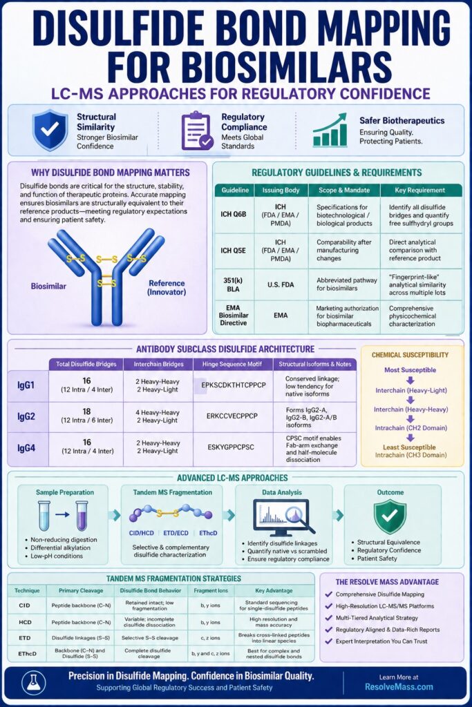
Regulatory compliance for disulfide characterization in biosimilars is primarily established through ICH Q6B and ICH Q5E guidelines, which require comprehensive determination of native disulfide bridges and free sulfhydryl residues. Regulatory agencies worldwide expect direct head-to-head analytical similarity evaluations between biosimilar candidates and innovator reference materials to verify batch-to-batch consistency and effective process control.
Discover regulatory frameworks and quality control standards in detail with our guide to ICH Q6B Guidelines for Biological Characterisation.
The International Council for Harmonisation (ICH) Q6B guideline specifically requires determination of the number and positions of free sulfhydryl groups and disulfide bridges whenever cysteine residues are present in a biotherapeutic product. Meeting these requirements involves evaluating both non-reduced and reduced peptide maps in combination with high-resolution mass spectrometry to verify that the covalent structure corresponds to the predicted theoretical architecture or the profile of the innovator reference product. Under ICH Q5E, which addresses comparability evaluations after manufacturing process modifications, biosimilar developers must demonstrate that changes involving cell lines, bioreactor conditions, or purification processes have not resulted in the formation of non-native disulfide linkages.
For insights into managing production variations during scale-up, read our deep dive on Cell Line Development for Biosimilars.
Analytical similarity is demonstrated through a tier-based strategy that involves comparing multiple reference product batches with the biosimilar candidate. Disulfide bond arrangements are considered Critical Quality Attributes (CQAs) because incorrect pairings can directly affect tertiary folding and biological activity. In addition, regulatory submissions require complementary free thiol assays, including Ellman’s reagent colorimetry or mass spectrometry-based differential labeling, to measure unpaired cysteine residues and confirm consistency throughout the manufacturing process.
Review analytical protocols for head-to-head biosimilar evaluation in our overview of Biosimilar Comparability Studies.
| Regulatory Guideline | Issuing Body | Scope & Mandate for Disulfide Mapping | Critical Analytical Requirements |
|---|---|---|---|
| ICH Q6B | ICH (FDA / EMA / PMDA) | Specifications for biotechnological/biological products | Identification of all covalent disulfide bridges and quantitative assessment of free sulfhydryl content. |
| ICH Q5E | ICH (FDA / EMA / PMDA) | Comparability of biotechnological products following manufacturing changes | Direct analytical comparison between biosimilar and reference product to verify structural identity following process modifications. |
| 351(k) BLA Framework | U.S. FDA | Abbreviated regulatory pathway for biosimilars | Head-to-head analytical similarity studies demonstrating “fingerprint-like” structural equivalence across multiple product lots. |
| EMA Biosimilar Directive | EMA | Marketing authorization for biosimilar biopharmaceuticals | Comprehensive physicochemical characterization using orthogonal mass spectrometry methods. |
Subclass-Specific Disulfide Architectures in Biosimilar Monoclonal Antibodies
Monoclonal antibody subclasses, including IgG1, IgG2, and IgG4, possess distinct interchain and intrachain cysteine linkage networks that influence hinge flexibility, quaternary structural rigidity, and the potential formation of structural isoforms. Accurate characterization of these subclass-specific disulfide architectures is therefore essential for confirming that a biosimilar candidate reproduces the precise covalent framework of its reference drug.
Monoclonal antibodies contain conserved intrachain disulfide bonds within each immunoglobulin domain, with two present in Light Chains and four in Heavy Chains, in addition to variable interchain linkages located in the hinge region and at the heavy-light interfaces.
Understand how charge variations impact antibody stability with our detailed report on Charge Variant Analysis in Biosimilars: Mass Spectrometry Approaches for Heterogeneity.
IgG1 Architectural Characteristics
An IgG1 molecule contains 16 total disulfide bonds, comprising 12 intrachain domain disulfides and 4 interchain disulfides. The interchain connections include two heavy-heavy chain hinge disulfides formed at conserved CPPC motifs and two heavy-light chain disulfides. In human IgG1, the C-terminal cysteine of the light chain, Cys214, forms an interchain disulfide bond with the fifth cysteine of the heavy chain, Cys220, located within the CH1 domain.
IgG2 Architectural Characteristics
An IgG2 molecule contains 18 total disulfide bonds, consisting of 12 intrachain disulfides and 6 interchain disulfides. The heavy-light interchain linkage connects the C-terminal cysteine of the light chain with the third cysteine of the heavy chain, Cys131. The IgG2 hinge region contains four inter-heavy chain disulfide bridges within an ERKCCVECPPCP sequence. This highly concentrated cysteine arrangement gives rise to structural isoforms, including IgG2-A, IgG2-B, and IgG2-A/B intermediates, which possess different interchain connectivity patterns and can consequently affect hydrodynamic radius and receptor-binding profiles.
IgG4 Architectural Characteristics
An IgG4 molecule contains 16 total disulfide bonds arranged in a pattern comparable to IgG1, but it contains a flexible CPSC hinge motif rather than the CPPC motif. This structural difference facilitates intrachain disulfide bond formation between heavy chain cysteines, resulting in non-covalently associated half-molecules and dynamic Fab-arm exchange in vivo.
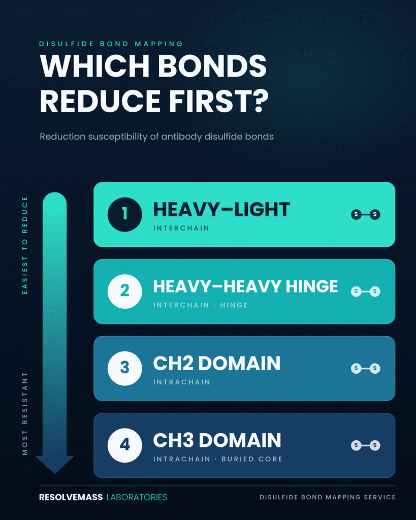
Chemical Susceptibility Hierarchy
Chemical reduction studies demonstrate a distinct hierarchy in the susceptibility of antibody disulfide bonds. Interchain disulfide bonds are more readily reduced than intrachain bonds because they generally exhibit greater solvent accessibility. Among the interchain linkages, heavy-light bonds undergo reduction more rapidly than heavy-heavy hinge bridges. Within the intrachain domains, CH2 domain disulfides are the most labile, whereas CH3 domain disulfides are deeply embedded within the hydrophobic core and therefore exhibit the greatest resistance to reduction.
| Antibody Subclass | Total Disulfide Bridges | Interchain Bridges | Hinge Sequence Motif | Structural Isoforms & Analytical Considerations |
|---|---|---|---|---|
| IgG1 | 16 (12 Intra / 4 Inter) | 2 Heavy-Heavy, 2 Heavy-Light | EPKSCDKTHTCPPCP | Conserved linkage pattern with a low tendency toward native structural isoform formation. |
| IgG2 | 18 (12 Intra / 6 Inter) | 4 Heavy-Heavy, 2 Heavy-Light | ERKCCVECPPCP | Exists as IgG2-A, IgG2-B, and IgG2-A/B structural isoforms that require high-resolution LC separation. |
| IgG4 | 16 (12 Intra / 4 Inter) | 2 Heavy-Heavy, 2 Heavy-Light | ESKYGPPCPSC | The CPSC motif promotes reversible inter-heavy chain dissociation and Fab-arm exchange. |
Advanced LC-MS Approaches in Disulfide Bond Mapping Service for Biosimilars
Advanced mass spectrometry workflows used in a Disulfide Bond Mapping Service for Biosimilars integrate non-reducing enzymatic digestion, differential alkylation approaches, and specialized tandem MS fragmentation techniques to distinguish native disulfide pairings from non-native variants. High-resolution LC-MS platforms allow accurate localization of intrachain and interchain cysteine linkages while also enabling the quantification of low-abundance scrambled species.
Non-Reducing Digestion versus Differential Alkylation
In direct non-reducing disulfide mapping, the intact biotherapeutic is enzymatically digested under non-reducing conditions, producing cross-linked dipeptides that preserve their covalent disulfide bonds. Comparing non-reduced and reduced chromatographic profiles allows disulfide-linked peptides to be identified through changes in their mass and retention behavior. However, complex biopharmaceutical molecules containing dense cysteine clusters may produce large, multiply linked peptides that are difficult to efficiently digest using a single enzyme.
To address these structural challenges, differential alkylation strategies use a two-step chemical labeling approach:
- Native free sulfhydryl groups are first blocked using an alkylating reagent, such as iodoacetamide (IAM), which attaches a carbamidomethyl tag (+57.021 Da) to unpaired cysteine residues.
- The remaining disulfide bonds are then completely reduced using dithiothreitol (DTT) or tris(2-carboxyethyl)phosphine (TCEP) and labeled with a second alkylating reagent, such as N-ethylmaleimide (NEM, +125.047 Da).
Mass spectrometry analysis distinguishes native disulfide bonds from free sulfhydryl groups by evaluating the mass delta differences associated with the two distinct labeling tags.
Mitigating In Vitro Disulfide Scrambling
Base-catalyzed thiol-disulfide exchange, commonly referred to as disulfide scrambling, is a major technical artifact that can arise during sample preparation. Under alkaline conditions, particularly at pH values above 7.5, unprotonated thiolate anions (R-S−) can attack existing disulfide bonds and generate artificial non-native pairings. These newly formed linkages may obscure the actual biological comparability between a biosimilar and its reference product. To minimize this artifact, sample preparation should be conducted under slightly acidic to near-neutral conditions, typically within the pH range of 6.0–6.5. Low-pH digestion procedures using acid-stable endoproteinases, including pepsin, endoproteinase Lys-C, or modified low-pH trypsin, reduce the formation of nucleophilic thiolate species while maintaining the native disulfide architecture.
Learn how stress testing reveals degradation pathways and structural vulnerabilities in our guide on Forced Degradation of Biosimilars.
Tandem MS Fragmentation Mechanisms
The selection of an appropriate mass spectrometry fragmentation strategy is essential for accurately characterizing complex and interconnected disulfide networks:
Collision-Induced Dissociation (CID) & Higher-Energy Collisional Dissociation (HCD): These slow-heating vibrational activation techniques preferentially break weaker peptide backbone amide bonds (b/y ions) while generally preserving disulfide linkages. CID/HCD spectra obtained from multi-linked dipeptides may contain highly dense and overlapping ion patterns, making the precise localization of disulfide linkages difficult.
Electron Transfer Dissociation (ETD) & Electron Capture Dissociation (ECD): These radical-driven electron-based fragmentation mechanisms specifically target disulfide linkages (S-S). They can selectively cleave the covalent sulfur-sulfur bond while largely preserving the peptide backbone. ETD can therefore separate cross-linked dipeptides into individual linear components, simplifying sequence assignment and structural interpretation.
Electron-Transfer/Higher-Energy Collision Dissociation (EThcD): This hybrid fragmentation approach combines ETD-mediated electron transfer with supplemental HCD collision energy. EThcD can cleave both disulfide linkages and peptide backbones within the same analysis, producing comprehensive c/z and b/y fragment ion series. This dual fragmentation capability provides extensive sequence coverage and enables confident characterization of complex and nested disulfide bonds.
| Fragmentation Technique | Primary Bond Cleavage | Disulfide Bond Behavior | Generated Fragment Ions | Key Advantage for Biosimilar Characterization |
|---|---|---|---|---|
| CID | Peptide backbone amide bonds (C-N) | Retained intact; low fragmentation efficiency | b-type and y-type ions | Standard sequencing approach for single-disulfide dipeptides. |
| HCD | Peptide backbone amide bonds (C-N) | Variable cleavage; incomplete disulfide dissociation | b-type and y-type ions | Provides high-resolution mass accuracy for relatively straightforward dipeptides. |
| ETD | Covalent disulfide linkages (S-S) | Selective and preferential S-S bond cleavage | c-type and z-type ions | Breaks cross-linked peptides into linear species to facilitate identification. |
| EThcD | Simultaneous backbone (C-N) and disulfide (S-S) cleavage | Complete gas-phase disulfide dissociation | Dual b/y and c/z fragment ion series | Optimal fragmentation strategy for resolving nested or highly intertwined disulfide bonds. |
Integrated Multi-Tiered Proteomic Platforms and Bioinformatics Curation
An integrated multi-tiered proteomic platform combines bottom-up peptide mapping, middle-down subunit characterization, and top-down intact mass spectrometry to achieve comprehensive structural verification of biopharmaceutical products. The integration of automated database-searching tools with expert manual data curation supports confident and unambiguous characterization of complex disulfide variants, including low-abundance species.
Explore broader bioanalytical strategies for therapeutic protein identification in our comprehensive resource on Proteomics Approach for Biosimilars.
Multi-Level Analytical Architecture
Bottom-Up Peptide Mapping: This approach enables high-resolution characterization of individual disulfide-linked peptide pairs and supports residue-level localization of both native and non-native cysteine pairings.
Middle-Down Analysis: This strategy uses domain-specific proteases, including IdeS, which cleaves below the hinge region, or GingisKHAN, to generate F(ab’)2, Fab, and Fc/2 fragments approximately 25–50 kDa in size. Middle-down LC-MS enables the characterization of hinge-region heterogeneity, subunit dissociation, and domain-specific disulfide scrambling while minimizing the need for extensive chemical manipulation.
Top-Down Mass Spectrometry: This technique examines intact, unreduced proteoforms using native MS or high-resolution Orbitrap mass analyzers. Top-down analysis verifies overall molecular stoichiometry and total disulfide integrity, providing an orthogonal confirmation of structural findings generated through lower-level analytical workflows.
Discover non-denaturing workflows for intact complex analysis in our article on Native Mass Spectrometry for Biosimilars.
Automated Data Processing and Spectrum-Level Curation
The interpretation of non-reducing LC-MS/MS data requires specialized bioinformatics pipelines, including PEAKS, UNIFI, and pLink-SS, that can compare experimental tandem mass spectra with databases containing combinatorial cross-linked peptide candidates. These search algorithms calculate theoretical mass-to-charge ratios (m/z) for paired peptide fragments and rank potential disulfide linkages according to cross-correlation scores and other matching criteria.
Because complex enzymatic digests can produce false-positive structural assignments, expert manual curation remains an essential component of the analytical workflow. Analysts examine isotopic envelopes, assess diagnostic fragment ions, including disulfide-cleaved peptide sub-ions, and verify retention-time alignment across orthogonal digestion strategies. This level of analytical scrutiny enables the quantification of low-abundance scrambled variants, including species present at levels as low as 0.1% relative abundance, thereby supporting regulatory compliance and process validation activities.
Examine methods for detecting high-molecular-weight species and non-native complexes in our guide to Aggregation Analysis in Biosimilars.
Conclusion
Implementing a robust Disulfide Bond Mapping Service for Biosimilars is essential for demonstrating higher-order structural comparability, meeting regulatory expectations, and supporting the safety and quality of therapeutic products. Through the application of advanced LC-MS/MS technologies, carefully controlled sample preparation procedures, and multi-tiered proteomic strategies, analytical programs generate the detailed structural evidence required to verify biosimilar quality.
Read more about comprehensive analytical workflows across diverse biological drug classes in Post-Translational Modifications (PTMs) in Biosimilars.
Establishing that a biosimilar candidate reproduces the disulfide connectivity, structural stability, and free thiol profile of its innovator reference product addresses key requirements outlined in ICH Q6B and ICH Q5E. High-resolution mass spectrometry platforms provide the sensitivity and analytical precision necessary to identify subtle structural differences and strengthen global biosimilar regulatory dossiers.
Learn how rigorous analytical strategies apply to specialized biotherapeutics through our case study on Insulin Biosimilar Characterization.
For specialized analytical support and comparability testing, sponsors can connect directly with technical experts through the ResolveMass Contact Page.
Frequently Asked Questions (FAQs)
Strict pH control is necessary because alkaline conditions can promote artificial thiol-disulfide exchange during sample preparation. At pH values above 7.5, thiolate anions (R-S−) become more prevalent and can attack existing disulfide bonds, producing rearranged cysteine pairings. Maintaining the sample within a slightly acidic to neutral range, typically pH 6.0–6.5, limits thiolate formation. This helps preserve the native disulfide architecture and prevents preparation-related structural artifacts.
Electron-Transfer/Higher-Energy Collision Dissociation (EThcD) combines two complementary activation mechanisms within a single fragmentation workflow. It can cleave covalent disulfide linkages (S-S) while also generating fragments from peptide backbone bonds (C-N). In contrast, CID and HCD may provide limited or inconsistent disulfide bond cleavage, particularly for complex cross-linked peptides. The combined c/z and b/y fragment ion series generated by EThcD supports confident sequence coverage and site-specific assignment of complex cysteine linkages.
Differential alkylation distinguishes free and disulfide-bound cysteine residues through sequential chemical labeling with two different mass tags. Initially, free sulfhydryl groups are blocked with a reagent such as iodoacetamide, which produces a characteristic mass addition of +57.021 Da. The remaining disulfide bonds are subsequently reduced and the newly exposed thiols are labeled with a second reagent, such as N-ethylmaleimide, producing a +125.047 Da mass shift. High-resolution mass spectrometry then differentiates the two cysteine populations based on their distinct labeling signatures.
IgG2 monoclonal antibodies contain 18 disulfide bonds, including four heavy-heavy chain hinge disulfides located within the ERKCCVECPPCP sequence. This highly concentrated cysteine arrangement can generate multiple structural isoforms, including IgG2-A, IgG2-B, and IgG2-A/B forms, with differences in interchain connectivity. These closely related species may be difficult to separate and characterize using conventional analytical workflows. High-resolution liquid chromatography combined with tandem mass spectrometry is therefore required for reliable isoform resolution and disulfide connectivity assessment.
ICH Q6B requires the characterization of free sulfhydryl groups and disulfide bridges in biotechnological and biological products containing cysteine residues. Analytical studies should establish the number, location, and relative or quantitative levels of these structural features. Free thiol measurements help confirm the extent of disulfide bond formation and identify unpaired cysteine residues that may contribute to structural heterogeneity. These data also support manufacturing process consistency and the overall assessment of product quality.
Middle-down mass spectrometry analyzes antibody subunits generated using domain-specific proteases, such as IdeS, which can produce F(ab’)2, Fab, and Fc/2 fragments of approximately 25–50 kDa. By examining these defined subunits, analysts can focus on hinge-region heterogeneity, subunit dissociation, and domain-specific disulfide alterations. The approach requires less extensive chemical manipulation than some conventional workflows and provides structural information at an intermediate level of detail. Consequently, middle-down analysis complements bottom-up peptide mapping and top-down intact mass spectrometry.
Disulfide bonds are fundamental to the correct tertiary and quaternary folding of many therapeutic proteins. Changes in cysteine pairing can influence antigen-binding properties, Fc-receptor-mediated effector functions, thermodynamic stability, and the tendency of the protein to aggregate. Structural alterations and aggregation may also increase the potential for unwanted patient immunogenicity. For these reasons, disulfide connectivity is treated as a Critical Quality Attribute (CQA that requires detailed analytical characterization and comparison with the reference product.
High-resolution Orbitrap mass spectrometers coupled with Ultra-High Performance Liquid Chromatography (UHPLC) provide the mass accuracy, chromatographic resolution, and sensitivity needed to detect low-level disulfide variants. Targeted tandem mass spectrometry approaches, including PRM and EThcD, can focus analytical detection on specific non-native disulfide-linked peptides. These workflows improve the ability to distinguish minor scrambled species from the dominant native forms. With optimized sample preparation and data analysis, variants present at approximately 0.1% relative abundance can be detected and quantified during process development.
The totality-of-evidence approach requires biosimilar developers to establish extensive structural, physicochemical, and functional similarity with the innovator reference product. Disulfide bond mapping contributes direct molecular-level evidence regarding cysteine connectivity and the preservation of the covalent structural architecture. When combined with other orthogonal analytical and functional studies, these results strengthen the overall comparability package. Such comprehensive characterization supports regulatory evaluations by agencies including the FDA, EMA, and Health Canada.
Reference:
- Lakbub, J. C., Shipman, J. T., & Desaire, H. (2018). Recent mass spectrometry-based techniques and considerations for disulfide bond characterization in proteins. Analytical and Bioanalytical Chemistry, 410(10), 2467–2484. https://doi.org/10.1007/s00216-017-0772-1
- Wu, S.-L., Jiang, H., Lu, Q., Dai, S., Hancock, W. S., & Karger, B. L. (2009). Mass spectrometric determination of disulfide linkages in recombinant therapeutic proteins using online LC-MS with electron-transfer dissociation. Analytical Chemistry, 81(1), 112–122. https://doi.org/10.1021/ac801560k
- Lakbub, J. C., Shipman, J. T., & Desaire, H. (2018). Recent mass spectrometry-based techniques and considerations for disulfide bond characterization in proteins. Analytical and Bioanalytical Chemistry, 410(10), 2467–2484. https://doi.org/10.1007/s00216-017-0772-1
- Fornelli, L., Ayoub, D., Aizikov, K., Liu, X., Damoc, E., Pevzner, P. A., Makarov, A., Beck, A., & Tsybin, Y. O. (2017). Top-down analysis of immunoglobulin G isotypes 1 and 2 with electron transfer dissociation on a high-field Orbitrap mass spectrometer. Journal of Proteomics, 159, 67–76. https://doi.org/10.1016/j.jprot.2017.02.013
Get In Touch With Us
About The Author


