

Introduction
Glycosylation Profiling for Biosimilar Characterization establishes the structural, physicochemical, and functional comparability of complex oligosaccharide structures attached to therapeutic biopharmaceuticals with those present in an innovator reference product. In regulatory submissions, Glycosylation Profiling for Biosimilar Characterization provides essential analytical evidence demonstrating that minor post-translational modifications do not adversely affect drug safety, systemic exposure, or biological efficacy. Therapeutic monoclonal antibodies (mAbs), antibody-drug conjugates (ADCs), and Fc-fusion proteins contain N-linked glycans attached to conserved sites, particularly Asparagine 297 (Asn297) within the CH2 domain of the immunoglobulin Fc region. Because recombinant expression platforms—including Chinese Hamster Ovary (CHO), murine myeloma (NS0, Sp2/0), and human embryonic kidney (HEK293) host cells—use different enzymatic pathways, even modest changes in cell culture conditions or downstream purification processes can modify carbohydrate micro-heterogeneity. Establishing analytical similarity therefore requires demonstrating that the glycoform distribution of the biosimilar candidate remains within the historical quality target product profile (QTPP) established from multiple innovator drug substance lots.
Explore our critical quality attributes in biosimilars to understand how key molecular attributes are identified and evaluated during biosimilar characterization.
Share via:
Quick Summary:
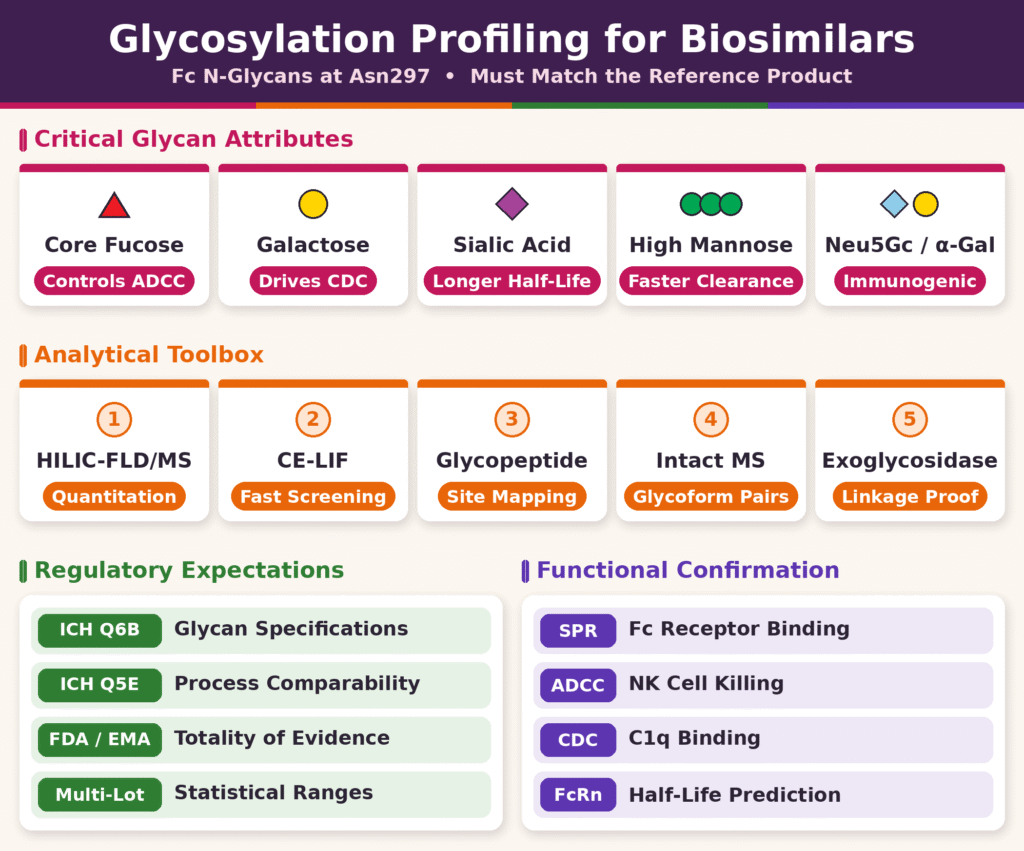
- Purpose: Glycosylation profiling shows that a biosimilar’s sugar structures, especially the N-glycans at Asn297 in the Fc region, match those of the reference product. Small differences in host cell line (CHO, NS0, HEK293) or process conditions can shift these glycans.
- Critical glycan attributes: Core fucose controls ADCC, since its absence can increase ADCC up to 50-fold. Galactose drives CDC through C1q binding. Sialic acid extends half-life. High-mannose species speed up clearance.
- Immunogenicity risk: Non-human epitopes such as Neu5Gc and α-Gal, typical of murine cell lines, can trigger immune or hypersensitivity reactions. They must be absent or tightly controlled.
- Released glycan methods: HILIC-UPLC-FLD/MS is the main quantitative tool and resolves isomers like G1F[6] and G1F[3]. CE-LIF gives ultra-fast, high-resolution separation, which makes it ideal for clone selection and batch release.
- Structural confirmation methods: Glycopeptide LC-MS/MS maps glycans to specific sites, separating Fc from Fab glycosylation. Intact and subunit MS confirm the overall glycoform pairings. Exoglycosidase arrays prove linkages and stereochemistry that mass alone cannot distinguish.
- Regulatory expectations: ICH Q6B requires characterizing glycans and their batch consistency, and ICH Q5E sets the comparability framework. The FDA and EMA take a “totality-of-the-evidence” approach based on multiple reference lots, applying tighter statistical limits to attributes linked to the drug’s mechanism of action.
- Functional correlation: Structural similarity alone is not enough. Glycan data must be linked to bioassays (SPR, ADCC, CDC/C1q, and FcRn binding) to show that any differences do not affect safety or efficacy.
Critical Quality Attributes in Glycosylation Profiling for Biosimilar Characterization
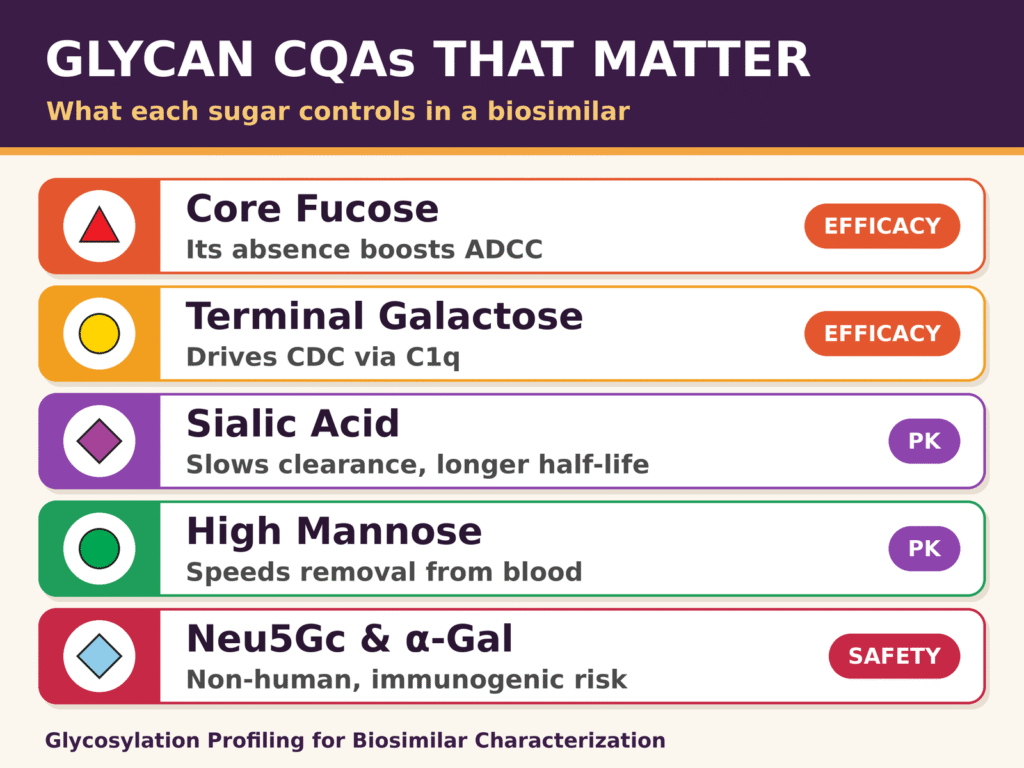
Critical Quality Attributes (CQAs) in Glycosylation Profiling for Biosimilar Characterization include specific carbohydrate structures such as core fucose, terminal galactose, sialic acids, high-mannose species, and non-human epitopes. These attributes can influence immune effector functions, circulating half-life, and immunogenic safety. Comprehensive characterization programs must therefore identify, quantify, and continuously monitor these glycosylation attributes throughout development to satisfy comparative analytical requirements.
- Core Fucosylation: Influences antibody-dependent cellular cytotoxicity (ADCC) by affecting Fc gamma receptor IIIa (FcγRIIIa) binding affinity.
- Terminal Galactosylation: Affects complement-dependent cytotoxicity (CDC) through its influence on complement C1q binding and tertiary structural stability.
- Sialylation: Influences circulating clearance rates and contributes to the modulation of anti-inflammatory mechanisms.
- High-Mannose Structures: Promote systemic elimination through endothelial mannose receptors while increasing FcγRIIIa affinity.
- Non-Human Epitopes: Create substantial immunogenicity concerns because of pre-existing human neutralizing antibodies.
Learn more about biosimilar comparability studies and how orthogonal analytical testing supports the assessment of critical quality attributes.
Core Fucosylation and FcγRIIIa Effector Function
Core afucosylation directly increases the affinity of monoclonal antibodies for the activating FcγRIIIa receptor present on natural killer (NK) cells, potentially producing up to a 50-fold increase in ADCC activity. Removal of the α-1,6-linked core fucose from the basal N-acetylglucosamine (GlcNAc) residue at Asn297 reduces steric hindrance at the Fc/FcγRIIIa interface. Even relatively small changes in the proportion of afucosylated glycoforms, including G0, G1, and G2, between a proposed biosimilar and its reference product may influence ADCC potency. Consequently, comparability exercises require carefully defined statistical tolerance intervals to evaluate these differences.
Terminal Galactosylation and Complement Activation
Terminal β-1,4-galactosyl residues facilitate antibody hexamerization following cell surface antigen binding, thereby supporting complement component C1q engagement and subsequent CDC activity. Fully galactosylated species, including G2F and G1F, exhibit greater C1q binding affinity than non-galactosylated forms such as G0F. Terminal galactosylation also contributes to the higher-order structural stability of the Fc domain and supplies the chemical substrate required for the attachment of terminal sialic acid residues.
Sialylation and Pharmacokinetic Clearance
Terminal sialic acid residues, including N-acetylneuraminic acid (Neu5Ac), protect underlying galactose units from recognition by hepatic asialoglycoprotein receptors. This protection can reduce rapid hepatic clearance and prolong circulating pharmacokinetic half-life. For Fc-fusion proteins, recombinant erythropoietin, and heavy-chain-modified biologics, maintaining appropriate sialylation levels is important for achieving the intended clinical efficacy. Additionally, α-2,6-linked sialic acid structures interact with specific immune lectin receptors and may contribute to anti-inflammatory activity.
High-Mannose Species and Accelerated Serum Elimination
High-mannose glycan variants, ranging from Man5 through Man9, can interact with endocytic mannose receptors expressed on macrophages and sinusoidal endothelial cells, resulting in accelerated systemic clearance. Although high-mannose species can exhibit increased FcγRIIIa binding because of reduced steric bulk around the Fc core, their potential effect on area-under-the-curve (AUC) exposure makes them important critical quality attributes. These species must therefore be appropriately controlled during bioreactor optimization and manufacturing process development.
Explore our aggregation analysis solutions for biosimilars to strengthen broader evaluation of molecular variants and product quality attributes.
Immunogenic Non-Human Epitopes: Neu5Gc and Alpha-Gal
Non-human glycan epitopes, particularly N-glycolylneuraminic acid (Neu5Gc) and galactose-α-1,3-galactose (α-Gal), can stimulate strong anti-glycan immune responses or acute hypersensitivity reactions in humans. Murine expression systems, including NS0 and Sp2/0, contain functional α-1,3-galactosyltransferase and CMP-Neu5Gc hydroxylase enzymes, which can result in the incorporation of these foreign residues into recombinant products. Biosimilar candidates manufactured using CHO or other mammalian expression systems must demonstrate the absence of, or strict control over, Neu5Gc and α-Gal to levels below applicable regulatory risk thresholds.
Discover how impurity profiling of biosimilars can support the identification and control of potentially significant product-related impurities and variants.
| Glycan Quality Attribute | Biological Mechanism of Action | Receptor or Target Binding | Clinical and Regulatory Implication |
|---|---|---|---|
| Afucosylation (G0, G1, G2) | Removes steric hindrance at the Fc interface. | FcγRIIIa on Natural Killer cells. | Critical CQA for ADCC-dependent biologics; requires precise matching. |
| Galactosylation (G1F, G2F vs G0F) | Enhances Fc hexamerization and complement activation. | Complement factor C1q. | Determines CDC potency and Fc conformational stability. |
| Sialylation (Neu5Ac content) | Blocks asialoglycoprotein receptor binding. | Asialoglycoprotein receptor / DC-SIGN. | Helps prevent premature liver clearance; critical for Fc-fusion proteins. |
| High Mannose (Man5 to Man9) | Promotes receptor-driven endocytosis. | Reticuloendothelial mannose receptors. | Shortens circulating pharmacokinetic half-life. |
| Non-Human Epitopes (α-Gal, Neu5Gc) | Triggers neutralizing anti-glycan antibodies. | Human circulating anti-α-Gal/Neu5Gc antibodies. | High risk of immunogenicity, serum sickness, or anaphylaxis. |
Advanced Analytical Methodologies for Glycosylation Profiling for Biosimilar Characterization
Advanced methodologies for Glycosylation Profiling for Biosimilar Characterization employ orthogonal analytical platforms, including liquid chromatography, capillary electrophoresis, mass spectrometry, and enzymatic digestion, to achieve comprehensive structural identification and quantitative evaluation of N- and O-linked oligosaccharides. Demonstrating meaningful analytical similarity requires multidimensional characterization of released glycan pools, site-specific glycopeptides, and intact biopharmaceutical isoforms.
Explore biosimilar characterization services for comprehensive analytical strategies covering structural, physicochemical, and molecular characterization requirements.
Released Glycan Analysis via HILIC-UPLC-FLD/MS
Released glycan analysis using Hydrophilic Interaction Liquid Chromatography coupled with Fluorescence Detection and High-Resolution Mass Spectrometry (HILIC-UPLC-FLD/MS) is widely used as a primary quantitative approach for biopharmaceutical glycoform profiling.
The analytical workflow starts with the enzymatic release of N-linked glycans from the protein backbone using Peptide-N-Glycanase F (PNGase F). The released oligosaccharides are subsequently derivatized at their reducing termini with fluorophores such as 2-aminobenzamide (2-AB), 2-aminobenzoic acid (2-AA), procainamide, or rapid NHS-carbamate tags such as RapiFluor-MS. Modern carbamate reagents streamline sample preparation through a short, simplified protocol and produce stable urea linkages that improve both fluorescence response and electrospray ionization efficiency.
HILIC separations employ polar stationary phases, commonly sub-2 μm amide-functionalized silica particles, together with high-organic-to-aqueous mobile phase gradients. Hydrophilic partitioning separates glycans according to size, charge, monomer composition, and glycosidic linkage, enabling baseline resolution of important positional isomers such as G1F[6] and G1F[3]. Online hyphenation of HILIC with electrospray electrospray ionization quadrupole time-of-flight (ESI-QTOF) or Orbitrap mass spectrometry allows simultaneous fluorometric quantification and mass-based structural confirmation.
Learn more about proteomics approaches for biosimilars and their role in generating detailed molecular characterization data.
High-Throughput Capillary Electrophoresis with Laser-Induced Fluorescence (CE-LIF)
Capillary Electrophoresis with Laser-Induced Fluorescence (CE-LIF) provides highly rapid and high-resolution separation of charged glycan variants according to differences in their charge-to-hydrodynamic volume ratio.
Before CE-LIF analysis, released glycans undergo reductive amination with negatively charged fluorophores, most commonly 8-aminopyrene-1,3,6-trisulfonic acid (APTS). Under high electric field strengths applied across narrow fused-silica capillaries, APTS-labeled glycans migrate rapidly and produce theoretical plate counts that exceed those typically achieved through conventional liquid chromatography. CE-LIF platforms can resolve sialylated species and neutral structural isomers in run times of less than two minutes per sample. This makes the technique particularly suitable for clone selection and high-throughput batch release testing.
Site-Specific Glycopeptide Mapping and Intact Mass Spectrometry
Site-specific glycopeptide mapping using reversed-phase ultra-performance liquid chromatography coupled to tandem mass spectrometry (RP-UPLC-MS/MS) determines the precise glycosylation site occupancy and micro-heterogeneity associated with individual amino acid residues.
Proteolytic digestion with trypsin, chymotrypsin, or Endoproteinase Lys-C breaks the biopharmaceutical into discrete peptide fragments while retaining their attached glycan moieties. RP-UPLC-MS/MS analysis maintains the regional sequence context and enables analysts to distinguish Fc-region glycosylation from Fab-region glycosylation in complex monoclonal antibodies and multi-specific constructs.
Explore proteomics-based biosimilar characterization to understand how peptide-level analytical strategies can support site-specific molecular assessment.
To assess global macro-heterogeneity without enzymatic digestion, intact and subunit mass spectrometry can be applied. Intact monoclonal antibodies, as well as fragments generated through immunoglobulin-degrading enzyme of Streptococcus pyogenes (IdeS) digestion followed by disulfide reduction, can be separated using short reversed-phase or size-exclusion columns. Deconvolution of high-resolution ESI mass spectra produces intact molecular weight distributions corresponding to primary heavy-chain glycoform pairings, including G0F/G0F, G0F/G1F, and G1F/G2F, thereby confirming overall glycan assembly.
Explore native mass spectrometry for biosimilars to learn how intact molecular-level analysis can complement glycosylation and higher-order structural characterization.
Linkage Analysis via Exoglycosidase Digestion Arrays
Exoglycosidase digestion arrays employ linkage-specific exoglycosidase enzymes to sequentially remove terminal monosaccharides. This approach confirms stereochemical configurations and assists in resolving structural isomers that may otherwise co-elute.
Because structural isomers can possess identical chemical compositions and molecular masses, mass spectrometry alone cannot distinguish specific glycosidic linkages, including α-2,3 versus α-2,6 sialic acids or α-1,3 versus β-1,4 galactosylation. Released glycan samples can therefore be treated with specialized enzyme panels containing α2-3,6,8,9 Neuraminidase, β1-4 Galactosidase, α1-3,4,6 Galactosidase, β-N-Acetylglucosaminidase, and α1-2,4,6 Fucosidase. These enzymes selectively remove their corresponding terminal sugars. Subsequent changes in HILIC retention times or Glucose Unit (GU) values provide evidence for the precise monomer sequence, linkage chemistry, and presence of non-human epitopes.
| Analytical Method | Sample Preparation | Primary Analytical Output | Key Technical Strengths | Method Limitations |
|---|---|---|---|---|
| HILIC-UPLC-FLD/MS | PNGase F release; reducing-end fluorophore labeling. | Relative quantitation of neutral/charged glycans; isomeric profiling. | Exceptional chromatographic resolution; direct online ESI-MS coupling. | Requires enzymatic release and fluorophore tagging. |
| CE-LIF | PNGase F release; APTS fluorophore derivatization. | Ultra-fast separation of sialylated and neutral glycan variants. | High peak capacity; run times under 2 minutes per sample. | Direct MS hyphenation is complex; relies on standard GU alignment. |
| Glycopeptide LC-MS/MS | Proteolytic digest (trypsin/Lys-C) preserving glycan-peptide linkage. | Site-specific micro-heterogeneity and domain occupancy. | Differentiates Fc vs Fab glycosylation sites. | Complex fragmentation spectra; ion suppression effects. |
| Subunit / Intact LC-MS | Intact protein, reduced chains, or IdeS-cleaved fragments. | Global mass distribution and main glycoform pairings. | Minimal sample preparation; verifies native chain assembly. | Lower resolution for low-abundance isomeric glycoforms. |
| Exoglycosidase Arrays | Released glycans incubated with specific hydrolase panels. | Verification of monosaccharide order, linkages, and non-human tags. | Definitive proof of glycosidic linkages and stereochemistry. | Multi-step incubations increase total analytical turnaround time. |
Regulatory Expectations and Analytical Similarity Guidelines
Regulatory expectations for biosimilar glycosylation profiling involve comprehensive multi-lot comparability assessments, adherence to international guidelines, and robust functional correlation to demonstrate that structural differences do not result in clinically meaningful changes in drug safety or efficacy. The US Food and Drug Administration (FDA), European Medicines Agency (EMA), and International Council for Harmonisation (ICH) apply stringent analytical similarity requirements under the 351(k) pathway and corresponding international regulatory frameworks.
Harmonized ICH Q6B and ICH Q5E Compliance
The ICH Q6B guideline (Specifications: Test Procedures and Acceptance Criteria for Biotechnological/Biological Products) requires sponsors to characterize carbohydrate structures, site occupancy, glycan profiles, and batch-to-batch consistency as important components of drug substance specifications. In biosimilar development, the principles outlined in ICH Q6B require both major and low-abundance glycoforms to be appropriately identified and quantified.
Review the ICH Q6B guidelines for biological characterization to better understand analytical expectations for biological product characterization.
The ICH Q5E guideline (Comparability of Biotechnological/Biological Products Subject to Changes in Their Manufacturing Process) establishes a regulatory framework for evaluating product quality following changes to bioprocessing or cell lines. Biosimilar developers apply the comparative testing principles described in ICH Q5E to determine whether the selected host cell line and cell culture parameters generate glycan distributions that remain consistent with the historical range observed for the innovator product.
Explore cell line development for biosimilars to learn how expression-system considerations can influence product quality and analytical comparability.
FDA and EMA Frameworks for Comparative Analytical Assessment
The FDA and EMA apply a stepwise “totality-of-the-evidence” approach centered on structural and functional analytical similarity assessments. Sponsors are expected to obtain multiple independent lots of the reference medicinal product manufactured over several years to establish the reference product’s historical quality target product profile (QTPP) and characterize its natural lot-to-lot variability.
Comparative glycan datasets generated through orthogonal analytical techniques, such as HILIC-UPLC-MS, CE-LIF, and glycopeptide mapping, are subjected to appropriate statistical evaluations. Quality attributes that are directly associated with primary mechanisms of action, such as core afucosylation for an ADCC-inducing therapeutic mAb, require narrow statistical equivalence limits. Attributes considered to have lower risk, including minor sialylated forms in non-systemic antibodies, are assessed using quality ranges established from the reference product dataset.
Explore the comparability exercise in biosimilar development to understand how analytical evidence can be integrated into a structured biosimilar development strategy.
Integrating Structural Glycan Data with Functional Bioassays
Establishing structural similarity alone is not sufficient for regulatory approval. Biopharmaceutical sponsors must establish a direct relationship between physicochemical glycan characteristics and in vitro biological bioassay results.
- Surface Plasmon Resonance (SPR): Determines real-time binding kinetics to Fc receptors (FcγRIIIa, FcγRIIa, FcγRI, and FcRn) and complement C1q.
- Cell-Based ADCC Bioassays: Measure functional immune cell activation and target cell lysis associated with changes in afucosylated glycoforms.
- CDC and C1q Binding Assays: Evaluate complement activation in relation to variations in terminal galactosylation.
- FcRn Binding and Pharmacokinetic Models: Evaluate neonatal Fc receptor binding affinity to support projections of systemic clearance.
Discuss your analytical requirements with ResolveMass biosimilar characterization experts to develop an orthogonal testing strategy aligned with your characterization and comparability objectives.
Conclusion
Comprehensive Glycosylation Profiling for Biosimilar Characterization is a fundamental requirement for demonstrating high analytical similarity and supporting regulatory approval of proposed biosimilar therapeutics. Integrating high-resolution separation technologies such as HILIC-UPLC-FLD/MS and CE-LIF with tandem mass spectrometry, exoglycosidase arrays, and functional bioassays enables extensive structural mapping and quantitative comparability assessment. Evaluating the direct biological consequences of critical glycosylation attributes—including core afucosylation, galactosylation, sialylation, and high-mannose variants—helps ensure that biosimilar development programs comply with regulatory expectations established by ICH Q6B, the FDA, and the EMA.
Explore ResolveMass analytical solutions for biosimilars and strengthen your biosimilar characterization strategy with comprehensive analytical testing.
To explore expert analytical testing, mass spectrometry characterization, and regulatory consultation services, visit the ResolveMass Contact Page.
Frequently Asked Questions
Core afucosylation refers to the absence of an α-1,6-linked fucose on the basal GlcNAc residue at Asn297. This structural characteristic can substantially increase monoclonal antibody interaction with the FcγRIIIa receptor on natural killer cells and enhance antibody-dependent cellular cytotoxicity (ADCC). Consequently, it is considered an important critical quality attribute that requires close comparison with the reference product.
Non-human glycan structures such as N-glycolylneuraminic acid (Neu5Gc) and galactose-α-1,3-galactose (α-Gal) can be recognized by pre-existing human anti-glycan antibodies. Their presence may contribute to immunogenic responses, hypersensitivity, or severe allergic reactions. Therefore, biosimilar characterization requires stringent assessment and control of these potentially immunogenic epitopes.
Released glycan profiling involves enzymatically removing glycans from the protein before analysis, allowing assessment of the overall glycan population and its relative distribution. Glycopeptide mapping instead retains the glycans on peptide fragments generated through proteolytic digestion. This approach provides site-specific information about glycosylation occupancy and micro-heterogeneity at individual amino acid residues.
The ICH Q6B guideline provides principles for characterizing the structure and carbohydrate composition of biotechnological and biological products. It addresses important characteristics such as glycan profiles, carbohydrate structures, and site occupancy. These assessments support the establishment of appropriate specifications, analytical acceptance criteria, and controls for glycoform consistency.
Exoglycosidase arrays use enzymes with specific linkage selectivity to remove terminal monosaccharides in a controlled sequence. This approach helps distinguish structural isomers that may have identical molecular masses but different glycosidic linkages. The resulting changes in glycan profiles provide evidence for terminal sugar identity, linkage configuration, and overall structural arrangement.
High-mannose glycan variants, including Man5 through Man9, can interact with endocytic mannose receptors on cells involved in the reticuloendothelial system. This receptor-mediated uptake can increase the rate at which a biopharmaceutical is removed from systemic circulation. Consequently, elevated high-mannose levels may reduce pharmacokinetic exposure and circulating half-life, making them an important process-control attribute.
Common traditional fluorescent labels include 2-aminobenzamide (2-AB) and 2-aminobenzoic acid (2-AA), which generally require longer reductive amination procedures. More recent workflows use reactive labeling reagents such as RapiFluor-MS, InstantPC, and procainamide for faster glycan derivatization. These labels provide strong fluorescence responses while also improving the signal obtained during mass spectrometry analysis.
Regulators assess glycan variability by comparing multiple lots of the reference product and biosimilar using appropriately qualified analytical methods. The resulting datasets are evaluated statistically to define reference-product quality ranges and natural lot-to-lot variability. Individual glycan attributes are then assessed to determine whether the biosimilar remains within an acceptable range of analytical similarity.
Biosimilar glycan comparability typically requires complementary analytical methods that examine different levels of glycan structure and function. HILIC-UPLC-FLD/MS evaluates released glycan profiles, CE-LIF supports rapid glycan separation, and RP-UPLC-MS/MS provides site-specific glycopeptide information. Intact/subunit mass spectrometry and functional assays such as ADCC, CDC, and SPR binding provide additional evidence of overall analytical and functional similarity.
Reference:
- U.S. Food and Drug Administration. (2015, April). Quality considerations in demonstrating biosimilarity of a therapeutic protein product to a reference product: Guidance for industry. https://www.fda.gov/media/135612/download
- Alhazmi, H. A., & Albratty, M. (2023). Analytical techniques for the characterization and quantification of monoclonal antibodies. Pharmaceuticals, 16(2), 291. https://doi.org/10.3390/ph16020291
- Wang, H., Wu, L., Wang, C., Xu, J., Yin, H., Guo, H., Zheng, L., Shao, H., & Chen, G. (2021). Biosimilar or not: Physicochemical and biological characterization of MabThera and its two biosimilar candidates. ACS Pharmacology & Translational Science, 4(2), 790–801. https://doi.org/10.1021/acsptsci.0c00225
- Kwon, J.-C., Kwon, O. H., Jeong, R. U., Kim, N., Song, S., Choi, I., Lee, J., & Horiuchi, T. (2021). Physicochemical and biological similarity assessment of LBAL, a biosimilar to adalimumab reference product (Humira®). Animal Cells and Systems, 25(3), 182–194. https://doi.org/10.1080/19768354.2021.1943709
- Galili, U. (2005). The α-gal epitope and the anti-Gal antibody in xenotransplantation and in cancer immunotherapy. Immunology and Cell Biology, 83(6), 674–686. https://doi.org/10.1111/j.1440-1711.2005.01366.x
- U.S. Food and Drug Administration. (2025, September). Development of therapeutic protein biosimilars: Comparative analytical assessment and other quality-related considerations: Guidance for industry. https://www.fda.gov/regulatory-information/search-fda-guidance-documents/development-therapeutic-protein-biosimilars-comparative-analytical-assessment-and-other-quality
Get In Touch With Us
About The Author


